Microscopes magnifications resolutions and calculations Light microscope uses


")


")


- Slides: 24
Microscopes magnifications, resolutions and calculations
Light microscope- uses light and lenses to enlarge tiny objects
Pollen grains – light microscope
Electron microscope - electromagnetic lenses are used to direct electrons onto the tiny specimen
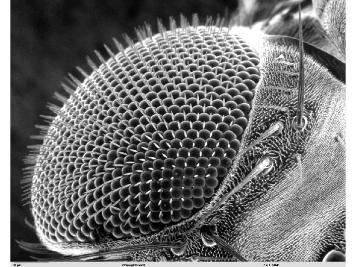
Pollen grains – scanning electron microscope 3 D
ABPI • http: //www. abpischools. org. uk/topic/cellbiol ogy/2
The difference between magnification and resolution. • Magnification is the degree to which the size of an image is larger than the object itself. • Resolution is the degree to which it is possible to distinguish between two objects that are very close together. The electron microscope has higher magnification and resolution
Radiolarian = single celled protozoan found in oceans part of zooplankton
Moth - eye = 800 micrometres
hydrothermal worm taken at 525 times magnification
How do we find the overall magnification of a light microscope? Eyepiece Objective lens Eyepiece Objective Magnification Overall Magnification X 10 X 4 40 X 10 100 X 10 X 40 400 X 100 1000
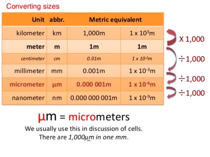
mm 1000 Micrometre 1000 nm
Magnification= size of image/ actual size of specimen (must be in same units)
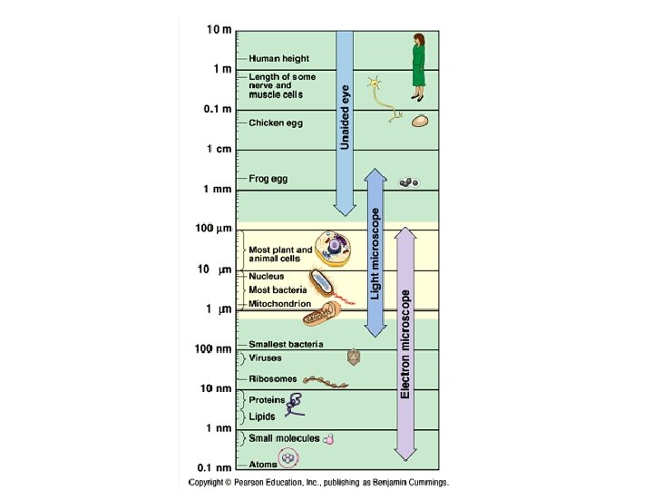
What can we see with a microscope?
Typical sizes • • • Examples of typical sizes: 10 -100µm: cells of eukaryotes 1 -10µm: organelles 1µm: bacteria (size vary) 100 nm: viruses (size vary) 10 nm: thickness of cell membranes 2 nm: DNA molecule in human choromosome 1 nm: molecules 0. 1 nm: atoms
Nanometre Micrometre Millimetre 5 1 0. 005 0. 000005 0. 001 0. 000001 1000 1 0. 001 1 000 1000 1 3000 3 0. 007 0. 000007 500 0. 5 7 500 000
Magnification= size of image/ actual size of specimen (must be in same units)
The diagram below is a drawing of an organelle from a ciliated cell as seen with an electron microscope. Calculate the actual length of the organelle as shown by the line AB in the diagram. Express your answer to the nearest micrometer (μm). Show your working. Answer =. . . mm A= 5. 1 I 102 mm = M 20000 = 102000 mm 20000
Magnification of Cheek cells Size of this skin cell = Size in mm = Actual size of cheek cells = 60μm First – change to mm To change μm to mm we need to ÷ by 1000 So 60μm = 0. 06 mm So Magnification = size of image / actual size = Magnification = /0. 06 mm =
The diagram below is a drawing of an alveolus together with an associated blood capillary. M= = I A 21 mm 1. 5 mm The line AB in the diagram represents an actual distance of 1. 5 µm. 21000 mm Calculate the magnification of the drawing. Show your working. = 1. 5 mm Answer = ×. . . 14000
ABPI • http: //www. abpischools. org. uk/topic/cellbiol ogy/2