Compound Light Microscope A Light Microscope Parts Body

Compound Light Microscope
 Arm Objective Lens")
A. Light Microscope Parts Body Tube Revolving Nosepiece Eyepiece (Ocular Lens) Arm Objective Lens Stage Clips Diaphragm Light Source Stage Coarse Adjustment Knob Fine Adjustment Knob Base

B. Functions Microscope Part Function Body Tube Transmits image from specimen to eyepiece Nosepiece Holds objective lenses Objective Lenses for magnifying – Scanning, Low, & High Power Stage Clips Hold slide in place on stage Diaphragm Adjusts amount of light passing through specimen Light Source Provides light
 Has lens to magnify Attaches")
Microscope Part Function Transmits image to eye; Eyepiece (Ocular) Has lens to magnify Attaches body tube to stage Arm & base Stage Coarse Adjustment Knob Fine Adjustment Knob Base Where slide/specimen is placed Focuses image under scanning power only Focuses image under low and high power Supports microscope

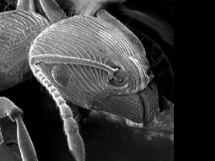
Electron Microscope

Cilia in rabbit trachea

Table Salt
")
Human Hair (split end)

Human Hair Follicle

Blood Cells

Pollen

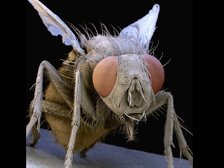
Fly’s Eye

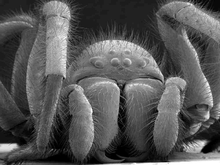
Spinnerets Of a spider

C. Image under the Microscope While viewing an image through a microscope, it appears larger, as well as upside down and backwards. Ex. The letter P as it would appear under a microscope. P P

D. Total Magnification To find the total magnification of the microscope while using a specific objective lens, you multiply the power of the eyepiece by the power of the objective lens. Ex. Eyepiece = 10 X; Objective Lens = 5 X Total Magnification: 10 x 5 = 50 X 5 X

E. Microscopic Measurement 1. In order to determine how large a specimen, such as a cell, is while viewing it under a microscope, you must first determine the diameter of the field of view. a. To do this, you must measure the diameter in mm.

Ex. The diameter of this field of view is 1. 5 mm.

Field of View is approximately 3. 5 mm Field of View is approximately 4. 5 mm
: 1 mm")
2. Now you must convert that measurement in mm to micrometers (µm): 1 mm = 1, 000 m

There’s an even smaller unit of measurement called a nanometer 1 m = 1, 000, 000 nm 1 µm = 1, 000 nm

To convert mm to µm, you must multiply your number by 1000. An easy way to do this is to scoop the decimal 3 places to the right. The diameter of the field of view is 1. 5 mm. 1. 5 0 0 = 1500 µm Field of View: 3. 5 mm x 1000 = 3500 m

3. Next you must estimate the number of cells that fit across the diameter. Many microscopes have a pointer that extends halfway across your field of view, which can help you in your estimation. Ex. How many cells could fit across the diameter ? 2

4. Finally, divide your field of view diameter by the number of cells that fit across in order to determine the length of one cell. Ex. Field of view diameter from part 2: 1500 µm Number of cells that fit across diameter: 2 Work: 1500/2 Length of one cell: 750 µm

Each cell is approximately 0. 5 mm = 500 m Approximately 4 cells can fit across the diameter Each cell is approximately 2 mm/4 = 0. 5 mm or 500 m

Ex. F. Low Power vs. High Power When switching from low power to high power, the magnification increases so the field of view decreases. Therefore, the cells appear larger and you see less of them in your field of view. However, their actual size is still the same. Low Power 100 X High Power 400 X

*Before switching to high power, make sure your specimen is centered and focused. (Image moves in opposite direction in which you move the slide. ) If you move the slide to the left……. F F The image of the letter appears to move……

Let’s see that all at once…… F F

If you move the slide away from you……. F F The image of the letter appears to move……

Let’s see that all at once…… F F

G. Making a Wet Mount 1. Place specimen on slide; Add a drop of water; Place edge of cover slip next to water on an angle and slowly lower cover slip to minimize water bubbles.

2. To add stain to a wet mount, place a drop of the stain next to the cover slip and draw it under the cover slip by placing a piece of paper towel against the other side of the cover slip. The paper towel will soak up the water, drawing the stain under the cover slip.
- Slides: 34