Lymphocyte Development and Antigen Receptor Gene Rearrangement Dr


 are created during maturation by somatic recombination")


 are attracted to the thymus from")


 in medulla")


 • B-1 cells 5 -10% of blood")
J")

- Slides: 31
Lymphocyte Development and Antigen Receptor Gene Rearrangement Dr. Eman Albataineh, Associate Prof. Immunology College of Medicine, Mu’tah university Immunology, 2 nd year students
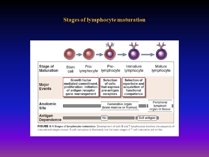
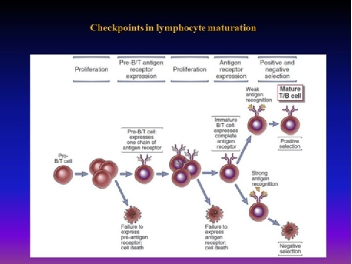
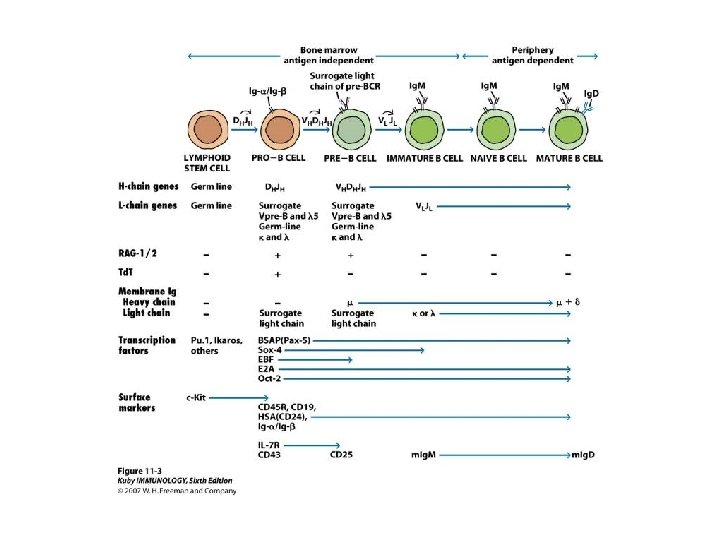
Steps of lymphocytes development • First; Pluripotent stem cells in bone marrow known as hematopoietic stem cells (HSCs), give rise to a common lymphoid progenitor (CLP) then give rise to pro-B cells, pro. T cells and NK cells. • Pro-T cells may commit to either the αβ or γδ T cell lineages. • Second; pro- B and pro-T cell proliferate in response to cytokines IL-7 • third; Functional antigen receptor genes start to be formed on cells (Per cells to immature cells) – For B cells in the bone marrow and in immature T cells in the thymus by a process of gene rearrangement. Any cell do not express the receptor not selected or become dead
• Pre- cells proliferate in response to signal transduction from formed receptor • Fourth; Selection events that preserve cells that have produced functional antigen receptor proteins and eliminate potentially dangerous cells that strongly recognize self antigens, cells that remain after selection called mature cells. • Fifth; Differentiation of mature B and T cells into functionally and phenotypically distinct subpopulations. B cells ( B-2 cells) develop into follicular (majority), marginal zone. in BM, and T cells develop into CD 4+ and CD 8+ αβ T lymphocytes in thymus. Then cells transported to peripheral LN. fetal liver stem cells give rise to B-1 cells
Functional receptor (TCR, BCR or antibody formation) are created during maturation by somatic recombination - The diversity is generated by random joining of different gene segments from a set of inherited (germline) DNA that are separated, but brought together (randomly) into rearranged, functional genes -somatic recombination happens in BM for B cells and in thymus for T cells -
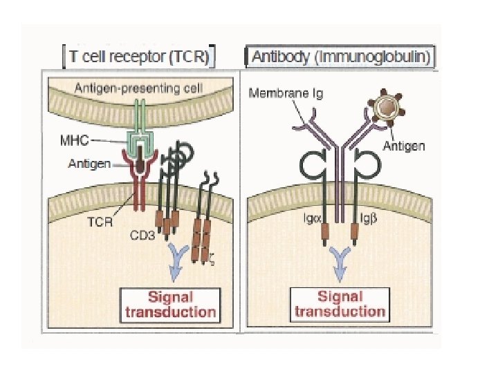
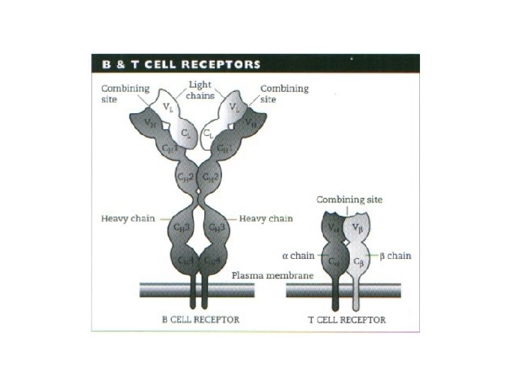
αß TCR • 90% of TCR is αß • TCR complex is the αß receptor plus the ζ chain and two CD 3 signaling proteins • Each chain constitute of one variable, one constant, hinge, transmembrane and cytoplasmic tail • covalently linked to each other by a disulfide bridge between extracellular cysteine residues • TCR that specifically recognizes peptide-MHC complexes • Hypervariable regions on both Vα and Vß are the same as those of antibody located on Ag-binding site and called CDR and they are 3 sites for each
BCR The B lymphocyte antigen receptor is a transmembrane antibody molecule (2 heavy and 2 light chains) associated with two signaling chains called Igα and Igβ - There is also hinge region, transmembrane part
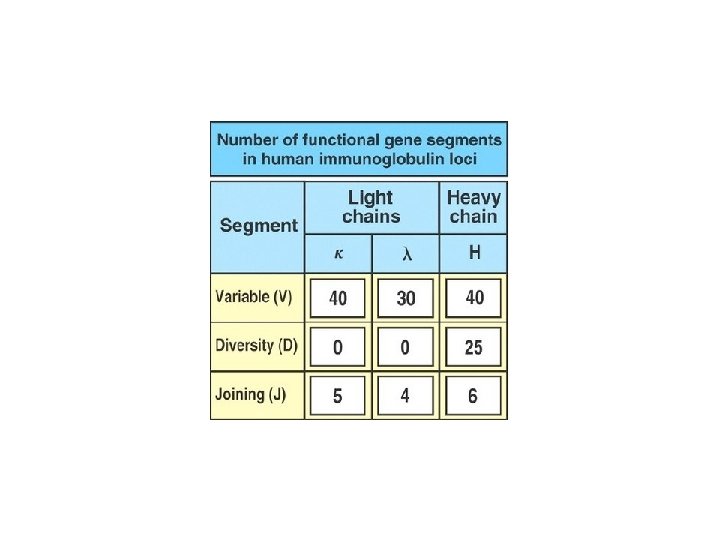
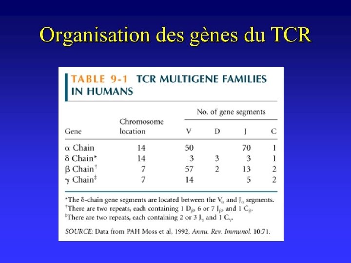
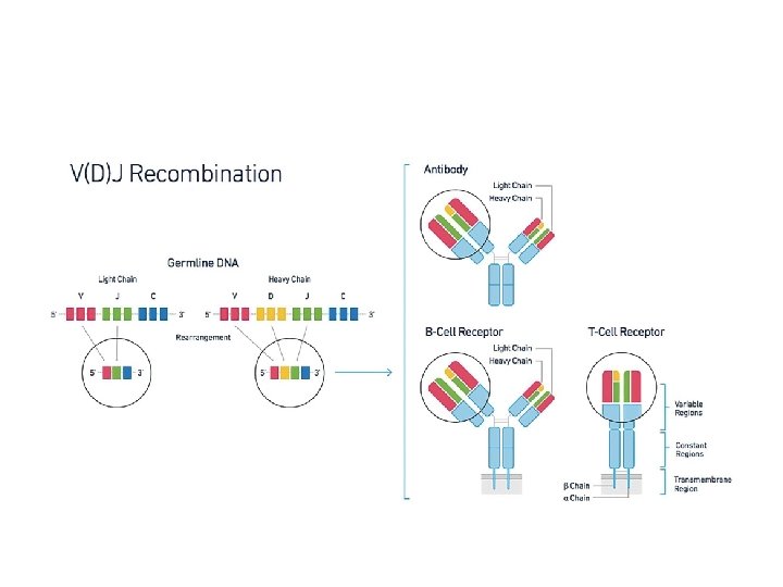
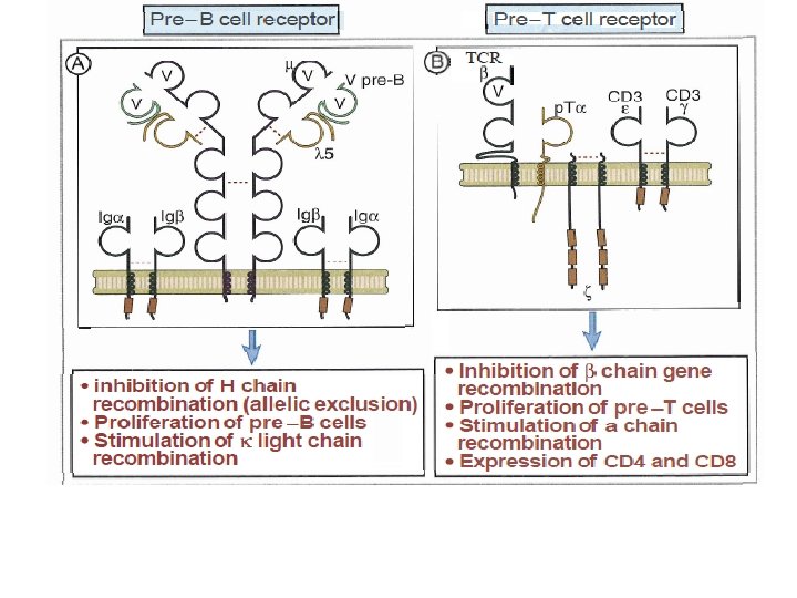
T & B Receptor formation or somatic recombination The T and B cell receptor gene segments are -V for variable, -D for diversity, - J for joining, - C for constant, for BCR two types (kappa and lambda) light chain and 5 for heavy chain (Cϒ for IGG, Cδ for IGD, Cε for IGE, Cμ for IGM and Cα for IGA). For TCR one for alpha and 2 for bet chain - V-D-J To make variable parts of BCR heavy chain and TCR Beta chain - V-J to make variable parts of BCR light chain and TCR alpha chain - Enzyme used to make these combinations is RAG enzymes. RAG-1 and RAG-2 are proteins at the ends of VDJ genes that separate, shuffle, and rejoin the VDJ genes - Joining the constant segment - (C μ for heavy chain of BCR then C lambda or kappa for BCR light chain - and C β then C alpha for TCR – Complete receptor formed on immature T & B cells
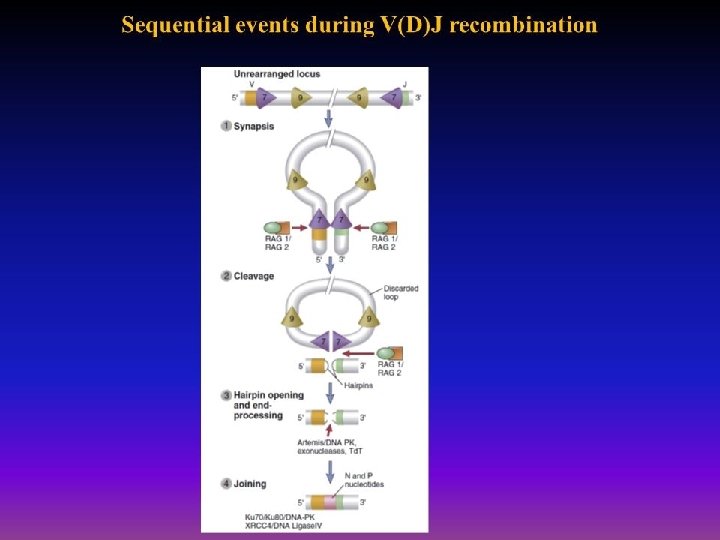
somatic recombination q DNA Recombination include – Synapse, making chromosomal loop – Cleavage by RAG enzyme – Hairpin opening and end-processing(addition or removal of bases) mediated by Artemis endonuclease, – Joining (Ligase) q Variability in binding sites Because of • Combinatorial diversity. rearrangement brings together multiple germline gene segments that may combine randomly, and different combinations produce different antigen receptors. • J gene segments in the TCRα and beta and immunoglobulin light and heavy –chain. This region encodes the CDR 3 loops in immunoglobulins and T-cell receptors that form the center of the antigen-binding site
T cell development • T cell precursors (prothymocytes) are attracted to the thymus from the BM by a chemotactic factor secreted by thymic epithelial cells. • The pro thymocytes are TCR - CD 3+CD 4 -CD 8 or "double-negative" cells (in subcapsular area). • Some Double-negative cells productively rearrange gamma and delta chain gene segments develop into gamma/delta T cels (γδ T cells 10%) The majority of double-negative cells will go on to rearrange alpha and beta chain gene segments 90%. • Tc. R beta and gamma genes are located on chromosome 7 and Tc. R alpha and delta genes are located on chromosome 14.
follow • in cortex; The TCR β chain protein is expressed on the cell surface first (by DNA recombination of VDJ beta segments with beta constant segments) in association with an invariant protein called pre-Tα and with CD 3 and ζ proteins to form the pre-T cell receptor (pre. TCR) complex • then alpha chain gene rearrangement is enhanced (VJ alpha with constant alpha) forming complete T cell receptor with CD 3 (Immature T cells). • At the same time both CD 4 and CD 8 are expressed and the cells called double positive immature T cells
Selection of immature T cells • Positive selection of double positive cells (CD 4+CD 8+) is the process that preserves T cells that recognize self MHC (with self peptides) with low avidity. • Negative selection of double positive is the process in which thymocytes whose TCRs bind strongly to self peptide antigens in association with self MHC molecules are deleted or convereted to Treg • Further check point for deletion self reactive T cells occurs In medulla, the epithelial cells express a nuclear protein called AIRE (autoimmune regulator) that induces the expression of a number of tissue-specific genes in the thymus. These genes are normally expressed only in specific peripheral organs. Their AIRE-dependent expression in the thymus makes many tissuespecific peptides available for presentation to developing T cells, facilitating the deletion (negative selection) of these cells
• Transforming into single positive (either CD 4 or CD 8) in medulla because one co-receptor is shut-off randomly, or as a result of Positive Selection of Thymocytes: Development of the Self MHC–Restricted T Cell Repertoire). Those that bind MHC 1 transformed into CD 8, and those bind MHC 2 transformed into CD 4
γδ T cells • • CD 4 -, CD 8 -, CD 3+ T cells, 5% in peripheral blood T cells Frequent in mucosal epithelium Can help in antibody class switch as alpha beta T cells Have a regulatory function, it sense tissue stress rather than antigen, and downregulate damaging immune response • Help in innate immune because --sense Ag directly without processing or MHC restriction. they help in viral infection also help in early life when alpha beta T cells and antigen processing is immature sense peptide and non-peptide Ag (mycobacterium)
B cells • Pro-B cells; the earliest stage in BM • Ag receptor expression is the first key to lymphocyte survival; • Early: a single chain receptor (Ig. H (the 2 IGM heavy chains with surrogate light chains and immunoglobulin alpha and beta) = pre-B cells; • Later: completed Ag receptors formed by formation of light chains kappa type, if fail use lambda light chain Light chain contain just V-J segments immature B cells = complete IGM BCR
Selection of immature B cells • Selection follows initial survival after immature lymphocytes express antigen receptors (Best understood for T cells, but also occurs for B cells) – Positive Selection (life, expansion, continued maturation) occurs if the Ig receptor binds self antigen with low avidity. Cells that not binding die
Negative selection – Negative selection in B cells not always occur as just receptor editing happen if the B cell receptor bind strongly to self. – Receptor editing is Changing the variable part on light chain; replacing VJ of light chain with new VJ Kappa or lambda. If editing in B cells fail; clonal deletion – only 5% of formed T cells and 10% of B cells selected. Most B cells migrate to peripheral LN where maturation happens ( mature B cell) by expressing IGD beside IGM
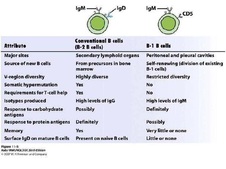
B 1 cells (CD 5+ B cells) • B-1 cells 5 -10% of blood B cells naturally found from fetal life, produce IGM, natural antibody present without immunization. has limited diversity give rapid antibody production against microbe. Act against carbohydrates, do not do isotype switch or do affinity maturation, no need to T cell help, and self renewing, present in the peritoneum and in mucosal sites. • Marginal zone B cells are a distinct population of B cells that mainly respond to polysaccharides. After activation, these cells differentiate into short-lived plasma cells that produce mainly Ig. M.
Allelic exclusion • After a B cell produces a functional immunoglobulin gene during V(D)J recombination, it cannot express any other variable region (a process known as allelic exclusion) thus each B cell can produce antibodies containing only one kind of variable chain • and it ensures that every B cell will express a single receptor, thus maintaining clonal specificity
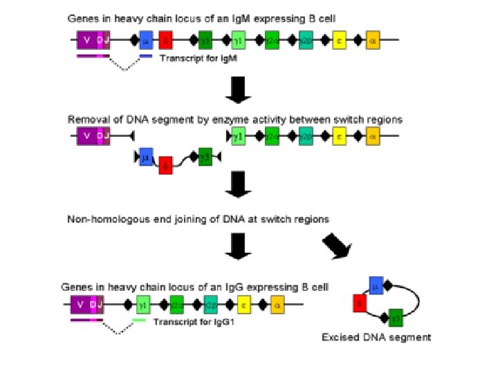
TCR and BCR In peripheral LN • The recombination process that creates diversity in BCR and TCR in peripheral LN is unique to that during the early stages of their development in primary lymphoid organs • T cell receptor revision (antigen receptor editing) is a process in the peripheral immune system which is used by mature T cells to alter their original antigenic specificity based on antigen. • In B cells in the lymph nodes, Activation-induced cytidine deaminase causes mutations that produce antibody diversity – Somatic hypermutation, in which the antibody genes are minimally mutated to generate antibody with higher affinity for a particular antigen than any of its close variants – Class switch recombination, in which B cells change their expression from Ig. M to Ig. G or other immune types