Overview of lymphocyte development Commitment Gene rearrangement Selection
ﻣﺘﻌﻬﺪ ﺷﺪﻥ Gene rearrangement ( )ﻧﻮﺗﺮکیﺒی Selection (")


 cells are")

 cells are a")

 cells are")


 Cells u u Follicular helper T (Tfh) cells are a")
 u u The primary function of regulatory T cells, also")
 Cells u u u Natural killer T (NKT) cells are")

 u u are a subset of T cells")
- Slides: 34
Overview of lymphocyte development Commitment ( )ﻣﺘﻌﻬﺪ ﺷﺪﻥ Gene rearrangement ( )ﻧﻮﺗﺮکیﺒی Selection ( )گﺰیﻨﺶ
B lymphocyte development stem cell u Pro B cells CD 19, CD 10, µ heavy chain rearrangement u Pre B cells Pre antigen receptor expression u immature B cells Ig. M BCR expression, independent of antigen and their encounter with it , receptor editing, cell death, functional unresponsiveness u Mature B cells u
u Two types of molecules produced by nonlymphoid thymic cells important for T cell maturation 1)Class-I and II MHC 2)Cytokines and chemokines
T cell subpopulation
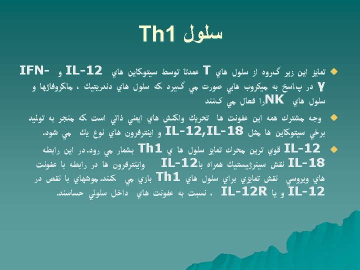
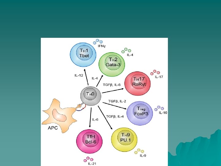
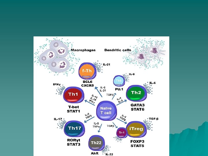
Th 1 cell u u u Th 1 cell Surface phenotype(αβTCR, CD 3, CD 4, IL-12 R, IFNγR, CXCR 3 Effector molecules secreted IFNγ, IL-2, LTα Function Promote protective immunity against intracellular pathogens. By secreting IFNγ they induce activation of macrophages and upregulation of i. NOS, leading to the killing of intracellular pathogens such as Leishmania major, Listeria monocytogenes and Mycobacterium spp. Their development is regulated by IL-12.
Th 1 Cells u u u T helper type 1 (Th 1) cells are a lineage of CD 4+ effector T cell that promotes cell-mediated immune responses and is required for host defense against intracellular viral and bacterial pathogens. Th 1 cells secrete IFN-gamma, IL-2, IL-10, and TNF-alpha/beta. These cytokines promote macrophage activation, nitric oxide production, and cytotoxic T lymphocyte proliferation, leading to the phagocytosis and destruction of microbial pathogens. In addition to the cytokines secreted by Th 1 cells, the expression of specific cell surface receptors, including IL-12 R beta 2, IL-27 R alpha, IFN-gamma R 2, CCR 5, and CXCR 3, can be used to distinguish Th 1 cells from other T cell subtypes Th 1 cell differentiation and expansion are driven by cytokines that signal through a subset of these receptors, including IL-27, IL-12, and IFN-gamma. Although Th 1 cells are critical for the clearance of intracellular pathogens, exaggerated Th 1 responses have been found to be associated with autoimmune diseases, including rheumatoid arthritis, multiple sclerosis, inflammatory bowel disease, and type 1 diabetes.
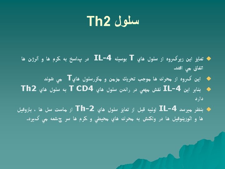
Th 2 Cells u u u Th 2 cell Surface phenotype ( αβ TCR, CD 3, CD 4, IL-4 R, IL-33 R, CCR 4) Effector molecules secreted IL-4, IL-5, IL-13, IL-9 , IL-10 and IL-17 E/IL-25 Function Promote humoral immune responses and host defence against extracellular parasites. However, they can also potentiate allergic responses and asthma. Th 2 differentiation occurs in the presence of IL-4 and either IL-2, IL-7, or Thymic Stromal Lymphopoietin (TSLP). IL-4 regulates clonal expansion of Th 2 cells, and along with IL-13, promotes B cell production of Ig. E and alternative macrophage activation. Other cytokines produced by Th 2 cells stimulate eosinophil activation and survival (IL-5), or promote mast cell activation (IL-9). In addition to the production of specific cytokines, Excessive Th 2 -type immune responses have been implicated in the development of chronic allergic inflammation and asthma.
Th 9 Cells u u T helper type 9 (Th 9) cells are a distinct subpopulation of CD 4+ effector T cell that preferentially secretes high levels of IL-9, CCL 17, CCL 22, and IL-10 (in mouse). Following activation, naive CD 4+ T cells differentiate into Th 9 cells in the presence of TGF-beta and IL 4. Under Th 9 -polarizing conditions, additional cytokines have also been shown to either enhance Th 9 differentiation (IL-1 beta, IL-6, IL-21, type I interferons), or promote IL-9 secretion (IL-25, IL-2). Although the signaling molecules and transcription factors involved in Th 9 differentiation are still being investigated, it is recognized that Th 9 cells are closely related to Th 2 cells Unlike Th 2 cells, however, Th 9 cells do not express IL-4, IL-5, or IL-13. Th 9 cells are important for host defense against parasitic helminth infections, but may also have detrimental effects including contributing to the development of chronic allergic inflammation, airway remodeling, and autoimmune disease.
Th 9 cell u u u Th 9 cell Surface phenotype (αβ TCR, CD 3, CD 4) Effector molecules secreted(IL-9, IL-10) Function Involved in host defence against extracellular parasites, primarily nematodes. Despite their production of anti-inflammatory IL-10, they promote allergic inflammation. Their role in other inflammatory diseases still remains unclear as this subset has only recently been characterized
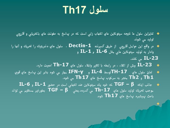
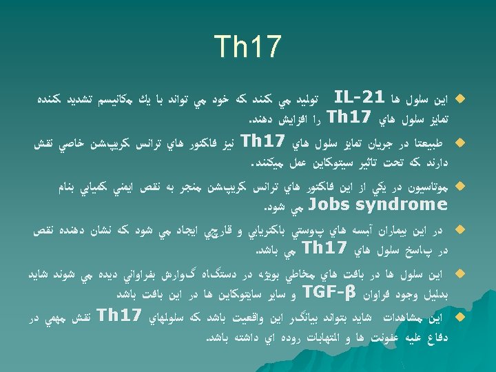
Th 17 Cells u u u T helper type 17 (Th 17) cells are involved in the immune response mounted against specific fungi and extracellular bacteria. IL-21 and IL-23 regulate the establishment and clonal expansion of Th 17 cells. Cytokines secreted by Th 17 cells stimulate chemokine secretion by resident cells, leading to the recruitment of neutrophils and macrophages to sites of inflammation. These cells, in turn, produce additional cytokines and proteases that further exacerbate the immune response. In contrast to mouse Th 17 differentiation, Th 17 polarization in humans requires IL-1 beta, IL 6, IL-21, and IL-23, but seems to be less dependent upon TGF-beta. One other notable difference is that human Th 17 cells secrete IL-26, an IL-10 family cytokine without a murine homologue. Cytokines produced by Th 17 cells can have both beneficial and pathogenic effects. While they play a central role in eliminating harmful microbes, persistent secretion of Th 17 cytokines promotes chronic inflammation and may be involved in the pathogenesis of inflammatory and autoimmune diseases, including rheumatoid arthritis, multiple sclerosis, and inflammatory bowel disorders
Th 17 cell u u u Th 17 cell Surface phenotype(αβTCR, CD 3, CD 4, IL-23 R, CCR 6, IL 1 R, CD 161 (human only) Effector molecules secreted IL-17 A, IL-17 F, IL-21, IL-22, CCL 20 Function Promote protective immunity against extracellular bacteria and fungi, mainly at mucosal surfaces. Also promote autoimmune and inflammatory diseases. Generated in the presence of TGFβ and IL-6 and/or IL-21 and are maintained by IL-23 and IL-1.
Th 22 u It is not antiinflammatory, nor is it necessarily proinflammatory. However, it is consistently described to enable epithelial innate immune responses, which can be detrimental or protective. u An example of a detrimental effect is epithelial hyperplasia in psoriasis (PS), which also can be induced by IL-22 in human artificial skin cultures. Furthermore, IL-22 synergizes with IL 17 in the induction of proinflammatory cytokines in human bronchial epithelial cells and colonic myofibroblasts. u Examples of protective effects of IL-22 were reported in Gramnegative bacterial pneumonia, in which IL-22 induces the secretion of antimicrobial substances in lung epithelial cells. In addition, IL-22, but not IL-17, protects from hepatitis-induced liver damage by preventing hepatocyte apoptosis. u In the skin, IL-22 induces antimicrobial peptides, promotes keratinocyte proliferation, and inhibits differentiation, which suggests a role in remodeling wound healing and in innate defense mechanisms. The role of IL-22 in common skin disorders such as atopic eczema (AE) and allergic contact dermatitis (ACD) is currently not known
Th 22 u u u u Th 22 cell Surface phenotype (αβTCR, CD 3, CD 4, CCR 10) Effector molecules secreted IL-22, IL-13, and TNF-alpha Function Identified in inflammatory skin diseases. Their role in host defence remains unclear as this subset has only recently been characterized. Their identity as an independent TH cell subset needs to be confirmed Similar to Th 17 cells, Th 22 cells express IL-22, CCR 4, and CCR 6, but in contrast, they also express CCR 10 and several fibroblast growth factors (FGFs). In addition, Th 22 cells do not express IL-17, CCL 20, IL-23 R, CD 161 (Th 17 markers), IL-4 (Th 2 marker), or IFN-gamma (Th 1 marker). Collectively, these characteristics distinguish Th 22 cells as a novel T helper cell lineage that is distinct from the Th 17, Th 2, and Th 1 subtypes. Activated naive CD 4+ T cells differentiate into Th 22 cells in the presence of IL-6 and TNFalpha. Expression of the CCR 4 and CCR 10 skin-homing receptors on Th 22 cells suggests that these cells are likely recruited to the skin, where they may contribute to host defense against microbial pathogens, and promote tissue repair or remodeling. Multiple studies indicate that Th 22 cells may also be involved in the pathogenesis of inflammatory skin disorders such as psoriasis, atopic eczema, and allergic contact dermatitis
T Follicular Helper (Tfh) Cells u u Follicular helper T (Tfh) cells are a distinct subset of CD 4+ helper T (Th) cells that regulate the development of antigen-specific B cell immunity. Tfh cell Surface phenotype αβ TCR, the development of antigen-specific B cell immunity. CD 3, CD 4, CXCR 5) Upon exposure to a foreign antigen, Tfh cells help B cells generate antibody-producing plasma cells and long-lived memory B cells. Tfh cells are identified by elevated expression levels of multiple surface proteins and Bcl-6, as well as enhanced IL-21 secretion. The high expression levels of these proteins correlate with Tfh cells' enhanced capacity to facilitate antibody production. In the T cell zone of secondary lymphoid tissue, antigen-presenting dendritic cells activate naïve CD 4+ T cells to produce IL-21. IL-6 and the autocrine action of IL-21 induce the activated CD 4+ T cells to express Bcl-6, the master transcription factor that controls Tfh cell differentiation. Bcl-6, a transcriptional repressor, suppresses the expression of factors that mediate the differentiation of Th 1, Th 2 and Th 17 cells, and clusters of mi. RNAs that negatively regulate molecules involved in Tfh functioning, such as the CXCR 5. Increased expression of CXCR 5 helps Tfh cells localize to B cell follicles, where they interact with germinal-centre B cells and secrete a variety of cytokines that stimulate B cells to generate antibody-producing plasma cells and memory B cells.
Regulatory T Cells (Tregs) u u The primary function of regulatory T cells, also known as suppressor T cells, is to maintain immune homeostasis. This involves suppression of successful immune responses and control of self versus non-self recognition. Failure of the latter results in autoimmune destruction of host cells and tissue. Like other T cells, regulatory T cells mature in the thymus where they are characterized by the variable expression of CD 8, CD 4, CD 25 and Fox. P 3. . taking advantage of the immunosuppressive potential of regulatory T cells is an important area for advancement in the fields of autoimmune disease and organ transplantation.
Natural Killer T (NKT) Cells u u u Natural killer T (NKT) cells are a specialized population of T cells that express a semiinvariant T cell receptor (TCR alpha beta) and surface antigens typically associated with natural killer cells. The TCR on NKT cells is unique in that it recognizes glycolipid antigens presented by the MHC I-like molecule CD 1. Most NKT cells, known as type I NKT cells, express an invariant TCR alpha chain and one of a small number of TCR beta chains. NKT cells can have either protective or deleterious effects due to their abilities to produce cytokines that promote either inflammation or immune tolerance. As a result, they contribute to antibacterial and antiviral immune responses, promote tumor-related immunosurveillance or immunosuppression, and inhibit or promote the development of autoimmune diseases. Like natural killer cells, NKT cells can also induce perforin-, Fas-, and TNF-related cytotoxicity, but this is generally not thought to be their primary function.
gamma delta T Cells u u Gamma delta T cells represent a small fraction (1 - 5 %) of the overall T cell population but are enriched (more than 50 % of the T cell population) in epithelial cell-rich compartments like skin, the digestive tract, and reproductive organ mucosa. Gamma delta T cells are a subset of T cells defined by the genetic composition of their T Cell Receptor (TCR). All T cells are derived from common progenitor thymocytes, and while the majority of T cells express TCR chain heterodimers encoded by the alpha and beta gene loci, gamma delta T cells express TCR chains encoded by the gamma and delta gene loci. Like helper T cells, gamma delta T cells secrete particular effector cytokines in a subtype-and context-specific manner, however, unlike alpha beta T cells, most delta gamma T cells lack CD 4 and CD 8 and share a number of markers associated with natural killer cells or antigenpresenting cells such as Fc gamma RIII/CD 16 and Toll-like receptors.
CD 8+ cytotoxic T lymphocytes (CTLs) u u are a subset of T cells that express an alpha beta T cell receptor (TCR) and are responsible for the direct killing of infected, damaged, and dysfunctional cells, including tumor cells. CD 8 expression and IFN-gamma production are the most commonly used markers for CTL identification. Additionally, Perforin and Granzyme B, which are required for CTL-mediated cell death, are commonly utilized as secondary markers It has been reported that CTLs also produce IL-10, an anti-inflammatory cytokine, at the peak of infection to prevent excessive tissue damage. Importantly, CTLs appear to be critical for cancer prevention as Perforin suppresses lymphoma in mice and mutations in Perforin are associated with lymphoma in humans.