Lecture 1 From Fertilization to Gastrulation Copyright 2019

- Slides: 30
Lecture 1: From Fertilization to Gastrulation Copyright © 2019 Wolters Kluwer • All Rights Reserved
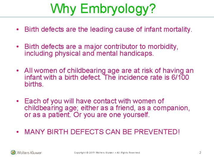
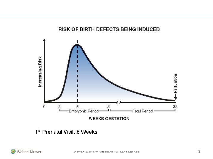
Why Embryology? • Birth defects are the leading cause of infant mortality. • Birth defects are a major contributor to morbidity, including physical and mental handicaps. • All women of childbearing age are at risk of having an infant with a birth defect. The incidence rate is 6/100 births. • Each of you will have contact with women of childbearing age; either as a friend, as a companion, or as a patient. Or you are one yourself. • MANY BIRTH DEFECTS CAN BE PREVENTED! Copyright © 2019 Wolters Kluwer • All Rights Reserved 2
1 st Prenatal Visit: 8 Weeks Copyright © 2019 Wolters Kluwer • All Rights Reserved 3
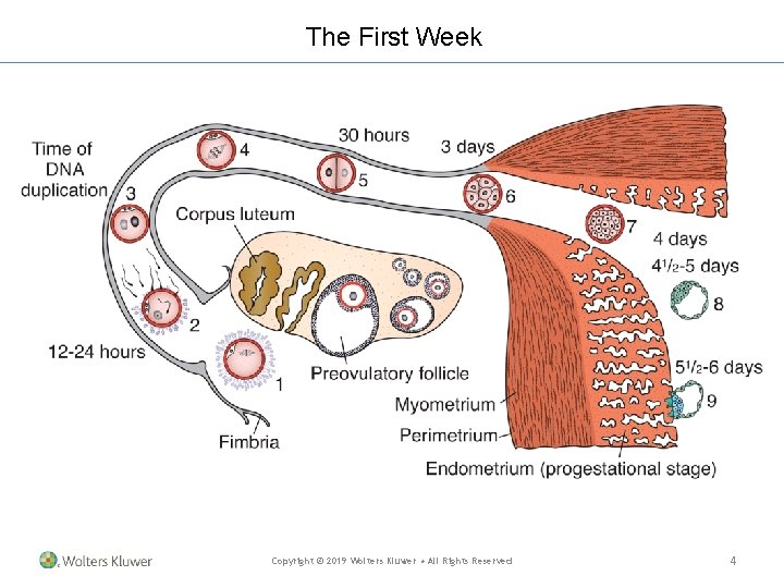
The First Week Copyright © 2019 Wolters Kluwer • All Rights Reserved 4
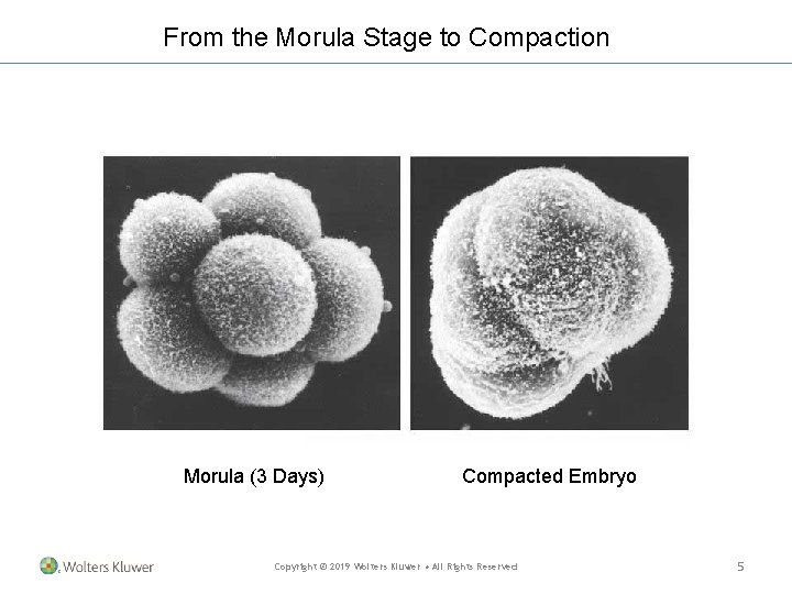
From the Morula Stage to Compaction Morula (3 Days) Compacted Embryo Copyright © 2019 Wolters Kluwer • All Rights Reserved 5
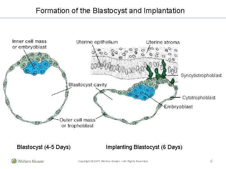
Formation of the Blastocyst and Implantation Syncytiotrophoblast Cytotrophoblast Blastocyst (4 -5 Days) Implanting Blastocyst (6 Days) Copyright © 2019 Wolters Kluwer • All Rights Reserved 6
Hydatidiform Moles • Formed from trophoblast • Paternal genome (genomic imprinting) • Secrete high levels of h. CG (syncytiotrophoblast) • May become invasive (choriocarcinomas) Copyright © 2019 Wolters Kluwer • All Rights Reserved 7
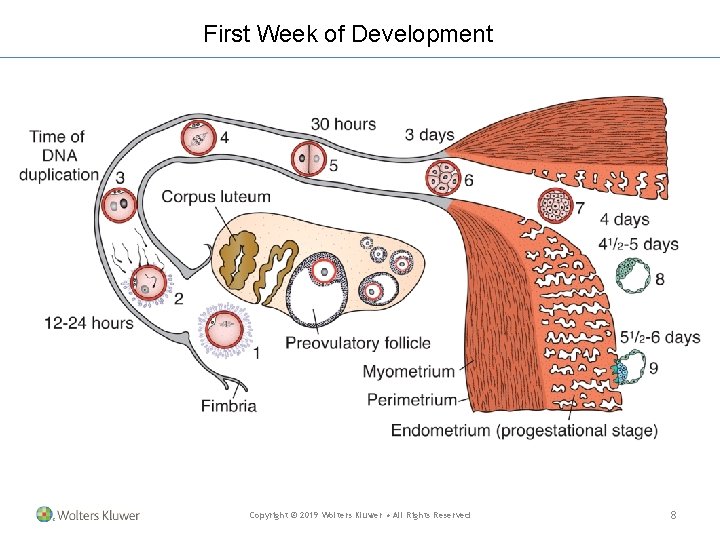
First Week of Development Copyright © 2019 Wolters Kluwer • All Rights Reserved 8
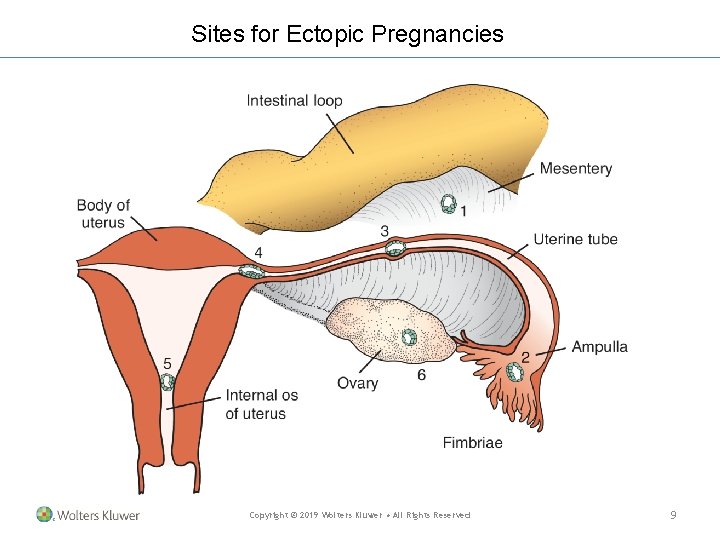
Sites for Ectopic Pregnancies Copyright © 2019 Wolters Kluwer • All Rights Reserved 9
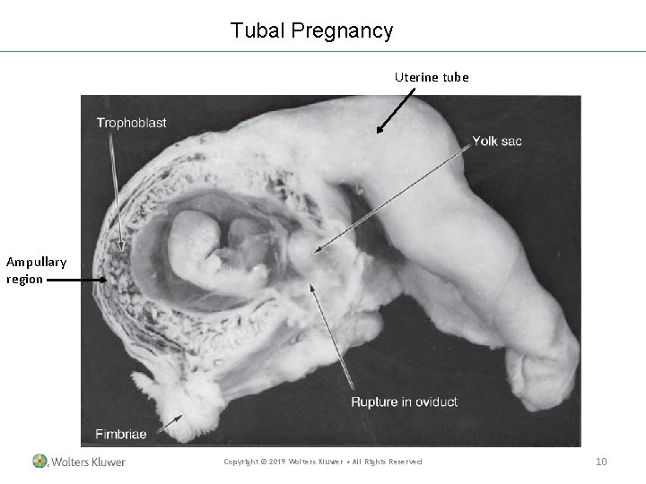
Tubal Pregnancy Uterine tube Ampullary region Copyright © 2019 Wolters Kluwer • All Rights Reserved 10
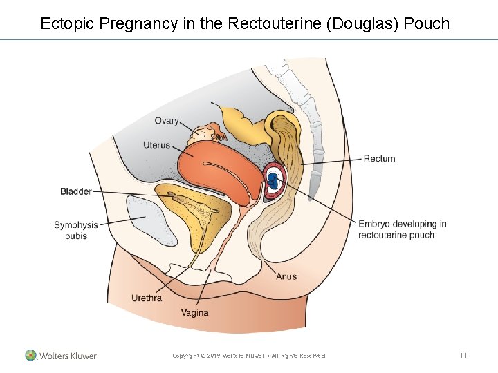
Ectopic Pregnancy in the Rectouterine (Douglas) Pouch Copyright © 2019 Wolters Kluwer • All Rights Reserved 11
Assisted Reproductive Technologies (ART) Fertility treatments in which both the eggs and sperm are handled in the laboratory (i. e. , in vitro fertilization and related procedures). Eggs and sperm are placed in a culture medium; fertilized eggs are placed in the uterus. Copyright © 2019 Wolters Kluwer • All Rights Reserved 12
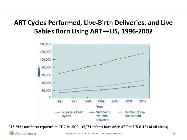
ART Cycles Performed, Live-Birth Deliveries, and Live Babies Born Using ART—US, 1996 -2002 115, 392 procedures reported to CDC in 2002; 45, 751 infants born after ART in US (1. 1% of all births) Copyright © 2019 Wolters Kluwer • All Rights Reserved 13
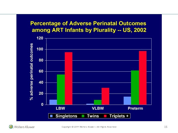
Adverse Outcomes Potentially Associated with ART • • Embryo effects Spontaneous abortions Multiple births Adverse perinatal outcomes: low birth weight, preterm delivery (even among singleton births) Birth defects Developmental disabilities Childhood malignancies Longer term outcomes? • Effects on puberty/future fertility • Effects on chronic disease risk Copyright © 2019 Wolters Kluwer • All Rights Reserved 14
Copyright © 2019 Wolters Kluwer • All Rights Reserved 15
Copyright © 2019 Wolters Kluwer • All Rights Reserved 16
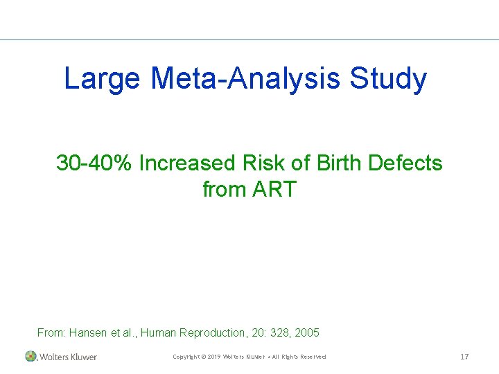
Large Meta-Analysis Study 30 -40% Increased Risk of Birth Defects from ART From: Hansen et al. , Human Reproduction, 20: 328, 2005 Copyright © 2019 Wolters Kluwer • All Rights Reserved 17
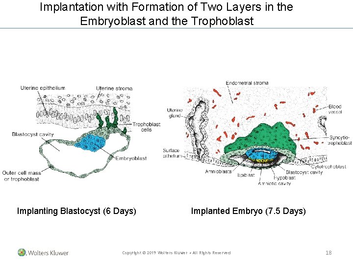
Implantation with Formation of Two Layers in the Embryoblast and the Trophoblast Implanting Blastocyst (6 Days) Implanted Embryo (7. 5 Days) Copyright © 2019 Wolters Kluwer • All Rights Reserved 18
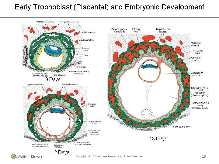
Early Trophoblast (Placental) and Embryonic Development 9 Days 13 Days 12 Days Copyright © 2019 Wolters Kluwer • All Rights Reserved 19
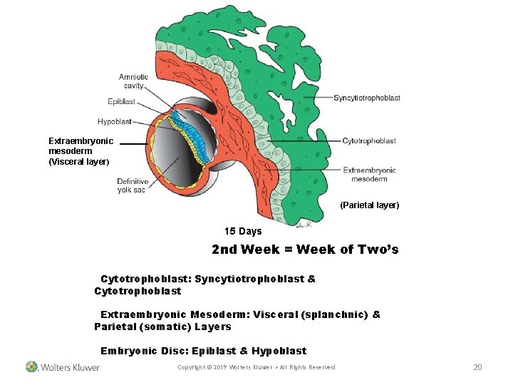
Extraembryonic mesoderm (Visceral layer) (Parietal layer) 15 Days 2 nd Week = Week of Two’s Cytotrophoblast: Syncytiotrophoblast & Cytotrophoblast Extraembryonic Mesoderm: Visceral (splanchnic) & Parietal (somatic) Layers Embryonic Disc: Epiblast & Hypoblast Copyright © 2019 Wolters Kluwer • All Rights Reserved 20
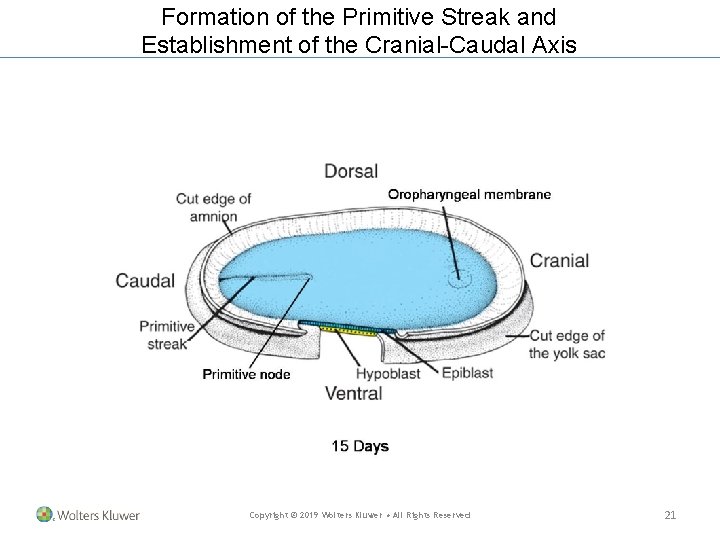
Formation of the Primitive Streak and Establishment of the Cranial-Caudal Axis Copyright © 2019 Wolters Kluwer • All Rights Reserved 21
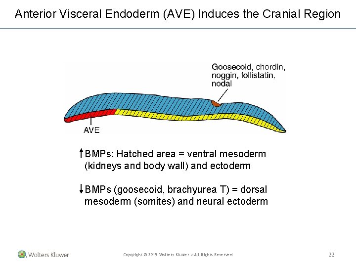
Anterior Visceral Endoderm (AVE) Induces the Cranial Region BMPs: Hatched area = ventral mesoderm (kidneys and body wall) and ectoderm BMPs (goosecoid, brachyurea T) = dorsal mesoderm (somites) and neural ectoderm Copyright © 2019 Wolters Kluwer • All Rights Reserved 22
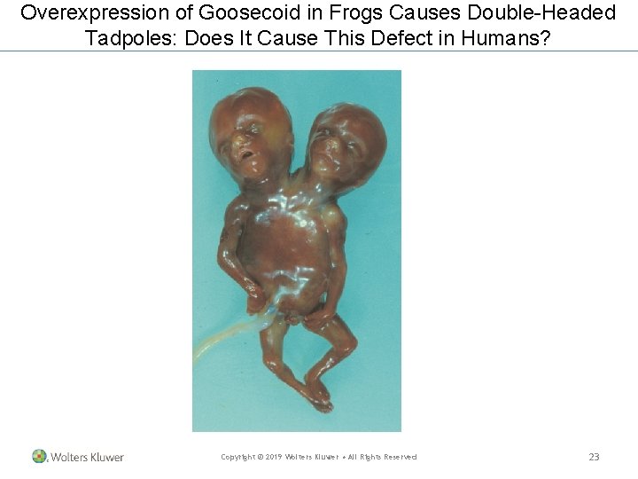
Overexpression of Goosecoid in Frogs Causes Double-Headed Tadpoles: Does It Cause This Defect in Humans? Copyright © 2019 Wolters Kluwer • All Rights Reserved 23
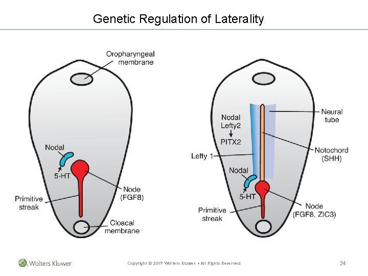
Genetic Regulation of Laterality Copyright © 2019 Wolters Kluwer • All Rights Reserved 24
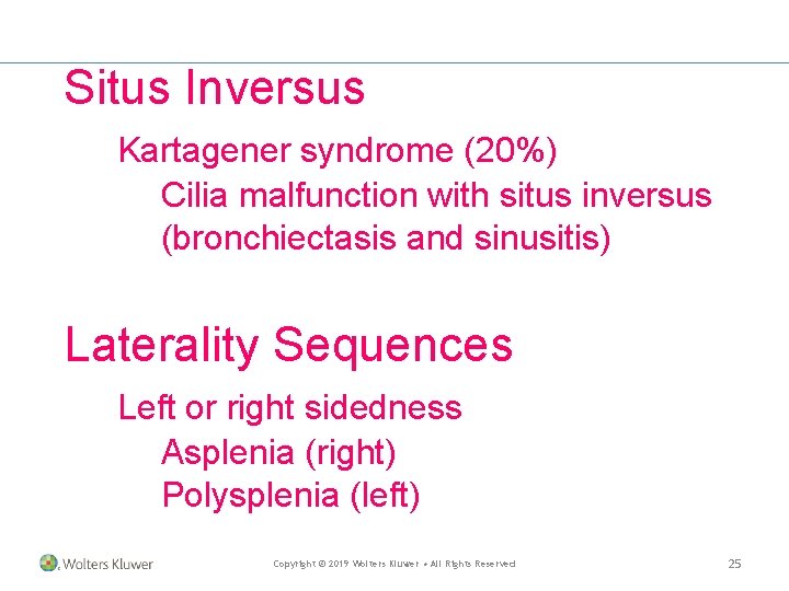
Situs Inversus Kartagener syndrome (20%) Cilia malfunction with situs inversus (bronchiectasis and sinusitis) Laterality Sequences Left or right sidedness Asplenia (right) Polysplenia (left) Copyright © 2019 Wolters Kluwer • All Rights Reserved 25
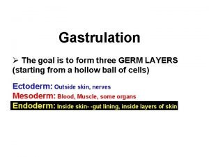
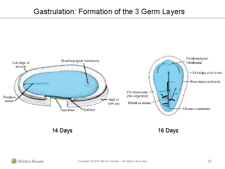
Gastrulation: Formation of the 3 Germ Layers 14 Days 16 Days Copyright © 2019 Wolters Kluwer • All Rights Reserved 26
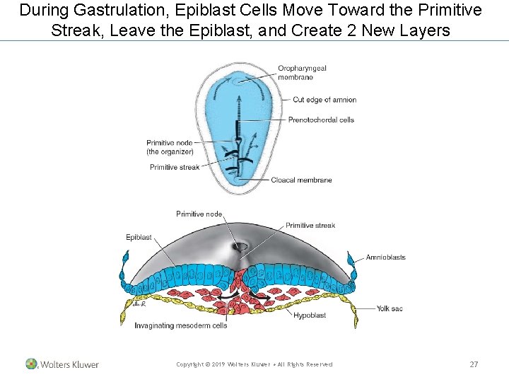
During Gastrulation, Epiblast Cells Move Toward the Primitive Streak, Leave the Epiblast, and Create 2 New Layers Copyright © 2019 Wolters Kluwer • All Rights Reserved 27
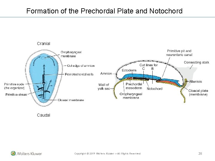
Formation of the Prechordal Plate and Notochord Cranial Caudal Copyright © 2019 Wolters Kluwer • All Rights Reserved 28
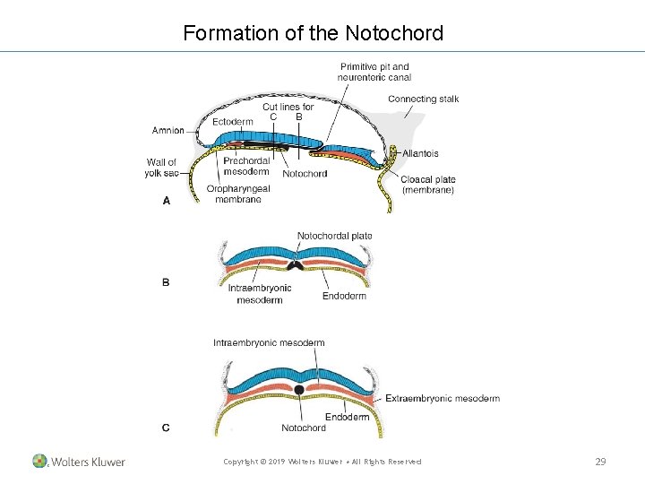
Formation of the Notochord Copyright © 2019 Wolters Kluwer • All Rights Reserved 29
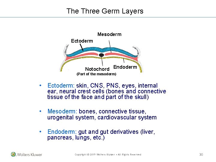
The Three Germ Layers Mesoderm Ectoderm Notochord Endoderm (Part of the mesoderm) • Ectoderm: skin, CNS, PNS, eyes, internal ear, neural crest cells (bones and connective tissue of the face and part of the skull) • Mesoderm: bones, connective tissue, urogenital system, cardiovascular system • Endoderm: gut and gut derivatives (liver, pancreas, lungs, etc. ) Copyright © 2019 Wolters Kluwer • All Rights Reserved 30