Fertilization Fertilization activates the egg Activation of the


 embryo")


 (c)")


- Slides: 38
Fertilization • Fertilization activates the egg • Activation of the egg triggers embryonic development
Acrosomal Reaction • The acrosomal reaction occurs in echinoderms Ex. sea urchins • Receptors on the vitelline egg layer are specific • The reaction is the fast block to polyspermy • A depolarization of the membrane stops other sperm from penetrating
The acrosomal and cortical reactions during sea urchin fertilization
Timeline for the fertilization of sea urchin eggs
Activation of The Egg • Increase in calcium also triggers an increase in metabolic reaction in the egg • Artificial activation of egg can occur by injecting calcium ions
A wave of Ca 2+ release during the cortical reaction
Mammalian Fertilization • Most mammals show internal fertilization • Sperm has to reach zona pellucida by penetrating follicle cells • An acrosomal reaction occurs and sperm cell enters egg • Zona pellucida hardens which blocks polyspermy • Centrosomes originate from sperm cell • Chromosomes share a common spindle during first mitotic division
Fertilization in Mammals
Early Embryonic Development 6 days 2 weeks
Figure 47. 6 Cleavage in an echinoderm (sea urchin) embryo
Cross Section of a Frog Blastula
Cleavage • Fast mitotic divisions without G 1 and G 2 phases • Results in smaller blastomeres • Polar planes of division occur with animal and vegetal poles • Holoblastic cleavage is complete division of eggs with little yolk ex: frogs • Meroblastic = incomplete division with moderate amt of yolk ex: birds
Gastrulation in a Frog Embryo
Body Symmetry Exhibit cephalization
Bilateral Symmetry – body plan in which only a single, imaginary line can divide the body into two equal halves; Ex: vertebrates Radial Symmetry – body plan in which body parts repeat around the center of the body; Ex: sea stars Spiral Symmetry – planes of division are diagonal (Ex: mollusks, segmented worms, arthropods)
• Organisms without body cavities are considered acoelomates Body covering (from ectoderm) (c) Tissuefilled region (from mesoderm) Acoelomates such as flatworms lack a body cavity between the digestive tract and outer body wall. Digestive tract (from endoderm) Figure 32. 8 c
• A pseudocoelom – Is a body cavity derived from the blastocoel, rather than from mesoderm Body covering (from ectoderm) (b) Pseudocoelomates such as nematodes have a body cavity only partially lined by tissue derived from mesoderm. Figure 32. 8 b Pseudocoelom Digestive tract (from ectoderm) Muscle layer (from mesoderm)
• A true body cavity is called a coelom and is derived from mesoderm; these animals are called coelomates Coelom (a) Coelomates such as annelids have a true coelom, a body cavity completely lined by tissue derived from mesoderm. Tissue layer lining coelom and suspending internal organs (from mesoderm) Digestive tract (from endoderm) Figure 32. 8 a Body covering (from ectoderm)
Figure 32. 7 A comparison of early development in protostomes and deuterostomes
Cleavage in a Frog Embryo
Sea urchin development, from single cell to larva
The Establishment of the Body Axes and the First Cleavage Plane in an Amphibian Becomes Posterior End Becomes Dorsal Side Becomes Anterior End
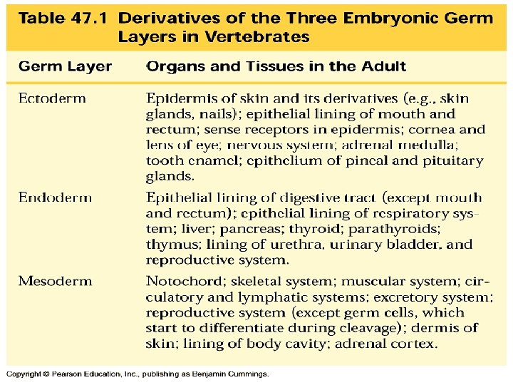
• Many different structures are derived from the three embryonic germ layers during organogenesis ECTODERM MESODERM ENDODERM p. 999 • Epidermis of skin and its derivatives (including sweat glands, hair follicles) • Epithelial lining of mouth and rectum • Sense receptors in epidermis • Cornea and lens of eye • Nervous system • Adrenal medulla • Tooth enamel • Epithelium or pineal and pituitary glands • Notochord • Skeletal system • Muscular layer of stomach, intestine, etc. • Excretory system • Circulatory and lymphatic systems • Reproductive system (except germ cells) • Dermis of skin • Lining of body cavity • Adrenal cortex • Epithelial lining of digestive tract • Epithelial lining of respiratory system • Lining of urethra, urinary bladder, and reproductive system • Liver • Pancreas • Thymus • Thyroid and parathyroid glands
Organogenesis • Folding, splitting and clustering of cells begins organogenesis • Ectoderm rolls into neural tube • Mesodermal blocks form somites along axis of notochord: – Gives rise to vertebrae and muscles of backbone • Neural plate folds into the neural tube and becomes central nervous system
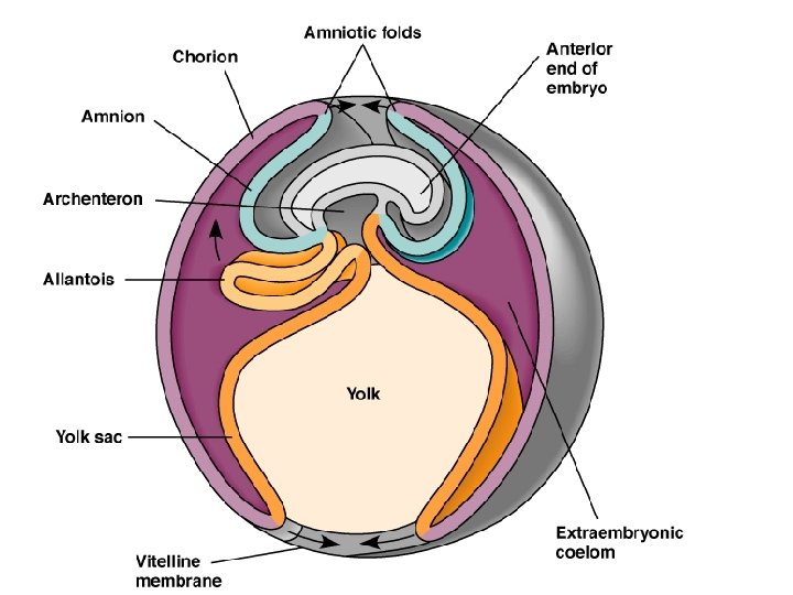
• Chorion - gas exchange and waste storage, lines the egg shell • Allantois – gas exchange and waste storage connects embryo to chorion • Yolk sac – food storage vitelline vessels embed into the yolk • Amnion – protective fluid filled sac
http: //eng-sci. udmercy. edu/courses/bio 123/Chapter 49/Chick. html
Human Embryo Brain Heart
Organogenesis in a Frog Embryo
Cleavage, gastrulation, and early organogenesis in a chick embryo
Organogenesis in a Chick Embryo About Eye 56 Hours Old Forebrain Blood Vessels Heart Somites Neural Tube
The Development of Extraembryonic Membranes in a Chick
Early development of a human embryo and its extraembryonic membranes
24 Hour Chick Embryo http: //www. uoguelph. ca/zoology/devobio/210 labs/24 hrwm. htm
48 Hour Chick Embryo http: //www. uoguelph. ca/zoology/devobio/210 labs/48 hrwm 1. htm
72 Hour Chick Embryo http: //www. uoguelph. ca/zoology/devobio/210 labs/72 hrwm. htm
http: //www. bioscience. drexel. edu/Homepage/Spring 2003/BIO%20268/Embryology/Chick/pages/C 6_W 006 T. htm