Figure 7 2 Cleavage of a frog egg


 and interior")
")
")
")
")


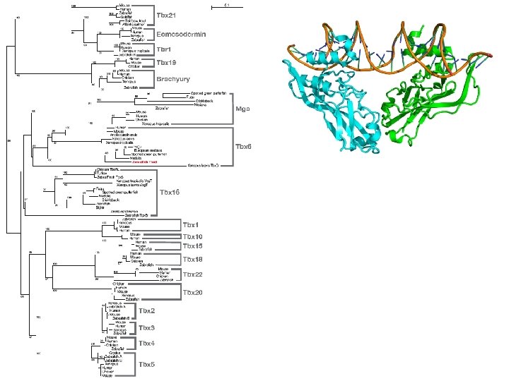
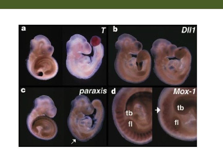
 T/Bra tbx 6 Chapman DL, Agulnik I, Hancock")


")
- Slides: 24
Figure 7. 2 Cleavage of a frog egg
Figure 7. 3 Scanning electron micrographs of frog egg cleavage
Figure 7. 5 Fate maps of the Xenopus laevis blastula exterior (A) and interior (B)
Figure 7. 6 Cell movements during frog gastrulation (Part 1)
Figure 7. 6 Cell movements during frog gastrulation (Part 2)
Figure 7. 6 Cell movements during frog gastrulation (Part 3)
Figure 7. 6 Cell movements during frog gastrulation (Part 4)
Figure 7. 7 Surface view of an early dorsal blastopore lip of Xenopus
Figure 7. 11 Protocadherin expression separates axial and paraxial mesoderm
Figure 12. 1 Mesodermal development in frog and chick embryos
Figure 8. 15 Development of a human embryo from fertilization to implantation
Figure 8. 23 Amnion structure and cell movements during human gastrulation
Figure 12. 1 Mesodermal development in frog and chick embryos
Primitive streak stage (7. 5 dpc) T/Bra tbx 6 Chapman DL, Agulnik I, Hancock S, Silver LM, Papaioannou VE. Tbx 6, a mouse T-Box gene implicated in paraxial mesoderm formation at gastrulation. Dev Biol. 1996 Dec 15; 180(2): 534 -42.
hin c. II
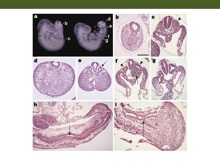
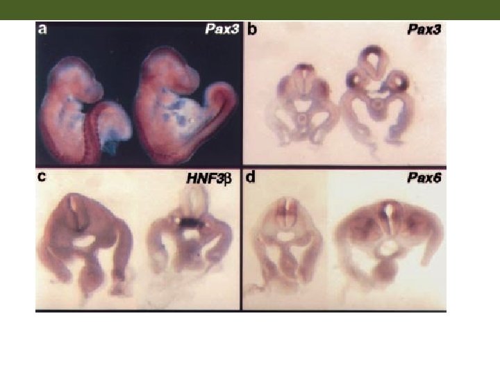
Summary Chapman DL, Papaioannou VE. Three neural tubes in mouse embryos with mutations in the T-box gene Tbx 6. Nature. 1998 Feb 12; 391(6668): 695 -7.
Figure 7. 6 Cell movements during frog gastrulation (Part 3)