Introduction to Virology Masheal Aljumaah OCT 2018 What
- Slides: 36
Introduction to Virology Masheal Aljumaah OCT 2018
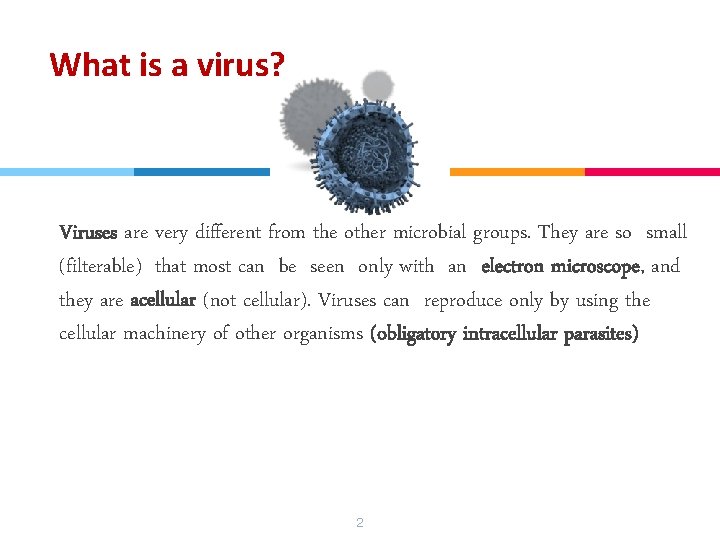
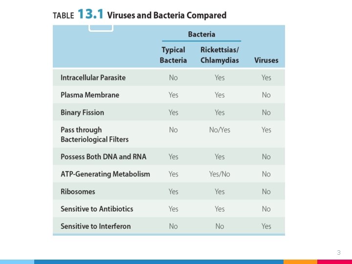
What is a virus? “ Viruses are very different from the other microbial groups. They are so small (filterable) that most can be seen only with an electron microscope, and they are acellular (not cellular). Viruses can reproduce only by using the cellular machinery of other organisms (obligatory intracellular parasites) 2
3
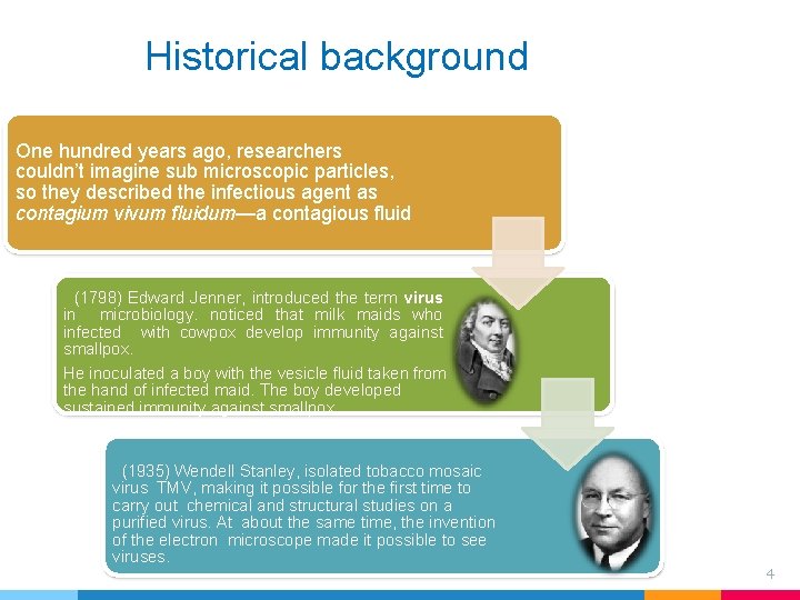
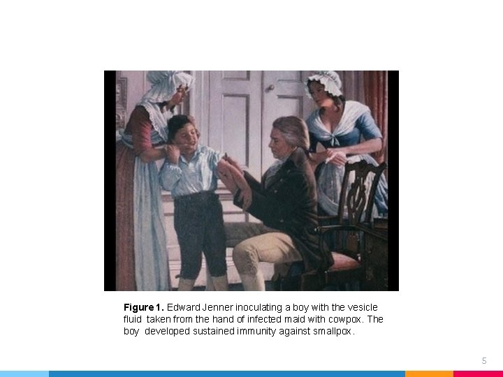
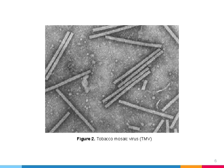
Historical background One hundred years ago, researchers couldn’t imagine sub microscopic particles, so they described the infectious agent as contagium vivum fluidum—a contagious fluid (1798) Edward Jenner, introduced the term virus in microbiology. noticed that milk maids who infected with cowpox develop immunity against smallpox. He inoculated a boy with the vesicle fluid taken from the hand of infected maid. The boy developed sustained immunity against smallpox. (1935) Wendell Stanley, isolated tobacco mosaic virus TMV, making it possible for the first time to carry out chemical and structural studies on a purified virus. At about the same time, the invention of the electron microscope made it possible to see viruses. 4
Figure 1. Edward Jenner inoculating a boy with the vesicle fluid taken from the hand of infected maid with cowpox. The boy developed sustained immunity against smallpox. 5
Figure 2. Tobacco mosaic virus (TMV) 6
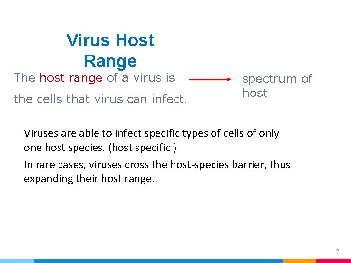
Virus Host Range The host range of a virus is the cells that virus can infect. spectrum of host Viruses are able to infect specific types of cells of only one host species. (host specific ) In rare cases, viruses cross the host-species barrier, thus expanding their host range. 7
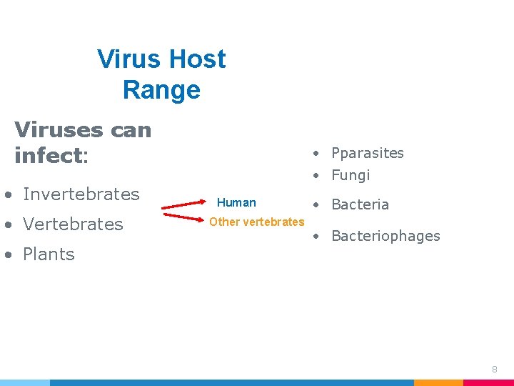
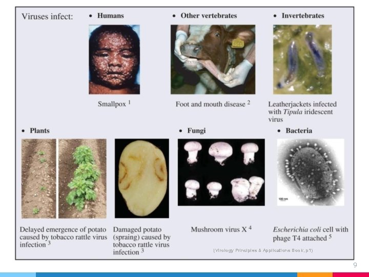
Virus Host Range Viruses can infect: • Invertebrates • Vertebrates • Plants • Pparasites • Fungi Human Other vertebrates • Bacteria • Bacteriophages 8
(Virology Principles & Applications Book, p 1) 9
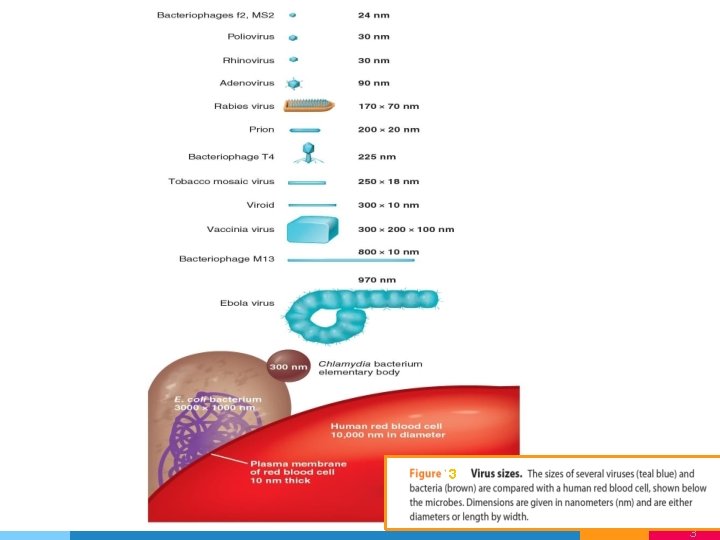
Viral Size Viral sizes are determined with the aid of electron microscopy. Different viruses vary considerably in size. Although most are quite a bit smaller than bacteria, some of the larger viruses (such as the vaccinia virus) are about the same size as some very small bacteria . Viruses range from 20 to 1000 nm in length. 10
3 1 3
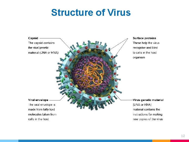
Structure of Virus 12
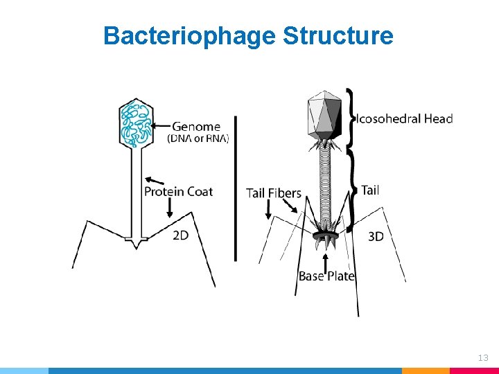
Bacteriophage Structure 13
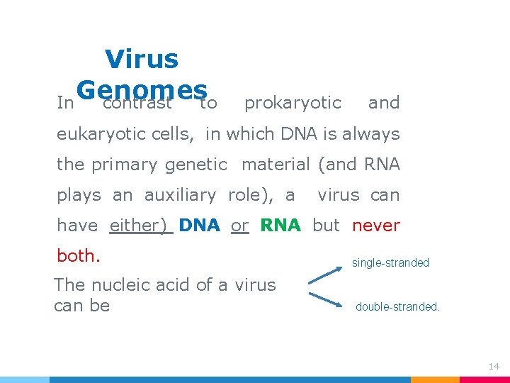
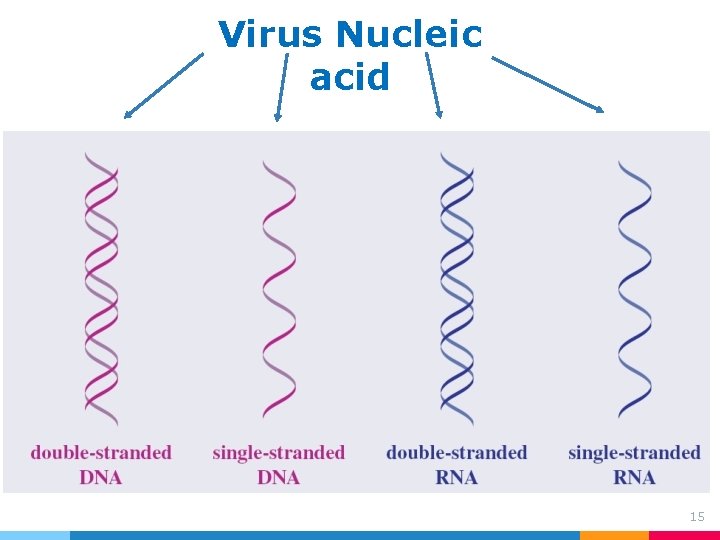
Virus Genomes In contrast to prokaryotic and eukaryotic cells, in which DNA is always the primary genetic material (and RNA plays an auxiliary role), a virus can have either) DNA or RNA but never both. single-stranded The nucleic acid of a virus can be double-stranded. 14
Virus Nucleic acid 15
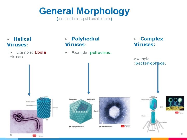
General Morphology (basis of their capsid architecture. ) Helical Viruses: ▷ ▷ Example: Ebola viruses Polyhedral Viruses: ▷ ▷ Complex Viruses: ▷ Example: poliovirus. example : bacteriophage. 16
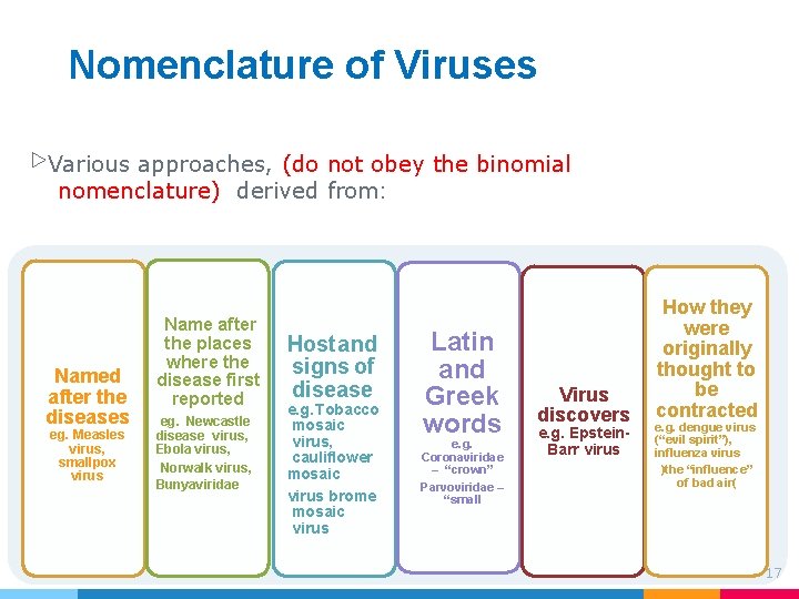
Nomenclature of Viruses ▷Various approaches, (do not obey the binomial nomenclature) derived from: Named after the diseases eg. Measles virus, smallpox virus Name after the places where the disease first reported eg. Newcastle disease virus, Ebola virus, Norwalk virus, Bunyaviridae Host and signs of disease e. g. Tobacco mosaic virus, cauliflower mosaic virus brome mosaic virus Latin and Greek words e. g. Coronaviridae – “crown” Parvoviridae – “small Virus discovers e. g. Epstein. Barr virus How they were originally thought to be contracted e. g. dengue virus (“evil spirit”), influenza virus )the “influence” of bad air( 17
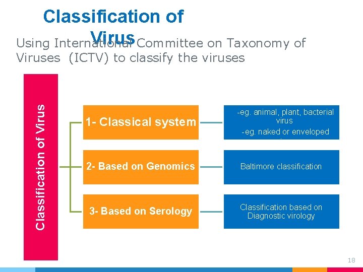
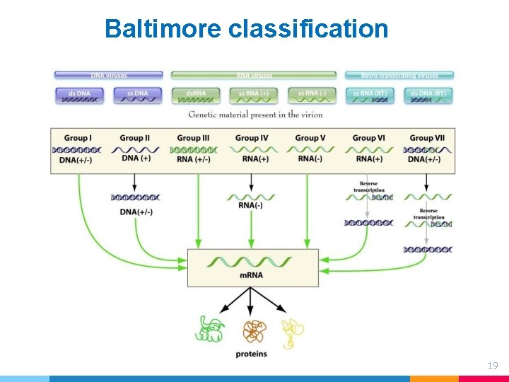
Classification of Virus Committee on Taxonomy of Using International Classification of Viruses (ICTV) to classify the viruses 1 - Classical system -eg. animal, plant, bacterial virus -eg. naked or enveloped virus 2 - Based on Genomics Baltimore classification 3 - Based on Serology Classification based on Diagnostic virology 18
Baltimore classification 19
4. How do we Detect and Measure Viruses? )Isolation, Cultivation, and Identification of Viruses ( 20
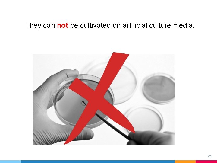
They can not be cultivated on artificial culture media. 29
“ The fact that viruses can’t multiply outside a living host cell complicates their detection, enumeration, and identification. Viruses must be provided with living cells instead of a fairly simple chemical medium (1. ( 22
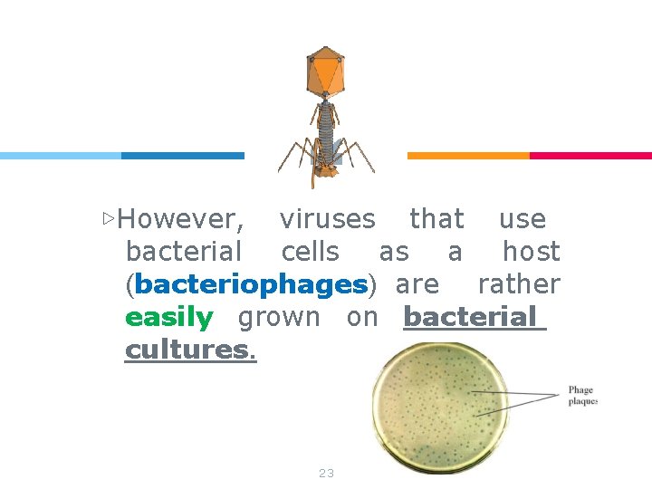
“ ▷However, viruses that use bacterial cells as a host (bacteriophages) are rather easily grown on bacterial cultures. 23
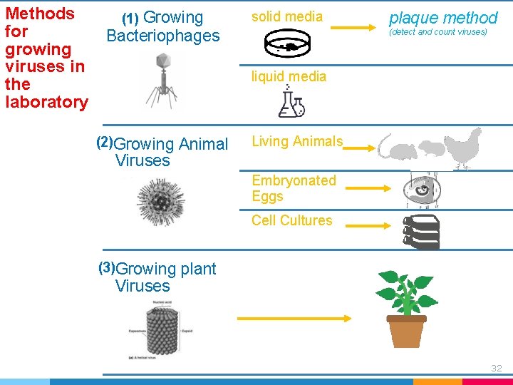
Methods for growing viruses in the laboratory (1) Growing solid media Bacteriophages plaque method (detect and count viruses) liquid media (2)Growing Animal Living Animals Viruses Embryonated Eggs Cell Cultures (3)Growing plant Viruses 32
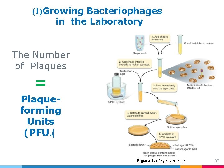
(1)Growing Bacteriophages in the Laboratory The Number of Plaques = Plaqueforming Units (PFU. ( Figure 4. plaque method. 33
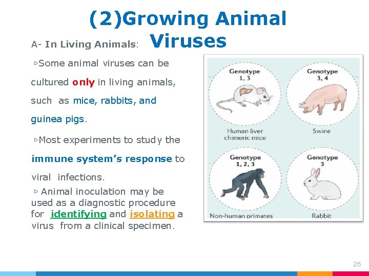
(2)Growing Animal A- In Living Animals: Viruses ▷Some animal viruses can be cultured only in living animals, such as mice, rabbits, and guinea pigs. ▷Most experiments to study the immune system’s response to viral infections. ▷ Animal inoculation may be used as a diagnostic procedure for identifying and isolating a virus from a clinical specimen. 26
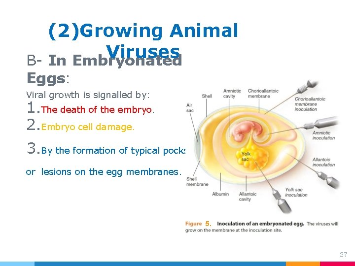
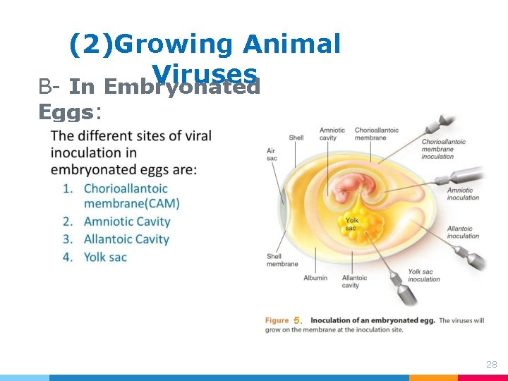
(2)Growing Animal Viruses B- In Embryonated Eggs: Viral growth is signalled by: 1. The death of the embryo. 2. Embryo cell damage. 3. By the formation of typical pocks or lesions on the egg membranes. 5. 27
(2)Growing Animal Viruses B- In Embryonated Eggs: 5. 28
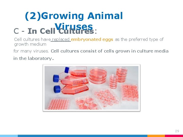
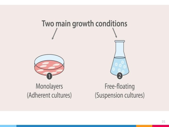
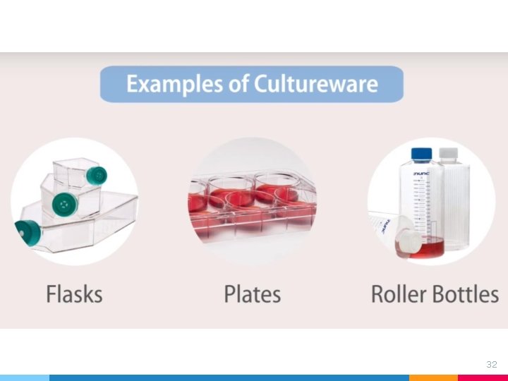
(2)Growing Animal Viruses C - In Cell Cultures: Cell cultures have replaced embryonated eggs as the preferred type of growth medium for many viruses. Cell cultures consist of cells grown in culture media in the laboratory. 29
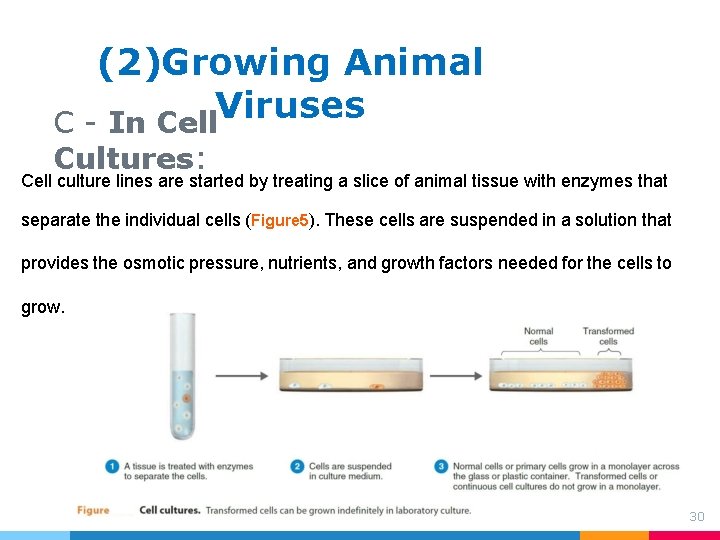
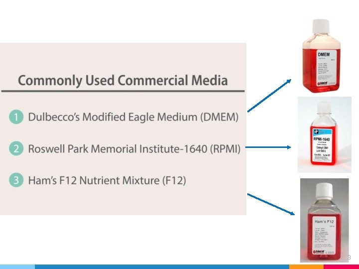
(2)Growing Animal Viruses C - In Cell Cultures: Cell culture lines are started by treating a slice of animal tissue with enzymes that separate the individual cells (Figure 5). These cells are suspended in a solution that provides the osmotic pressure, nutrients, and growth factors needed for the cells to grow. 30
31
32
33
43
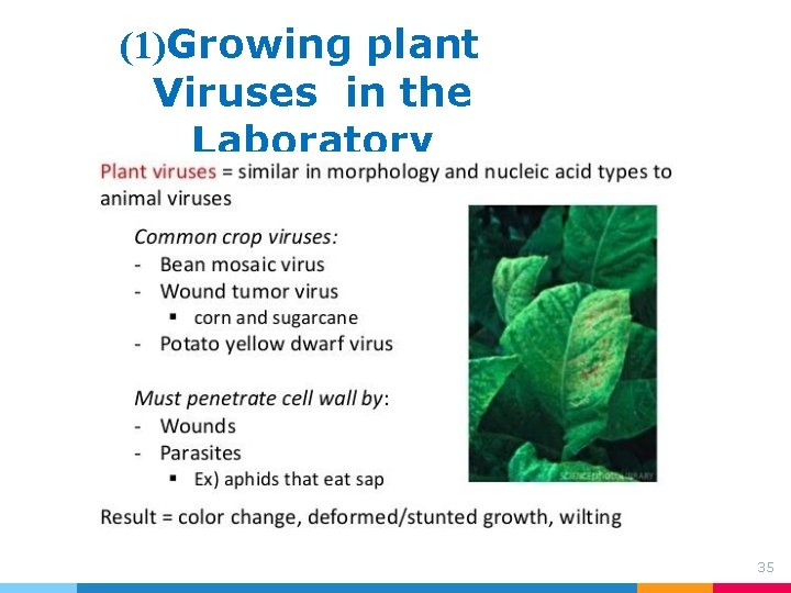
(1)Growing plant Viruses in the Laboratory 35
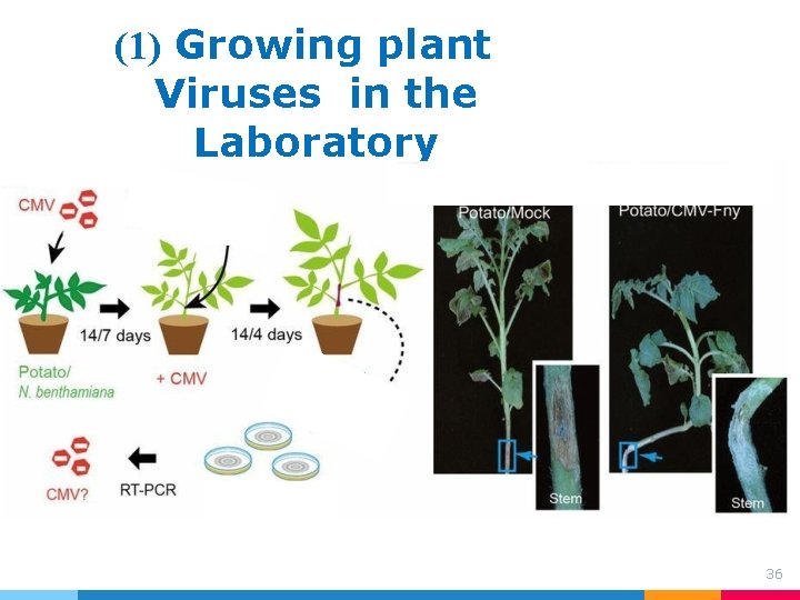
(1) Growing plant Viruses in the Laboratory 36