Intraarticular fractures DR Mohamed M Azmy MD The


- Slides: 24
Intraarticular fractures DR / Mohamed M. Azmy , MD
• The articulating surface of each bone is smooth. • Disruption of any component of the joint can result in altered joint function because of the pathological processes of arthrofibrosis or osteoarthrosis
Intraarticular Knee fracture
Knee arthritis following unsuccesseful intraarticular knee fracture
• closed reduction and external immobilization were commonly unsuccessful in the early treatment of displaced intraarticular fractures. • To avoid the complications of closed treatment, perfect anatomical restoration and freedom of joint movement could only be obtained by simultaneous open reduction and internal fixation.
Principles of ttt • Plaster cast immobilization of intraarticular fractures results in joint stiffness. • Plaster cast immobilization of intraarticular fractures combined with open reduction and internal fixation results in much greater stiffness. • Depressed articular fragments are impacted and will not be reduced by closed manipulation and traction.
• Anatomical reduction and stable fixation of articular fragments is necessary to restore joint congruity. • Immediate motion is necessary to prevent joint stiffness and to ensure articular healing and recovery. This requires stable internal fixation.
Evaluation of the patient and the injury • Because the etiology of many of these injuries is a highenergy mechanism, it is important to evaluate the patient fully for associated musculoskeletal and nonorthopedic injuries. • All fractures have the potential to compromise blood circulation to the surrounding skin, or even the limb itself, so the vascular status distal to the injury must be evaluated. This is best done by palpation of the pulses distal to the injury.
• The presence and location of any abrasions, joint effusion, skin blistering, and soft-tissue swelling should be noted. Point tenderness at ligamentous insertions may be the only clue to ligamentous disruption. • Muscular compartments should be evaluated for any evidence of compartment syndrome.
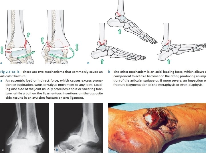
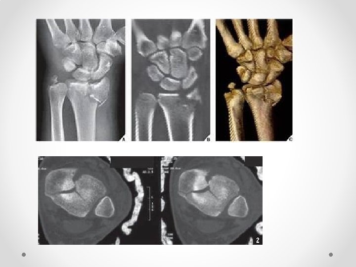
Evaluation of bone injury Initial x-ray analysis includes two views obtained in planes 90º to each other and centered over the zone of injury (Fig 2. 3 -3 a). The remainder of the limb will be examined by xrays, if clinically indicated. To ensure adequate detail, the area of interest should not be covered by dense bandaging or splintage.
• The addition of computed tomography—along with 2 -D and 3 -D reconstruction—provides additional information about the number and position of the articular fragments, the presence of impacted articular segments, the location of metaphyseal fracture lines, and the overall morphology of the injury.
• MRI is required in cases with ligamentous injuries
Scientific basis of treatment of articular fractures • anatomical reduction and stable fi xation with interfragmentary compression of an intraarticular fracture, followed by continuous passive motion (CPM), leads to true hyaline cartilage healing.
Anatomic restoration of joint surface leaded to sound healing with no arthritis
• Instability, caused by fracture, ligament, or meniscal injury can also lead to cartilage degeneration and may be important in determining outcome • So , limb malalignment should be avoided , and ligament reconstruction should be done.
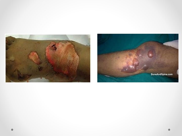
• If the soft-tissue envelope around the joint is swollen or traumatized with abrasions or degloving, open surgery within the first few days may be contraindicated.
• A detailed plan of the different steps and surgical tactic is mandatory before starting any internal fixation of an intraarticular fracture.
• Bone defects left within the metaphysis can be filled with autogenous or allogenic bone grafts or bone substitutes to provide early structural support to the articular surface and to stimulate reconstitution of metaphyseal bone stock.
Steps of reduction of iuntraarticular knee fracture With elevation of depressed articular fragments
Complications • • • Osteoarthritis. Joint stiffness. Associated joint instability. Osteonecrosis of devascularised articular fragments. Limb malalignment.