Identification of people alive cadavers and human remains


: arrows indicate")
: bracket showing")


 described")

 margin of the pubic face exhibits two characteristics that")
 from young to old. a. Right angle with the")


 is proportional to height There")


, 60% of badly")


 -Dental/clinical data (but not only): -INTERPOL forms -Dental files")


- Slides: 59
Identification of people alive, cadavers and human remains Agata Thannhauser
• Traffic accidents, natural disasters and crime entail numerous fatal victims, whose identity is often unknown. Depending on the circumstances of finding the bodies the particular methods are used to identify. These methods differ in degree of credibility. The list of identification techniques of dead bodies was put forward at the 5 th Interpol’s Meeting on the identification of victims of mass disasters and natural disasters, held in 1993 at Interpol’s headquarters in Lyon.
comparison of the genetic code of DNA comparison of fingerprints examination of teeth and other dental records radiology comparison of medical records (history of medical and surgical treatments)
comparison of characteristics - scars, tattoos, etc comparison of physical attributes identification of personal items, jewelry, etc identification on the basis of identity documents found with the body recognition by witnesses, family members or friends
Identifying unknown people
Problems • Physical appearance - short-term changes in facial expression occurring while blinking, speaking, expressing emotion or communicating. Also important are slow changes in the appearance caused by aging or changing image
• The geometry of the acquisition - usually the face in the picture is in an unknown location, it is not known which way it is facing and it is not known what is the size (scale)
• Conditions of imaging - human face lighting can have a big impact on the appearance on the photo
• Distortion of compression - they are often quite unexpected worsening of image quality caused by the compression (in order to send or archive)
IDENTIFICATION METHOD MEDICAL EVIDENCE NON-MEDICAL EVIDENCE
Establishing identity based on Human remains in decomposition post mortem transformed Remains in pieces Skeleton or single bones
Identification of personal items, jewelry, etc • Final places on the list of techniques of body identification, as presented at the 5 th Interpol’s Meeting, belong to identification of personal items, jewelry, etc, identification on the basis of identity documents, and identification by witnesses, family members or friends. • Such a low degree of reliability attributed to personal items and documents of identity items from the fact that, in forensics, such things have been proven to easily change owners, either intentionally or accidentally. • A low degree of reliability is also attributed to identification by family members or friends. The reason for this is making common, unintentional mistakes in identifying the dead body due to stress or corpse condition which prevent recognition even by closest relatives.
Identification of personal items, jewelry, etc One of the methods used to identify is the identification based on things found on or next to the remains. However it is necessary to remember , that this is only an auxiliary method –we can never confirm the identification based on this method. In the case of unidentified human corpses we describe the clothing very accurately (including size, color, texture of the material) shoes - including size, brand name, individual characteristics like tailor's alterations.
BIOLOGICAL PROFILE DETERMINATION OF SEX DETERMINATION OF AGE DETERMINATION OF STATURE
ACCURACY OF SEXING…. KROGMAN • • • ENTIRE SKELETON… 100% PELVIS & SKULL……. 98% PELVIS………………. . 95% SKULL………………… 90% LONG BONES………. . 80%
Classic Traits of the male versus female pelvis Trait Males Females Size Large and rugged Small and gracile Illium High and vertical Low and flat Pelvic inlet Heart shaped Circular or elliptical Pubic shape Narrow and rectangular Broad and square Subpubic angle V-shaped U-shape Obturator foramen Large and ovoid Small and trangular Greater sciatic notch Narrow Wide Preauricular sulcus Rare Well developed Shape of sacrum Long and narrow Short and broad
Determining the Sex of Unknown Human Skeletal Remains
Determining the Sex of Unknown Human Skeletal Remains Female pubis (ventral aspect): arrows indicate the ventral arc Male pubis (ventral) arrows show location where ventral arc would be located, this feature is absent in males VENTRAL ARC
Determining the Sex of Unknown Human Skeletal Remains Female pubis (dorsal view): bracket showing area of ischiopubic concavity Male pubis (dorsal) bracket indicating absence of concavity
Determining the Sex of Unknown Human Skeletal Remains Female sacroauricular joint with pre-auricular sulcus. Arrows showing pits in the sulcus Male sacroauricular joint. Bracketed area demonstrating absence of preauricular sulcus.
Characteristics of male and female skulls Traits Males Females Size Large and rugged Small and smooth Mastoid Large, projecting Small, nonprojecting Browridges Large Small, none Frontal Slanted High, rounded Nuchal area Rugged with hook Smooth, hook uncommon Supraorbital margin Rounded Sharp Chin Broad Pointed
Determining the Sex of Unknown Human Skeletal Remains Male nuchal crest with an inion hook Female nuchal region – note the absence of rugosity
Determining the Sex of Unknown Human Skeletal Remains Male mastoid Female mastoid
Determining the Sex of Unknown Human Skeletal Remains Difference in the frontal Slanted with large browridges in males High and rounded without browridges in females
Other bones are not usually as good an indicator regarding sex
SUMMARY 1. Because males generally are larger and more robust than females, the skeletons of the sexes can be distinguished by simply observing size and rugosity. 2. The are a number of a shape characteristics of the female pelvis, related to childbirth, that differntiate it from the pelvis of males. 3. The ishium-pubic index, which quantifies the longer pubic bone of females when compared to males, is the only commonly used metric method for distinguishing the sexes. 4. The skulls of males generally are larger and more rugged, with large mastoid processes and browridges, than the skull of females. 5. A number of metric measurements of the skull can be used with discriminant functions to differentiate the sexes. 6. A number of measurements of the postcranial bones can be used to separate males from females.
Determination of age from bones • Ages 0 -5: teeth are best – forensic odontology • Ages 6 -25: epiphyseal fusion – fusion of bone ends to bone shaft epiphyseal fusion varies with sex and is typically complete by age 25 • Ages 25 -40: very hard, can use pubic symphysis • Ages 40+: periodontal disease, arthritis, breakdown of pelvis, occupational stress, unique clues
AGE ESTIMATION METHOD Fusion states of cranial sutures. . The use of cranial suture closure for estimating age has a long and controversial history. In the mid 1920 s, T. Wingate Todd and D. W. Lyon published a series of articles describing changes in the cranial vault sutures related to age. They discovered that these lines were open in young people but had a tendency to close through time until, by old age, they were completely obliterated. Despite this promising start, their research was flawed because they discarded specimens from their study that exhibited anomalous closures. Over 13% of Whites and 34% of Blacks that were originally part of their sample were excluded during analysis. Although these attempts to choose only specimens that fit a preconceived notion of "normal" were well intentioned, modem scientific theory renders this unacceptable. In addition, even though they culled their sample to obtain more homogeneous results, Todd and Lyon still encountered a high amount of variability, which made age estimates based on their method prone to considerable error. Thus, as time passed, many forensic (and other) anthropologists simply did not use this method CRANIAL AND PALATE SUTURES
AGE ESTIMATION METHOD Fusion states of cranial sutures. open beginning fusion Buikstra and Ubelaker (1994) recognize four stages of closure: open, mini-mal, significant, and obliteration. Open refers to sutures where there is no evidence of closure. The gap between the bones is easily discernable and appears as a "bottomless" groove separating the bones. - Minimal is characterized by the closing of these gaps by bridges of bone that vary from a single connection to bridges that encompass less than 50% of the entire suture. CRANIAL AND PALATE SUTURES
AGE ESTIMATION METHOD Fusion states of cranial sutures. Significant closure means that there is more than 50% fusion between the bones, but with some parts still open. significant fusion Finally, obliteration refers to complete fusion between bones with no discernible gap; the sutures either appear as lines drawn on the bones or are no longer visible obliteration CRANIAL AND PALATE SUTURES
AGE ESTIMATION METHOD PUBIC SYMPHYSIS In his pioneering article, T. Wingate Todd (1920) described the alterations that occur in the pubic symphysis over time and how these can be used to estimate age at death. Todd described 10 stages of changes to this structure that, occur during aging, with time ranges of uneven lengths (e. g. , 18 to 19, 30 to 35, 50+) for each stage. Since his initial research, the value of using pubic face changes to evaluate age at death has received much attention. In 1955, Sheila Brooks, feeling that the age ranges for each stage were too high, modified Todd's time ranges down by several years in each category. Although the schedules and stages vary, all the above mentioned techniques look at the same features originally described by Todd: • the bone of the pubic face, • the ventral and dorsal margins, • the upper and lower extremities, • ossific nodules
• The bone of the pubic face undergoes several changes with age. • • • When young, it exhibits ridges separated by furrows that run transversely across the surface over time, the furrows fill in, usually starting posteriorly and proceeding anteriorly. Eventually, the face becomes flat with a granular look to the bone, which is replaced with fine-textured bone which again becomes more granular. Finally, in old age the face becomes pitted and eroded
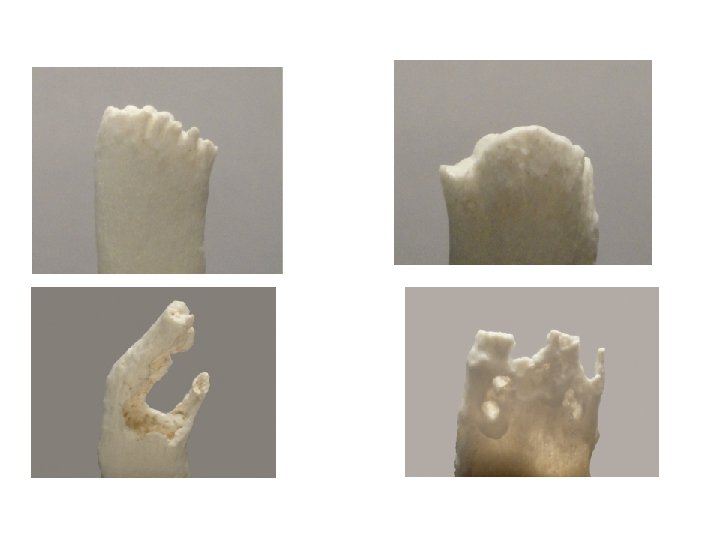
• The ventral (anterior) margin of the pubic face exhibits two characteristics that develop over time: a bevel and a rampart. In youth, the ventral margin forms a right angle with the pubic face. However, during the second decade of life as the above – described ridges degenarate and the furrows fill in, the cortical bone of the ventral surface begiins to invade the pubic face. This causes the sharp corner between the ventral margin and pubic surface to become blunted, until the margin bevels from the anterior surface of the pubis onto the pubic face. As time progresses, bone is deposited on the ventral bevel, forming what Todd called a rampart. This new bone causes the ventral margin to take the form of a right angle again between the anterior surface of the pubis and the pubic face. This feature usually starts inferiorly and begins to grow upward; at a slightly later time, it starts superiorly and moves downward. Eventually, the 'two ramparts grow together, forming a single well defined structure.
Changes in the ventralmargin (arrows) from young to old. a. Right angle with the pubic face b. Ventral bevel c. Rampart d. Fully developed
• In addition to ventral changes, the dorsal margin similarly alters over time. Initially, the pubic suffice is slightly curved from front to back As the ridges break down and the furrows fill in, the bone of the ventral pubic face builds up; this causes the surface to extend backward, forming a plateau.
• This margin usually extends farther backward than the posterior surface of the pubic bone, causing its dorsal edge to be fairly sharp. The upper and lower margins of the pubic face, called extremities by Todd, also undergo changes. During youth, the extremities are not easily distinguished, because the bone of the pubic face blends with both the inferior and superior surfaces of the pubis. With aging, these margins become more defined, starting first with the lower extremity. Eventually, the upper extremity also becomes defined (albeit not as distinctly as the lower), and both extremities are distinguished easily by viewing the pubic face.
• Three last osteological features that appear with increasing age are ossific nodules, the rim, and lipping. Ossific nodules are "blobs" of bone that can be seen in early adulthood. These appear to aid in the formation of both the upper extremity and the superior part of the ventral rampart. The rim is formed by the appearance of both the upper and lower extremities, as well as ventral and dorsal margins. This rim, which can be very distinct, usually is composed of cortical bone surrounding the more rough bone of the surface. The last feature is lipping. As age progresses, both the ventral and dorsal margins begin to curl outward, forming distinct lips. These structures are more visible on the dorsal surface, where they extebd and thicken the edge already formed by the dorsal plateau.
STERNAL RIB ENDS Four features of rib ends change over time according to a varied but discernable timetable: • surface bone, • surface contour, • rim edge, • rim contour
STERNAL RIB ENDS SURFACE BONE The surface bone of the rib end begins as smooth cortical bone, becomes granular over time and eventually becomes porous
STERNAL RIB ENDS SURFACE CONTOUR As these changes occur, the surface contour of the rib end transforms. This feature starts out billowy , but, as time goes on, this becomes less noticeable and eventually the end becomes more flat. With increasing time, the surface contour becomes indented, starting first as V-shaped and eventually becoming U-shaped.
STERNAL RIB ENDS RIM EDGE The next feature that changes with time is the rim around the sternal end. In youth, the end does not display this structure; however, as time goes on, a rim with a rounded edge begins to form as the costal cartilage starts to ossify As more times passes, this rim becomes thinner and assumes a jagged edge.
STERNAL RIB ENDS RIM CONTOUR The final feature of the rib end that changes with time is the rim contour. In youth, this feature is fairly straight, with contour only supplied by the coarseness of the bone. As time passes, this structure becomes more wavy until, in advanced age, the rim begins to send fingers of bone medially toward the sternum.
SUMMARY 1. The age at death of person can be estimated from the human skeleton by knowing changes that occur in both the growing and deteriorating skeleton. 2. Adults can be aged by observing changes in the pubic face, the sternal rib ends and the amount of suture closure observed in the skull. 3. Generally, the bone of the pubic face changes over time, from undulating surface covered by youthful-looking cortical bone to a flat area covered by pitted and old-looking bone. 4. Changes to the sternal ends of ribs include loss of smooth bone, development of a rim, and medial growth of fingers of bone along the costal cartilage. 5. The amount of fusion of the palatal as well as cranial sutures (observed both inside and outside the skull) can yield a rough estimate of age at death.
CALCULATION OF STATURE Long bone length (femur, tibia, humerus) is proportional to height There are tables that forensic anthropologists use.
ODONTOLOGY
• 1992 flight Lion-Strasburg: 87 victims; 50 bodies identified by odontologists • 1994 ship M/S Estonia (852 victims): 60% of Finnish victims identified with odontological methods • 1995 Petrinja, Croatia: mass grave, 46 civil victims. 16% identified via odontological techniques • 1996 Spain: 28 charred bodies in a bus: 57% identified with odontological data • 1998 Croatia: 64% of 1000 corpses from mass graves identified with odontological findings • Thailand Tsunami Victim Identification committee (TTVI) between 12 january and 31 march 2005 identified 1112 victims (962 dental identifications, 71 fingerprints, 10 anthropological and 3 DNA, 66 multiple methods) • Retrospective survey in Scandinavian countries: forensic odontology was used in 92% of 292 charred bodies
In the Linate aircraft mass disaster (8 -10 -2001; 118 victims), 60% of badly preserved victims were identified by forensic odontology
Tsunami 26 december 2005 … 226500 victims. . . AM data 3615 ID 2732 - genetics (-) - fingerprints (+) - odontology (+++) (80% Canadians 79% Germans 80% Australians……. ) mass disaster
Odontological identification is a fast, cheap, powerful way of identification Availability of mass dental care in civilized countries and conservation of dental records Odontological identification based on morphologigal features is possible even without any dental records Teeth are the most resistant part of the skeleton to time, physical and chemical agents Once erupted, teeth do not change their morphology except for attrition, decalcifications, fractures and restoration Teeth are the only part of the skeleton that is visible during life
Cultural modification……. geographic origin information 1. Teeth avulsion 2. Teeth position modification 3. Enamel notches (pointing) or grooves 4. Sharpening 5. Horizontal filing (“tooth amputation”) 6. Occlusal profile modification 7. Teeth color modification 8. Jewel setting
Post-mortem examination -external examination -occlusion -presence of teeth (am or pm loss, impacted, third molars, etc. ) -dental age -abnormal conditions -pathologies
-conservative -prosthetic -orthodontic implants
ANTE-MORTEM DATA (FROM RELATIVES, DENTISTS) -Dental/clinical data (but not only): -INTERPOL forms -Dental files (different nomenclature in different countries) -Radiographs (opg, intraoral rx, skull rx) -Pictures (not only clinical pic)
ANTE-MORTEM DATA Each annotation in the form should refer to the original source used and to its date The longest the interval between ante-mortem and post-mortem data, the biggest the chance that the feature has changed Therefore it is mandatory that operators know the possible evolutions of each oral pathology and therapy
RADIOLOGY The radiological technique involves taking a photographic picture of the skull at a 1: 1 scale. and pasting in into the skull radiograph. It is also possible to calculate anthropometric indicators of the skull and the radiograph. This technique is valid only in those cases where a radiograph of the skull has been made when a person was alive, and it is presumed that the skull tested belongs to the person whose radiograph is available. Individual characteristics are the shape and size of nose’s side cavities, especially sinusitis, and they are therefore suitable for comparative studies.
THANK YOU FOR YOUR TIME