Identification and Structure Determination of Higher Order Glycosphingolipids


 To separate and detect all")


")


+ Quad Fragments Quad Scan d 18: 1 340 u =")

+ Gal d")


+ 368 u = C 24: 0 d 18: 1 Glc")
+ 384 u = h 24: 0 Gal Glc Gal d")

+ 340 u = C 22: 0 d 18: 1 Glc")
+ d 18: 1 368 u = C 24: 0 Glc")
+ 384 u = h 24: 0 d 18: 1 Glc")

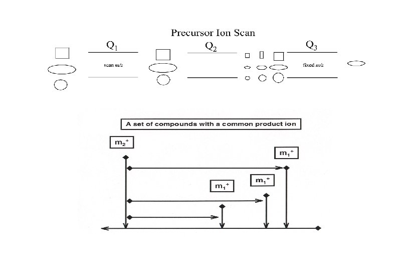
 Mass Analyzer (Q 1, Q 2, Q 3)")


- Slides: 33
Identification and Structure Determination of Higher Order Glycosphingolipids via LC-MS/MS M. Cameron Sullards, Ph. D. Georgia Institute of Technology: School of Chemistry and Biochemistry and School of Biology, Atlanta, GA 30332 -0363
Implications for disease: • Energy excess /deficit • Aberrant cell structure/function • Transport defects • Incorrect signaling • Developmental abnormalities Gene m. RNA Protein Post-translational modification(s) Metabolites Cellular functions Membrane (lipid) functions: • Energy • Structure • Transport • Signaling Agonists & other extracellular signals, including nutrients & xenobiotics Membrane lipids and regulation of cell structure/function
LIPID MAPS
LIPID MAPS Lipid Metabolites And Pathways Strategy GENOMICS PROTEOMICS METABOLOMICS “LIPIDOMICS”
Lipid Metabolites and Pathways Strategy LIPID MAPS GOALS (1) To separate and detect all of the lipids in a specific cell and to discover and characterize any novel lipids that may be present. (2) To quantitate each of the lipid metabolites present and to quantitate the changes in their levels and location during cellular function. (3) To define the biochemical pathways for each lipid and develop lipid maps which define the interaction networks.
LIPID MAPS CORES Macrophage Biology PI Edward A. Dennis - UCSD Christopher Glass - UCSD LC/Mass Spec Robert C. Murphy - Colorado Bioinformatics Lipid Synthesis/Characterization Michael Van. Nieuwenhze - UCSD Walter Shaw - Avanti Polar Lipids Steven White - UC Irvine Shankar Subramaniam - UCSD LIPID MAPS Fatty Acids/Eicosanoids Edward A. Dennis - UCSD Sphingolipids/Gangliosides Alfred H. Merrill - Georgia Tech Neutral Lipids Robert C. Murphy - Colorado Sterols David W. Russell - UTSW Glycerophospholipids H. Alex Brown - Vanderbilt Other Lipids/Structural Lipidomics Christian Raetz - Duke
Sphingolipids are the most structurally complex and diverse lipids of eukaryotes Glu cosylceram id e (Glc. Cer) N-Acetylgalactosamine Lactosylcera m id e (La c. Cer) Galactose OH HO O 1 Ac. NH O 3 OH O 4 O HO 2 C OH H 2 O 1 O H OH 4 HO O 1 O OH H NH 1' Cer O Ceramide (N-acylsphingosine) O HO HO OH OH Glucose OH 3 D-erythro-sphingosine OH OH H N Ac N-Acetyl. Neuraminic acid GM 3 GM 2 GM 1 (CH 3)3 NCH 2 O-P(O 2 H)-O-Cer Sphingomyelin J. L. W. Thudichum 1884
Sphin. GOMAP© (Download available at www. sphingomap. org)
MS/MS Methodology Identify structure specific dissociations unique to various classes SL’s (ie. Cer, Glc. Cer, Lac. Cer, Gb 3, and Gb 4) Utilize precursor ion & neutral loss scans to identify individual headgroup, base, and fatty acid combinations of endogenous SL’s Optimize ionization and dissociation conditions for all SL’s Quantify SL’s using internal standards and LC-MS/MS
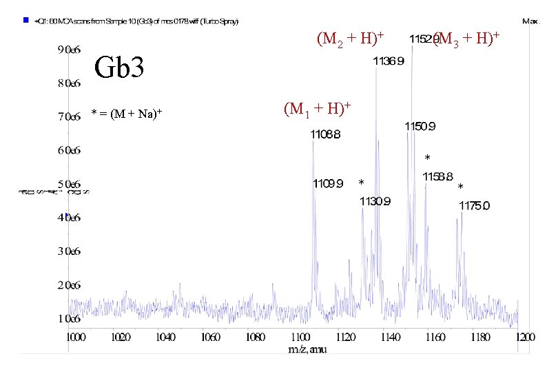
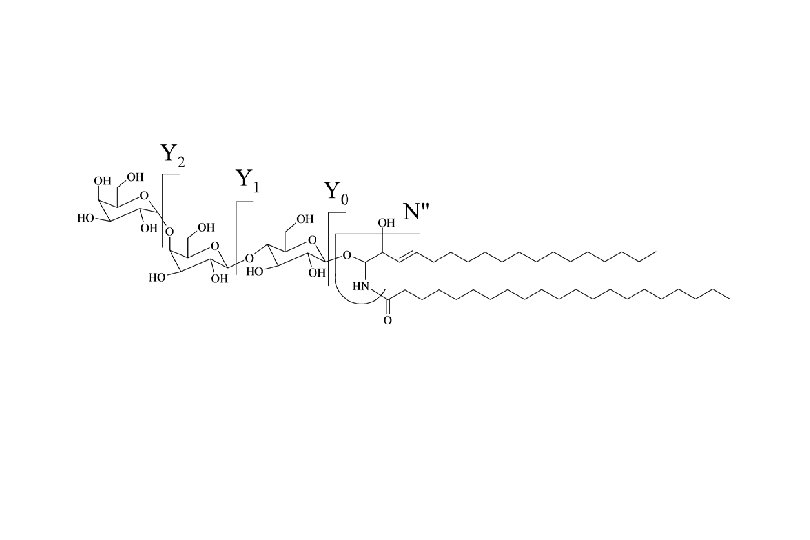
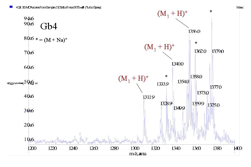
Globoside Gb 3 Globoside Gb 4
4000 Q TRAP System – Ion Path TM Skimmer Q 0 Q 1 Q 2 Q 3 LIT Orifice Curtain Plate LINAC Exit Lens
(M 1 + H)+ Quad Fragments Quad Scan d 18: 1 340 u = C 22: 0 Glc Gal
Quad Fragments LIT Scan d 18: 1 340 u = C 22: 0 (M 1 + H)+ Glc Gal
Quad Fragments LIT Scan w/ Q 0 Trapping (M 1 + H)+ Gal d 18: 1 340 u = C 22: 0 Gal Glc
4000 Q TRAP System – Ion Path TM Skimmer Q 0 Q 1 Q 2 Q 3 LIT Orifice Curtain Plate LINAC Exit Lens
MS/MS/MS 604. 6 1108. 7
(M 2 + H)+ 368 u = C 24: 0 d 18: 1 Glc Gal
(M 3 + H)+ 384 u = h 24: 0 Gal Glc Gal d 18: 1
C 24: 1 h 24: 0 d 18: 1 C 22: 0 h 24: 1 C 24: 1 1135. 1 h 22: 0 C 16: 0 1024. 9 C 18: 0 1053. 0 C 20: 0 1081. 1 1125. 1
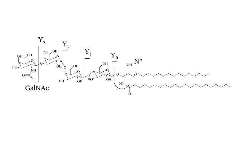
(M 1 + H)+ 340 u = C 22: 0 d 18: 1 Glc Gal Gal. NAc
(M 2 + H)+ d 18: 1 368 u = C 24: 0 Glc Gal Gal. NAc
(M 3 + H)+ 384 u = h 24: 0 d 18: 1 Glc Gal Gal. NAc
C 24: 0 d 18: 1 h 24: 0 C 22: 0 h 24: 1 C 16: 0 1228. 0 C 18: 0 C 20: 0 1256. 1 1284. 1
LC ESI MS/MS Sample In (Autosampler) Mass Analyzer (Q 1, Q 2, Q 3) Ion Source Inlet System HPLC Detector Relative Ion Abundance SM Data System Lac. Cer Data out Glc. Cer 0 2 4 6 Time (min) 8 10
h 24: 0 Gb 3 C 22: 0 d 18: 1 h 24: 1 h 22: 0 C 24: 1 C 16: 0 C 18: 0 C 20: 0 C 24: 0
C 22: 0 Gb 4 C 24: 0 d 18: 1 h 24: 0 C 24: 1 C 16: 0 h 24: 1 C 18: 0 h 22: 0 C 20: 0
ced Conclusions more highly abundant fragment ions enabling detailed structural analysis Precursor ion scans reveal critical information regarding low abundance globosides with high sensitivity in crude lipid extracts ction n LC identification of complex mixtures of globosides headgroup, long chain base, and fatty acid combinations in globosides
Acknowledgements Prof. Alfred H. Merrill, Jr. Meeyoung Park Anu Koppikar http: //www. sphingomap. org Matreya NIH/NIGMS/Lipid MAPS http: //www. lipidmaps. org