Hydatid cyst Dr Dhiresh Kumar Maharjan 12 th


- Slides: 30
Hydatid cyst Dr Dhiresh Kumar Maharjan 12 th May 2020
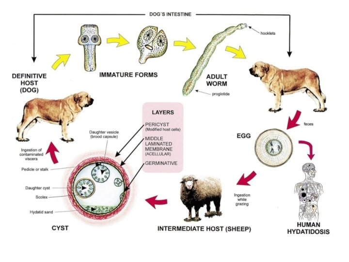
• Hydatid disease in people is mainly caused by infection with the larval stage of the dog tapeworm Echinococcus granulosus. • It is an important pathogenic, zoonotic and parasitic infection (acquired from animals) of humans, following ingestion of tapeworm eggs excreted in the faeces of infected dogs.
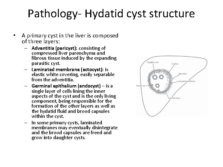
Pathology- Hydatid cyst structure • A primary cyst in the liver is composed of three layers: – Adventitia (pericyst): consisting of compressed liver parenchyma and fibrous tissue induced by the expanding parasitic cyst. – Laminated membrane (ectocyst): is elastic white covering, easily separable from the adventitia. – Germinal epithelium (endocyst) – is a single layer of cells lining the inner aspects of the cyst and is the only living component, being responsible for the formation of the other layers as well as the hydatid fluid and brood capsules within the cyst. – In some primary cysts, laminated membranes may eventually disintegrate and the brood capsules are freed and grow into daughter cysts.
Clinical features • The hydatid cyst is unique and localized in the right lobe of the liver in 65%. • The most frequent extrahepatic locations are the lungs, the spleen and the peritoneum. • Liver (55– 70%) is the obvious first site after entry through the gut and passage in the portal circulation. Most cysts tend to be located in the right lobe.
• As the cysts enlarge local pressure causes a mass effect on surrounding tissue producing symptoms and signs. • These may be generalized with upper abdominal pain and discomfort or more specific. Such as; obstructive jaundice. • Biliary rupture may occur through a small fissure or bile duct fistula. • A wide perforation allows the access of hydatid membranes to the main biliary ducts, which can cause symptoms simulating choledocholithiasis. • Alternatively, it may produce a picture very similar to ascending cholangitis with fever, pain and jaundice.
Complications of hydatid cyst of the liver • • Infection Rupture to the biliary tree Rupture to the peritoneal cavity Rupture to the pleural cavity.
Other complications • Portal hypertension • Acute abdominal pains with a sudden decrease in the volume of cyst and release of daughter vesicles during vomiting (hydatimesis) or in stool (hydatidentery) are highly suggestive of the opening of the cyst in the digestive tract
Diagnosis: Non complicated cysts • Hydatid cyst of the liver is frequently silent and only diagnosed incidentally during abdominal investigation for other pathology. • The clinical signs appear gradually with the increase volume of the cyst.
Complicated cysts • Cyst rupture can be associated with anaphylaxis. • Urticaria and erythema occur in cases of generalized anaphylactic reaction. • With biliary rupture the classic triad of jaundice, biliary colic and urticaria occurs.
Investigations • Imaging • Serologic and Immunologic studies.
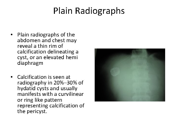
Plain Radiographs • Plain radiographs of the abdomen and chest may reveal a thin rim of calcification delineating a cyst, or an elevated hemi diaphragm • Calcification is seen at radiography in 20%– 30% of hydatid cysts and usually manifests with a curvilinear or ring like pattern representing calcification of the pericyst.
Imaging techniques • Ultrasonography (US) is the screening method of choice.
Ultrasonography (US) • Ultrasonography is the screening method of choice. • Findings usually seen are: – Solitary Cyst – anechoic univesicular cyst with well defined borders and enhancement of back wall echoes in a manner similar to simple or congenital cysts – Separation of membranes (ultrasonic water lily sign) due to collapse of germinal layer seen as an undulating linear collection of echoes. – Daughter cysts - probably the most characteristic sign with cysts within a cyst, producing a cartwheel or honeycomb cyst.
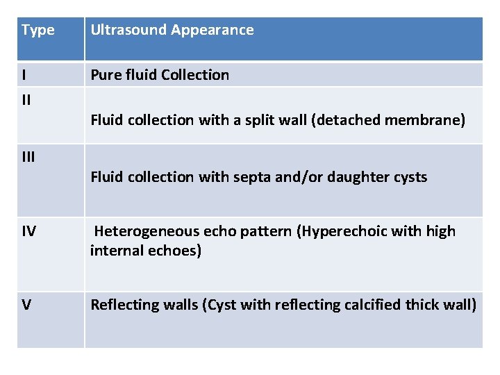
Type Ultrasound Appearance I II Pure fluid Collection III Fluid collection with a split wall (detached membrane) Fluid collection with septa and/or daughter cysts IV Heterogeneous echo pattern (Hyperechoic with high internal echoes) V Reflecting walls (Cyst with reflecting calcified thick wall)
USG abdomen: Gharbi Classification
CT Scan abdomen CT scan is an important preoperative diagnostic tool to determine vascular, biliary or extra hepatic extension, to recognize complications, such as rupture and infections, and therefore to assess resectability.
Serology and immunological tests • Detection of Ig. G antibodies implies exposure to the parasite, while in active infection high titers of specific Ig. M and Ig. A antibodies are observed. • ELISA is used most commonly, but alternate techniques are counter -immuno-electrophoresis and bacterial co-agglutination. • Elisa techniques have a high sensitivity above 90% and are useful in mass scale screening. • The counter-immuno-electrophoresis has the highest specificity (100%) and high sensitivity (80 – 90%). • CASONI TEST has been used most frequently in the past but is at present considered only of historical importance and has largely been abandoned because of low sensitivity.
Treatment • The principles of hydatid surgery are – Total removal of all infective components of the cysts; – the avoidance of spillage of cyst contents at time of surgery; – management of communication between cyst and adjacent structures; – management of the residual cavity; – Minimize risks of operation.
Medical treatment • Albendazole therapy (10 mg/kg body weight) for 8 weeks.
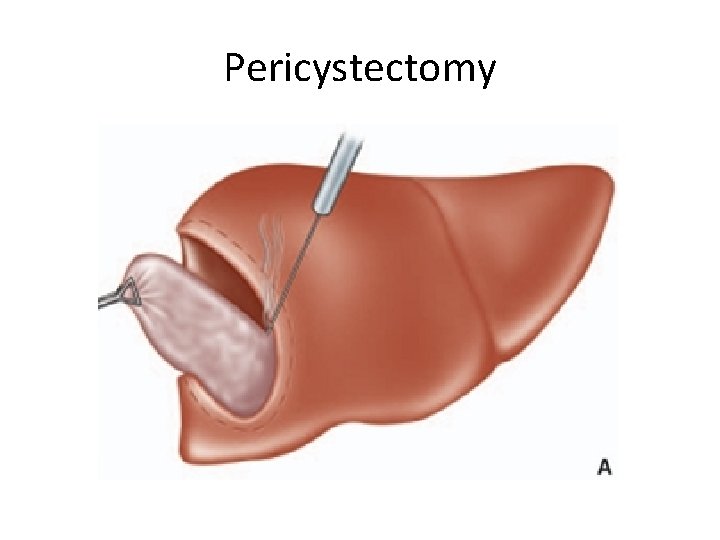
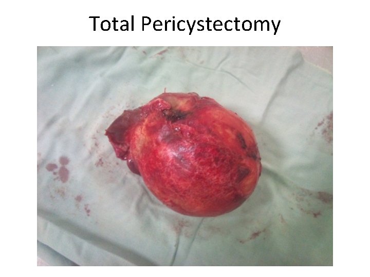
• • • Marsupialization Cystectomy, Pericystectomy Lobectomy Hepatectomy. Laparoscopic management of hydatid cysts
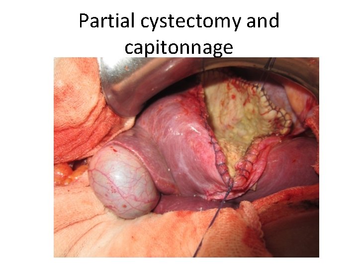
Partial cystectomy and capitonnage
Pericystectomy
Total Pericystectomy
Percutaneous treatment of hydatid cyst • PAIR (puncture, aspiration, injection, and reaspiration) is a percutaneous treatment technique for hydatid disease. • This technique was proposed in 1986 by the Tunisian team. • In this minimally invasive method, a needle is introduced into the cyst under ultrasound guidance.
Indications for PAIR • Nonechoic lesion greater than or equal to 5 cm in diameter • Cysts with daughter cysts and/or with membrane detachment • Multiple cysts if accessible to puncture • Infected cysts • Patients who refuse surgery. • Patients who relapse after surgery. • Patients in whom surgery is contraindicated • Patients who fail to respond to chemotherapy alone • Children over 3 years. • Pregnant women
Contraindications for PAIR • • • Non cooperative patients Inaccessible or risky location of the liver cyst Cyst in spine, brain, and/or heart Inactive or calcified lesion Cyst communicating with the biliary tree
• Patients should be followed clinically after PAIR treatment. • Recurrence is increased in more complicated cysts, including those with multiple daughter cysts. • Must be preceded by Albendazole therapy (10 mg/kg body weight) for 8 weeks.