HCC Locoregional Treatment and Evaluation of Tumour Response


- Slides: 19

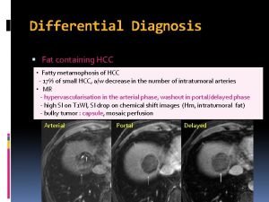
HCC Loco-regional Treatment and Evaluation of Tumour Response Prof. Lourens Bester Head of Radiology Interventional Radiologist University of Notre Dame Sydney

Introduction. • Transarterial radioembolisation* with yttrium 90 microspheres (SIRT) is an increasingly popular therapy for both primary and secondary liver malignancies. • The consequent biological changes are distinct from those of other transarterial therapies as reflected in the often baffling post-treatment imaging features. • Therefore the post-TARE/SIRT image interpretation is challenging. • In this review I am going to attempt to provides a description of the different findings seen in post-treatment scans, with the aim of facilitating appropriate radiological interpretation. * TARE = SIRT

Tumour Response and Staging Criteria • Apart from standard size criteria, response can also be evaluated using other aspects such as tumor necrosis, reduction in vascularity, reduced metabolic activity (PET) and functional ( Diffusion) magnetic resonance imaging (f-MRI) • Post treatment assessment can be difficult and depend on what criteria is used. • Assement of response using WHO, American Association for the Study of Liver Disease (AASLD) and the European Association for the Study of the Liver (EASL) come to different conclusions. • For Instance in HCC patients treated with SIRT, objective response (OR) was seen in 23% of patients using m-RECIST criteria and in 59% with EASL criteria. • Clearly highlighting the differences in assessment using different staging systems.

Tumour size, Necrosis and Vascularity. • Tumours may shrink, remain the same, or even increase in size. • Necrosis, oedema, and hemorrhage may cause an initial increase in the size of a tumour often leadig to confusion. • The median time to a positive response being 118– 120 days when using size only, 29– 30 days using necrosis only and 31– 34 days using size and necrosis. • In practice, rather than complete necrosis of all the treated tumours, patchy necrosis with variable residual enhancing areas is the more frequently encountered picture with no predictive value.

Tumour Size and Necrosis.

Reduction in tumour vascularity. Cancer Imaging. 2013; 13(4): 645– 657 .

Metabolic activity on FDG-PET. • The sensitivity of FDG-PET in primary HCC is poor. • This observation can be explained by the poor FDG uptake of well differentiated HCC with only poorly differentiated hepatomas accumulating FDG significantly. • PET/CT is reserved as a troubleshooter in differentiating viable neoplastic tissue from edema, hemorrhage or necrosis after treatment. • PET give an earlier response (6 -8 weeks) than the CT and MRI.

Metabolic activity - PET CANCER BIOTHERAPY & RADIOPHARMACEUTICALS Volume 20, Number 2, 2005

Diffusion-weighted MR imaging. • DWI is also reserved as a troubleshooter in differentiating viable neoplastic tissue from edema, hemorrhage or necrosis after treatment. • Post-SIRT enhancement can represent viable tumour, peritumoral hypereamia or post-treatment granulation tissue. • Hypovascular tumors are especially difficult to assess for response as we rely heavily on postcontrast enhancement to identify residual tumour. • Rhee et al. found by evaluating ADC values, diffusion-weighted MRI has been able to detect tumor response accurately within 42 days of SIRT.

Diffusion-weighted MR imaging. T 2 2 T 1+G Cancer Imaging. 2013; 13(4): 645– 657. ADC

Benign Post-treatment findings: Not to be interpreted as progression of the disease. • Peritumoral oedema and hypervascularity. • Rim enhancement. • Ill-defined geographic areas of hypoattenuation. • Volumetric changes. • Capsular retraction. • Perihepatic fluid.

Peritumoral Hyperaemia and Volumetric Changes With diffusion-weighted MRI peritumoral oedema can be differentiated from viable tumour. Peritumoral edema is transient but can persist for 3– 6 months after therapy. J Vasc Interv Radiol 2009

Rim Enhancement. Riad Salem Nortwestern Memorial Chicago

Geographic changes Post SIRT 1 Month 3 Month’s This radiologic finding has no known clinical significance. This benign finding can mimic progressive infiltrative tumour.

Geographic and Volumetric Changes 1 Month Post SIRT 3 Month’s Post SIRT

Perihepatic Fluid and Capsular Retraction. Baseline CT scan pre-SIRT CT scan 3 months post-SIRT . Sharma RA et al. Annals of Oncology 2006; 17 (Sup 6): vi 78 Abstract P-191. Data on file; Sirtex Medical Limited.

Complications • REILD. • Biliary. • Non Targeted flow.

REILD Scarred but survived Atassi et al, Radio. Graphics 2008; 28: 81 -99 Demise

Conclusion. • Reduction in tumour size, necrosis and lack of tumor enhancement as seen at CT and MRI are primary surrogates of a favorable response. • PET/CT and f-MRI are used to solve certain diagnostic dilemmas that may be encountered during follow-up imaging e. g. peritumoral edema, peritumoral hypereamia, perilesional rim enhancement and geographic areas of hypodensity. • Radiologists need to be aware of these benign findings that are unique to post SIRT treated livers. • Post treatment complications are rare but the reporting radiologist need to know what to look for.
 Osteochondroma gross pathology
 Adrenal tumour
 Mobile phone brain tumour
 Natural response and forced response
 What is natural response
 A subsequent
 Oberfinanzdirektion frankfurt
 Hcc genealogie
 Slidetodoc
 Gebouwinspectie met drone
 Hawkmail hcc
 Tace hcc
 Garantieproducten beleggen
 Shumihito
 Model saigontech
 Hcc bclc
 Hcc coding cheat sheet
 Hcc recapture rate
 Hcc differential equations
 Home care worker orientation manual

