Good Morning Case Patient elderly Ho Hypertensive on









- Slides: 46
Good Morning
Case – Patient, elderly H/o – Hypertensive, on antihypertensives, fall and unconscious, hyperpyrexia, deep coma, paralysis, pinpoint pupils.
Brainstem • Introduction It is that part of neural axis which extends rostrally from cranial end of SC to caudal part of diencephalon.
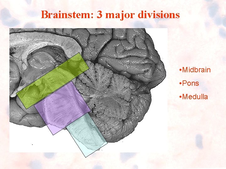
Brainstem: 3 major divisions • Midbrain • Pons • Medulla
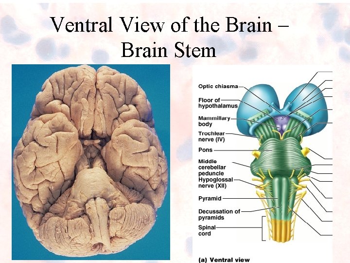
Ventral View of the Brain – Brain Stem
The Brain Stem: General Functions – Produces automatic behaviors necessary for survival – Passageway for all fiber tracts running between the cerebrum and spinal cord – Important cranial nerve nuclei • 10 of the 12 pairs of cranial nerves attach to it • Heavily involved with the innervation of the face and head
Learning Objectives • To know and learn about gross anatomy, both external and internal structure of pons • To know about its applied anatomy in brief
PONS (Bridge)
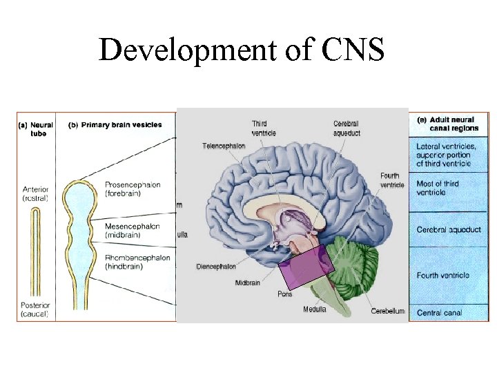
Pons • Developmentally part of hindbrain • Also called as Metencephalon • 2. 5 cm -interconnecting the midbrain with medulla. • It does not, however, interconnect them
Development of CNS
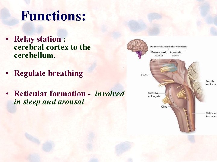
Functions: • Relay station : cerebral cortex to the cerebellum. • Regulate breathing • Reticular formation - involved in sleep and arousal
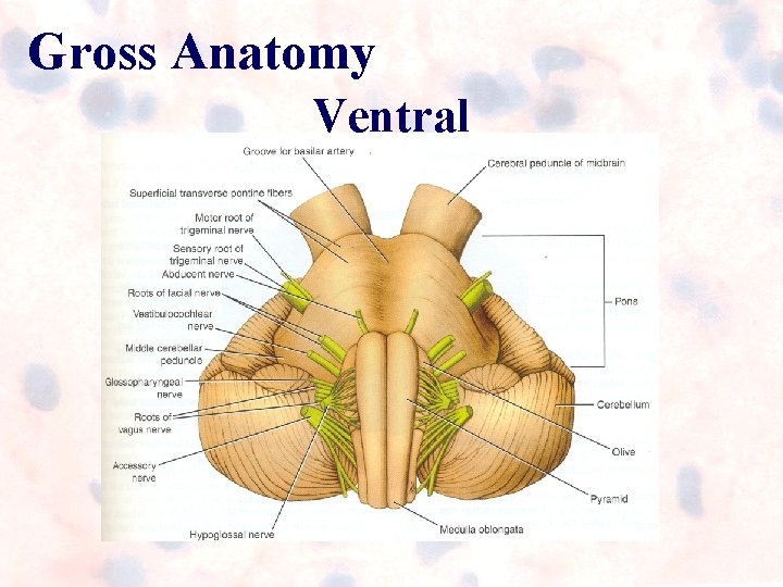
Gross Anatomy Ventral
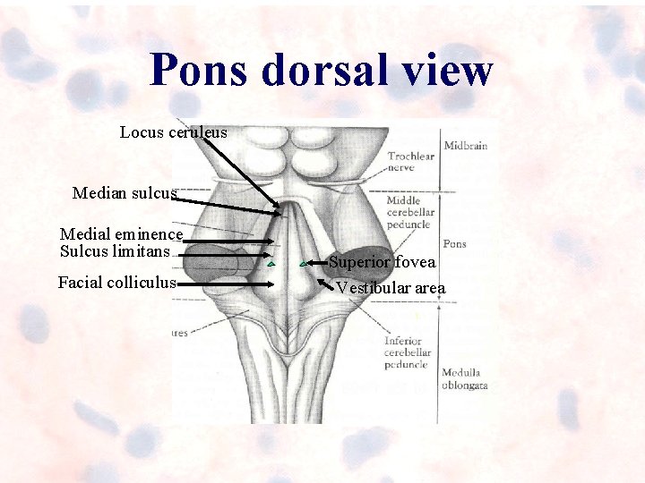
Pons dorsal view Locus ceruleus Median sulcus Medial eminence Sulcus limitans Facial colliculus Superior fovea Vestibular area
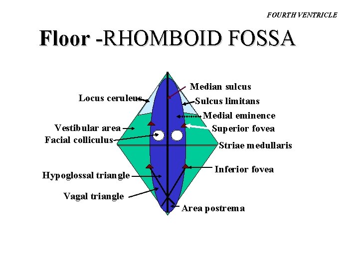
FOURTH VENTRICLE Floor -RHOMBOID FOSSA Locus ceruleus Vestibular area Facial colliculus Hypoglossal triangle Median sulcus Sulcus limitans Medial eminence Superior fovea Striae medullaris Inferior fovea Vagal triangle Area postrema
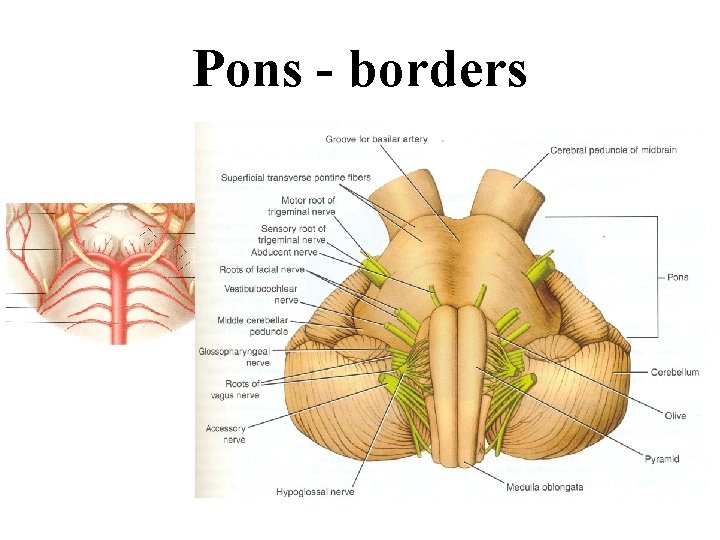
Pons - borders
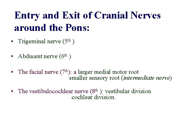
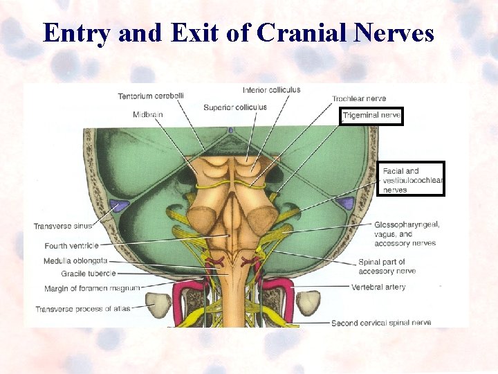
Entry and Exit of Cranial Nerves around the Pons: • Trigeminal nerve (5 th ) • Abducent nerve (6 th ) • The facial nerve (7 th): a larger medial motor root smaller sensory root (intermediate nerve) • The vestibulocochlear nerve (8 th ): vestibular division cochlear division.
Entry and Exit of Cranial Nerves
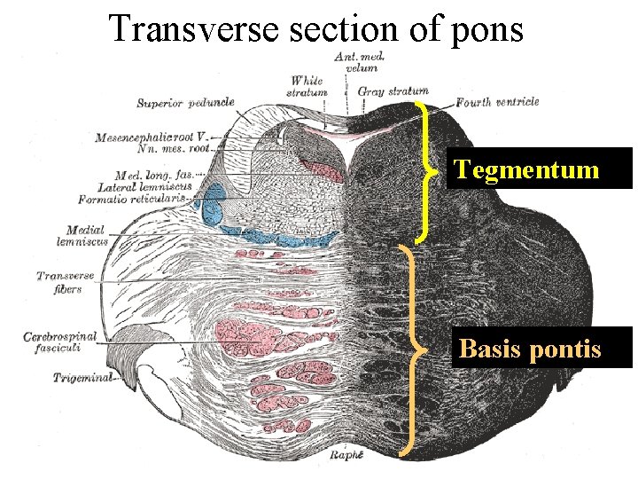
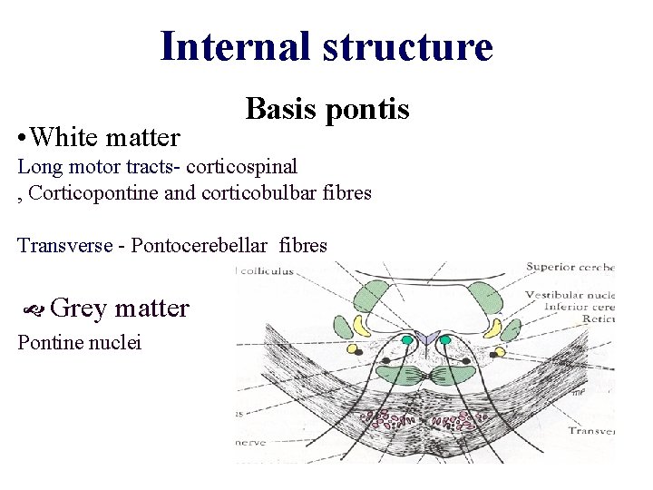
Transverse section of pons Tegmentum Basis pontis
Internal structure • White matter Basis pontis Long motor tracts- corticospinal , Corticopontine and corticobulbar fibres Transverse - Pontocerebellar fibres Grey matter Pontine nuclei
Internal structure………. Tegmentum Cranial nerve nuclei; sensory tracts Reticular formation MLF (nuclei of extra ocular muscle connections)
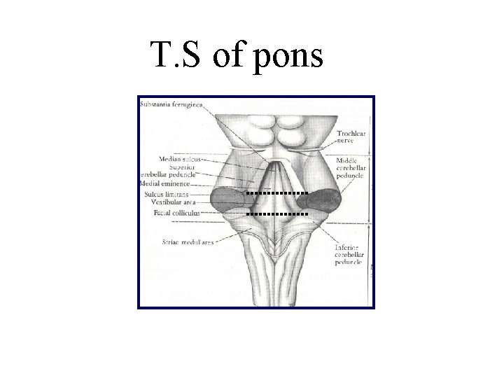
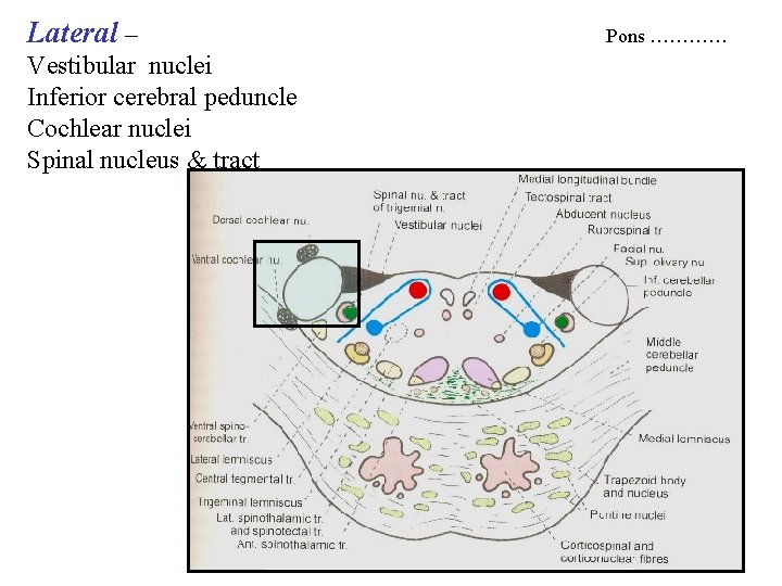
T. S of pons
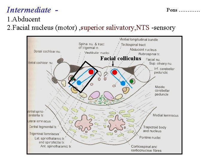
Cross section at the level of facial colliculus
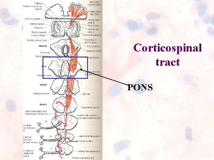
Corticospinal tract PONS
Cross section at the level of facial colliculus
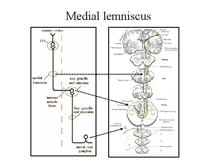
Medial lemniscus
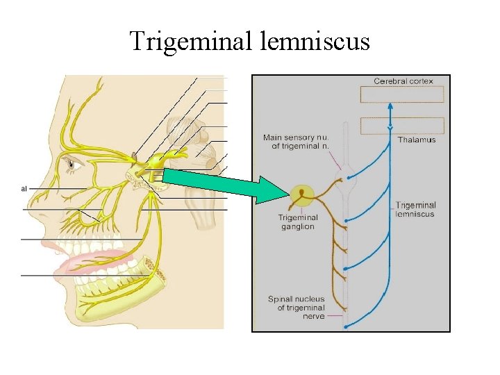
Trigeminal lemniscus
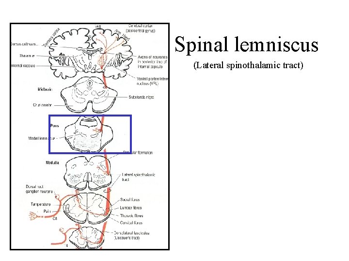
Spinal lemniscus (Lateral spinothalamic tract)
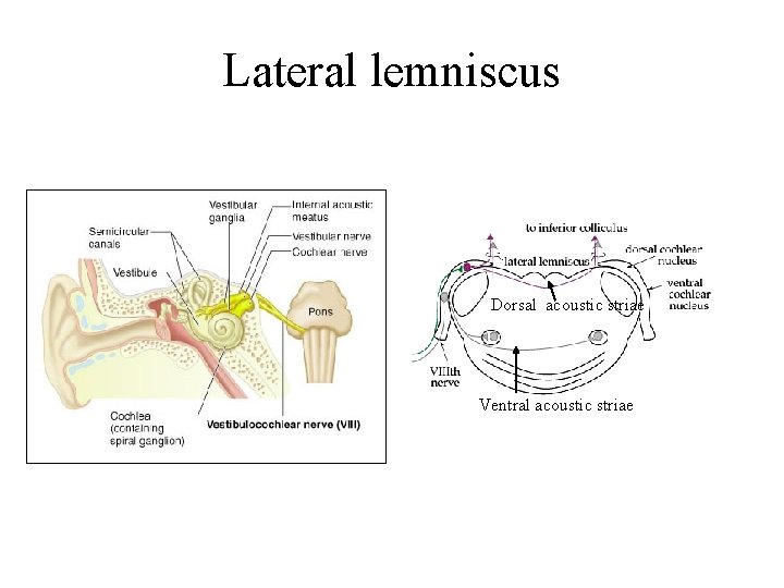
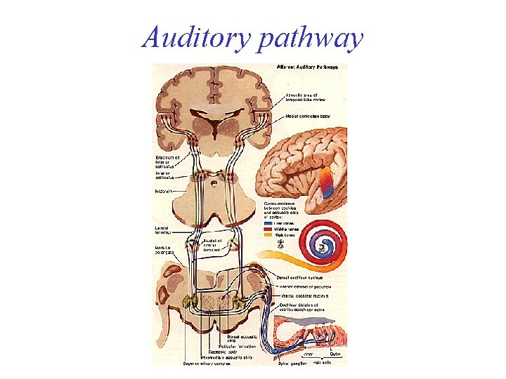
Lateral lemniscus Dorsal acoustic striae Ventral acoustic striae
Auditory pathway
Medial – MLF – adjustment of balance Tectospinal – spino visual reflex Rubrospinal – motor coordination of arm Pons …………
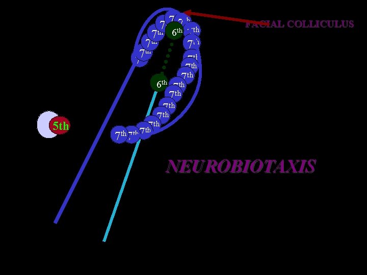
Intermediate - Pons ………… 1. Abducent 2. Facial nucleus (motor) , superior salivatory, NTS -sensory Facial colliculus
th th 7 th th 6 7 7 th 7 7 thth 7 6 th 7 th 5 th FACIAL COLLICULUS th 7 7 th NEUROBIOTAXIS
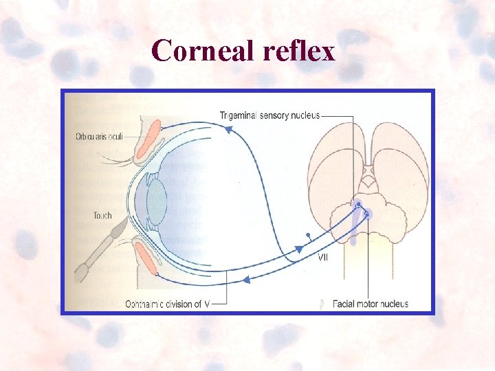
Corneal reflex
Lateral – Vestibular nuclei Inferior cerebral peduncle Cochlear nuclei Spinal nucleus & tract Pons …………
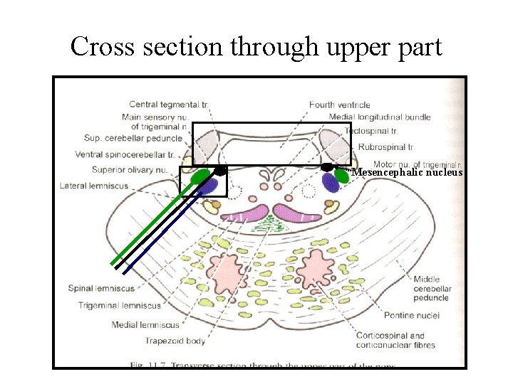
Cross section through upper part Mesencephalic nucleus
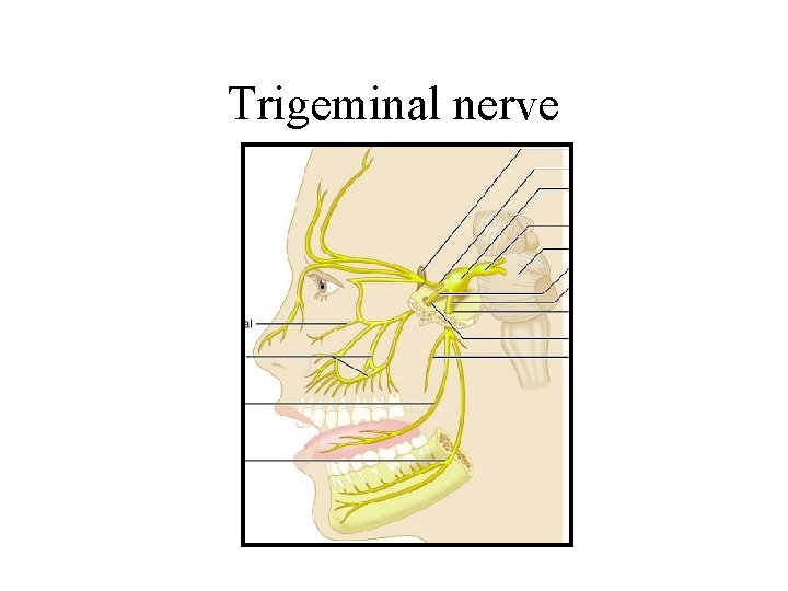
Trigeminal nerve
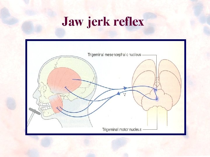
Jaw jerk reflex
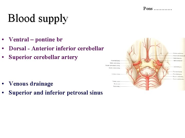
Pons ………… Blood supply • Ventral – pontine br • Dorsal - Anterior inferior cerebellar • Superior cerebellar artery • Venous drainage • Superior and inferior petrosal sinus
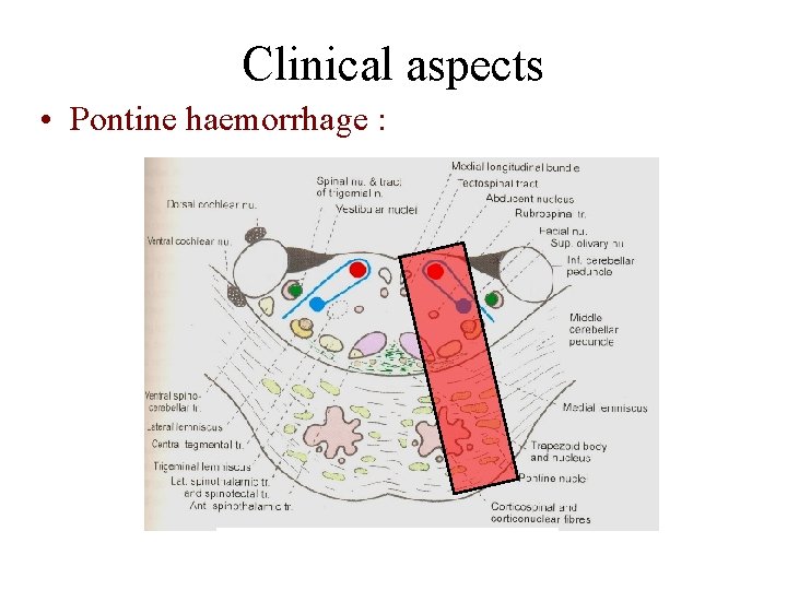
Clinical aspects • Pontine haemorrhage :
AICA syndrome • • • Facial nucleus Solitary nucleus Cochlear nucleus Vestibular nucleus Trigeminal – spinal nucleus and tract Spinothalamic tract- contralateral pain, temperature • Reticular formation(sympathetic fibres)
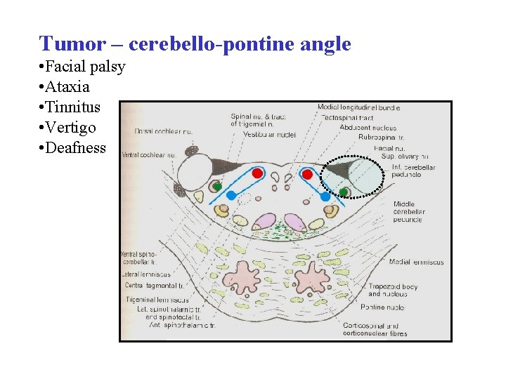
Tumor – cerebello-pontine angle • Facial palsy • Ataxia • Tinnitus • Vertigo • Deafness
FOURTH VENTRICLE
Millard gubler syndrome • Facial Nu – LMN palsy • Abducent Nu – Medial squint • Pyramidal tract – Contralateral hemiplegia
Astrocytoma of pons • Usually occurs in childhood