General Histology Cell junctions A cell junction is


Functional classification according to secretory mechanisms: A- Merocrine/Eccrine Secretion")


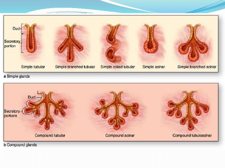
 Histological classification according to duct system: A. Simple (the ducts are not branched)")


- Slides: 19
General Histology
Cell junctions: • A cell junction is a structure of protein complexes within a tissue of a multicellular organism. They are especially abundant in epithelial tissues. • Functions: 1. They provide contact between neighboring cells, between a cell and the extracellular matrix. 2. They built up the paracellular barrier of epithelia and control the paracellular transport.
• There are three types of cell junctions: 1. Tight junctions 2. Gap junctions 3. Adhering junctions
1. Tight junction: v. Tight junctions are the closely associated areas of two cells whose membranes join together forming a virtual impermeable barrier to fluid. v. They are formed by claudin and occludin proteins, joining the cytoskeletons of the adjacent cells. v They prevent the passage of molecules and ions through the space between cells. So materials must actually enter the cells (by diffusion or active transport) in order to pass through the tissue.
2. Gap junctions: A gap junction is made up of membrane proteins form a tunnel between cells called connexons that allows different molecules and ions to pass freely between cells, and transfer chemical and physical signals.
3. Adherens junctions: v They can appear as bands encircling the cell or as spots of attachment to the extracellular matrix. v This type of junctions is good for keeping epithelial surface from sliding out of position.
Glandular Epithelium : v Characteristics: • They are formed by cells specialized to produce a fluid secretion. • They synthesize, store and secrete extracellular products that are not used by the cell itself but are of importance to other parts of organism • Glands can be either multicellular glands (e. g. salivary glands, pancreas) or unicellular glands consist of isolated epithelial cells (e. g. goblet cells)
v Classification of the glands according to the presence of duct: 1 - Exocrine glands : 2 - Endocrine glands • They maintain connection • They are ductless and the with the surface connection with the epithelium via the tubular surface was obliterated ducts through with the during development, so secretory product pass to release their secretory reach the surface (skin, product (hormones) into digestive tract) the bloodstream.
1. Exocrine Glands: v Structure: Histologically, composed from two parts: A. Secretory portion: contains the cells responsible for the secretory process. B. System of ducts: transport the secretion to the exterior of the gland.
Classification of exocrine glands : (1)Functional classification according to secretory mechanisms: A- Merocrine/Eccrine Secretion : • The secretory product is released by exocytosis where secretory granules leave the cell without any further loss of cell substance; (e. g. pancreas, salivary glands)
B- Apocrine Secretion • The apical part of cytoplasm of the cells is lost together with the secretory product; (e. g. female mammary gland)
C- Holocrine Secretion • Breakdown and discharge of the entire secretory cell and its product; (e. g. sebaceous glands of the skin)
(2) Histological classification according to duct system: A. Simple (the ducts are not branched) B. Compound (with a branching duct system) (3) Histological classification according to secretory portion: A. Tubular (shaped like a tube); e. g. glands of intestine, stomach B. Acinar or alveolar (flask-shaped with narrow centrally placed lumen); e. g. pancreas, parotid salivary gland C. Tubuloacinar (combination of the tube ends with a sac-like dilatation); e. g. submandibular and sublingual salivary glands
2. Endocrine Glands: v Structure: According to histological structure there are three main types of endocrine glands: 1. Trabecular 2. Follicular 3. Disseminated
1. Trabecular type – made from the cords of the cells. e. g. parathyroid gland, adrenal glands.
2. Follicular type : The cells form spherical structures – e. g. thyroid gland
3. Disseminated type – the endocrine cells are placed in groups or separately in another organ – e. g. Leydig cells in testis, Langerhans islets of pancreas