Cell junctions Intercellular connections mechanical Tight junctions Maculae


, soma (‘body’).")


 �Receptor �Intracellular signal �Target protein �Response Figure 6 -3:")


 Gate closed Ligand-gated ion channel receptor Ions Plasma")
- Slides: 24
Cell junctions
Intercellular connections mechanical Tight junctions Maculae adherens Gap junctions
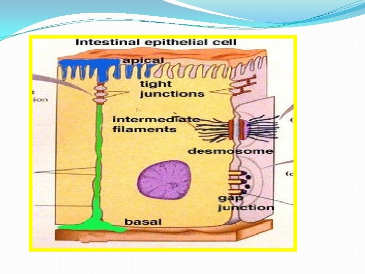
�Connections between the cells of tissue. �Hold the cells together v. Types 1. Tight junctions or zonulae occludens. 2. Desmosomes 3. Gap junction
Cell-to-Cell Interactions Cells within a tissue are connected to each other by cell junctions 1. tight junctions – create sheets of cells 2. anchoring junctions – connect the cytoskeletons of adjacent cells 3. communicating junctions – permit small molecules to pass between cells a. gap junctions – in animal cells b. plasmodesmata – in plant cells 4
�Tight junctions �Ridge like structures between adjacent cells. �Made up of proteins �Cells are bound together. �Found in epithelial cells vintestinal mucosa vrenal tubles �Form selective barrier Egs. leaky and tight junctions.
�They are important in preventing molecules leaking between the cells. �They block the movement of integral membrane proteins between the apical and basolateral surfaces of the cell �It is made up of proteins called claudins and occludins.
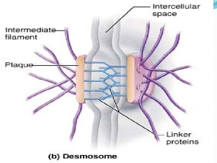
�Desmosome or maculae adherens fasten cells together strengthening the tissue. �Desmo (‘bound’), soma (‘body’). �Desmosomes are localized patches that hold two cells tightly together. They are common in epithelia (e. g. , the skin). �Fibrils arise from this portion to the interior of the membrane. �Hemi desmosome – half desomoses on the basal sides of the cell.
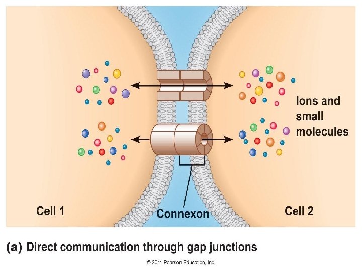
• Gap junctions or communicating junctions. • Gap junctions form channels between cells. • Allow ions and small molecules to pass directly from one cell to another. These channels are formed by membrane proteins called connexin. • Six connexins in the cell membrane form a channel called a connexon
�Functions �. Gap junctions permit electrical signals to pass directly from one cell to another. �Gap junctions are found in heart and smooth muscle cells and between some neurons. �To regulate the membrane potential between cells and allow electrical impulses to pass between cells. �Electrical impulses passing between cardiac muscle cells creating the heart beat. �Also gap junctions provide the contractions of the smooth muscle cells of the uterus during birth
Cell communication
Cell Communication There are four basic mechanisms for cellular communication: 1. direct contact 2. paracrine signaling 3. endocrine signaling 4. synaptic signaling 14
Direct contact
Paracrine signaling – signal released from a cell has an effect on neighboring cells 16
Long-distance signaling Blood vessel Endocrine cell Hormone travels in bloodstream to target cells Target cell (c . ) Hormonal signaling. Specialized endocrine cells screte hormones into body fluids, often the blood.
Communication between cells requires: ligand: the signaling molecule receptor protein: the molecule to which the receptor binds -may be on the plasma membrane or within the cell
How do Cells Communicate? �.
signal pathways �Signal molecule (ligand) �Receptor �Intracellular signal �Target protein �Response Figure 6 -3: Signal pathways
Second Messengers �Many signaling pathways involve small, nonprotein, water-soluble molecules or ions, called second messengers. �These molecules rapidly diffuse throughout the cell. �Second messengers participate in pathways initiated by both G-protein-linked receptors and tyrosine-kinase receptors. �Two of the most important are cyclic AMP and Ca 2+.
�Hormones act on specific receptors. �The receptors are proteins. �When the Ligand binds the receptor, the receptors sends a signal within the cell to modify the activity of the cell. �Different types of receptors �Intra cellular and cell surface receptors �Ion channel receptors , enzymatic and G-protein receptors.
GENERAL PRINCIPLES OF CELL COMMUNICATION Extracellular signal molecules bind to specific receptors An intracellular signaling molecule whose concentration increases (or decreases) in response to binding of an extracellular ligand to a cell-surface receptor.
�Ion channel receptors Signal molecule (ligand) Gate closed Ligand-gated ion channel receptor Ions Plasma Membrane Gate open Cellular response Gate close Figure 11. 7