Cell junction And Cell Communication Cell junctions Specific


, soma (‘body’). ØAdjacent membranes")


 q. Chemical (endocrine, paracrine, autocrine) q")
 Ø channels grow from each cell and join up Ø permits")


 ØReceptor ØIntracellular signal ØTarget protein ØResponse Figure 6 -3:")

 Gate closed Ligand-gated ion channel receptor Ions Plasma")


- G K Pal")
- Slides: 34
Cell junction And Cell Communication
Cell junctions
Specific learning objectives At the end of the class You must know. . § Define Cell junctions and cell communication. § Classify the Cell junctions. § Explain the types of Cell junctions. § Define the cell communication. § Classify the cell communication. § Explain the types of cell communication.
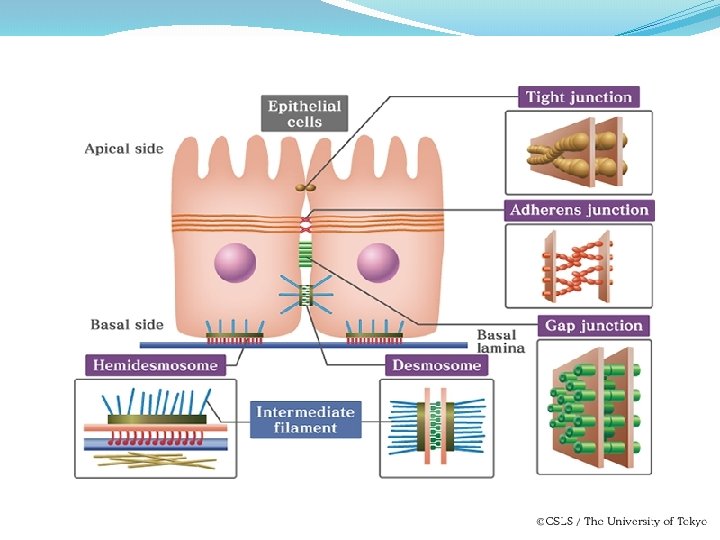
INTERCELLULAR JUNCTION 1. Tight junction 2. Gap junction 3. Desmosomes
Tight junction Ø Ridge like structures between adjacent cells. Ø Made up of proteins Ø Cells are bound together. Ø Found in epithelial cells vintestinal mucosa vrenal tubles Ø Form selective barrier Eg. Blood brain barrier.
Ø They are important in preventing molecules leaking between the cells and providing control passage (selective barrier) for materials through the epithelial sheet. Ø It is made up of proteins called claudins and occludins.
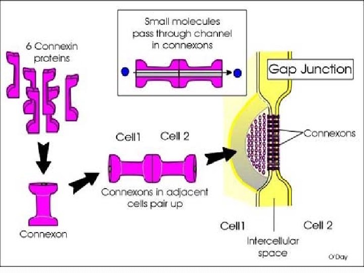
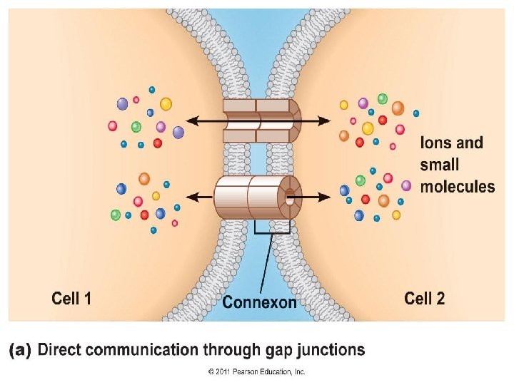
Gap junction ØGap junctions form channels between the cells. Ø Allow ions and small molecules to pass directly from one cell to another. Ø These channels are formed by membrane proteins called connexin. Ø Six connexins in the cell membrane form a channel called a connexon
Functions Ø Gap junctions permit electrical signals to pass directly from one cell to another. Ø Gap junctions are found in heart and smooth muscle cells and between some neurons. Ø To regulate the membrane potential between cells and allow electrical impulses to pass between cells. Ø Electrical impulses passing between cardiac muscle cells it lead conduction of the beat. Ø Also gap junctions provide the contractions of the smooth muscle cells of the uterus during birth
Plasma membranes of adjacent cells Microvilli Intercellular space Basement membrane Intercellular space Channel between cells (connexon) Figure 3. 5 c
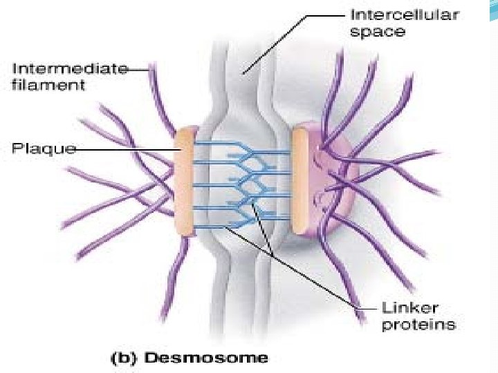
Desmosome Ø Fasten cells together strengthening the tissue. ØDesmo (‘bound’), soma (‘body’). ØAdjacent membranes become thickened and the space in between is filled with filament like material containing Cadherin and some other membrane protein. ØFibrils arise from this portion to the interior of the membrane. ØHemi desmosome – half desomoses on the basal sides of the cell.
Plasma membranes of adjacent cells Microvilli Intercellular space Basement membrane Intercellular space Plaque Intermediate filament (keratin) Linker glycoproteins (cadherins) (b) Desmosomes: Anchoring junctions bind adjacent cells together and help form an internal tension-reducing network of fibers. Figure 3. 5 b
INTERCELLULAR COMMUNICATION
Three Communication Systems q Electrical (gap junctions ) q. Chemical (endocrine, paracrine, autocrine) q Neural
ELECTICAL (GAP JUNCTIONS) Ø channels grow from each cell and join up Ø permits movement of small molecules between adjacent cells Ø The chemical messenger moves from cell to cell without entering the ECF Ø Eg. flow of Ca 2+ between heart muscle cells permits synchronous contraction of cells to generate heart beat
Chemical signaling between cells q Autocrine: The cell respond to a signal that it (itself) has secreted. (within cytoplasm or on the cell membrane). Eg: PAF of Platelets. q Paracrine: Chemical signal diffuses in an area and act on a nearby cell with receptors for that signal. Eg: Glucagon from alpha cells of Islets of Langerhans, which inhibit beta cells. q. Endocrine: the chemical signals are secreted and enter the blood circulation where they are transported to their target
Cell signalling can act over long or short distances
Paracrine signaling Signal released from a cell has an effect on neighboring cells 21
Long-distance signaling Blood vessel Endocrine cell Hormone travels in bloodstream to target cells Target cell (c . ) Hormonal signaling. Specialized endocrine cells screte hormones into body fluids, often the blood.
Neural communication
Long Distance Communication Neurons and Neurohormones
Communication between cells requires q. Ligand: the signaling molecule q. Receptor protein: the molecule to which the receptor binds- may be on the plasma membrane or within the cell
How do Cells Communicate? �.
Signal pathways ØSignal molecule (ligand) ØReceptor ØIntracellular signal ØTarget protein ØResponse Figure 6 -3: Signal pathways
�Hormones act on specific receptors ØThe receptors are proteins. ØWhen the Ligand binds the receptor, the receptors sends a signal within the cell to modify the activity of the cell. ØDifferent types of receptors ØIntra cellular and cell surface receptors ØIon channel receptors , enzymatic and G-protein receptors.
�Ion channel receptors Signal molecule (ligand) Gate closed Ligand-gated ion channel receptor Ions Plasma Membrane Gate open Cellular response Gate close Figure 11. 7
Seroid hormones bind to Intra cellular receptors
Pathway involving c. AMP as a secondary messenger
A signaling molecule may induce different responses in different cell types
SUMMARY
REFERENCES 1. Comprehensive Textbook of Medical physiology (Vol 1 first edition)- G K Pal 2. Text book of medical physiology (Vol 2 6 th edition)- A K Jain.