Fractures of the Spine in Children MD SINA


")


,")


- Slides: 29
Fractures of the Spine in Children , MD SINA HOSPITAL Vahid Farsio
Important Pediatric Differences Anatomical differences n Radiologic differences n Increased elasticity n
Anatomy – C 1 n n 3 ossification centers at birth – body and 2 neurocentral arches Neurocentral synchondroses (F) fuse at about 7 years of age n. Copley. Cervical spine disorders in infants and children. J Am Acad Orthop Surg. 1998; 6: 204.
Anatomy – C 2 n n 4 ossification centers at birth – body, 2 neural arches, dens Neurocentral synchondroses (F) fuse at age 3 -6 years Synchondrosis between body and dens (L) fuses age 3 – 6 years Thus no physis / synchondrosis should be visible on open mouth odontoid view in child older than 6 years n. Copley. Cervical spine disorders in infants and children. J Am Acad Orthop Surg. 1998; 6: 204.
Anatomy – Lower Cervical Vertebrae C 3 – C 7 n Neurocentral synchondroses (F) fuse at age 3 -6 years n Ossified vertebral bodies wedge shaped until square at about age 7 n. Copley. Cervical spine disorders in infants and children. J Am Acad Orthop Surg. 1998; 6: 204.
Epidemiology of Spinal Trauma in Children n Spinal injury is rare in children n Pediatric vertebral injuries occur 60 -80% of the time in the cervical region (30 -40% of all vertebral injuries in adults) n Overall incidence of spinal injury in children is 1 -2% n Motor Vehicle Accidents are the leading cause of pediatric SCI (60% of cases)…with falls and sports injuries (football and diving) thereafter n M: F ratio of 2: 1
n Pediatric vs. Adult Spine Anatomy ……. . Not just little adults! Children younger than 8 yrs are more susceptible to C- Children younger than 8 yrs are more susceptible to Cspine injuries because; n Larger head to body proportion Higher fulcrum……. “point of maximal mobility” (C 2 -3 at birth, C 3 -5 at 8 -12 yrs old to C 5 -6 at 12 yrs old and adults) n Weaker cervical musculature n Increased ligamentous laxity leading to greater mobility of the cspine n Immature joints and Ossification centers n Horizontal facet joints that facilitate sliding of the upper C-spine n Spinal columns are more elastic than the spinal cord (tolerating n
Key History and PE Components n History n n n Cause…. MVA, Sports (Football/Diving), Falls Mechanism…. . Hyperflexion (Clay shoveler’s or Teardrop Fx’s), hyperextension (Hangman’s Fx), Rotational (Jumped Facets), Compression or axial loading (Jefferson/Burst Fx) Symptoms…. . Numbness, tingling, or weakness during any time since accident even if resolved Predisposing conditions…. . 15% Down’s Syndrome pts have atlantoaxial instability, Achondroplasia (Cervicomedullary Junction stenosis) Physical Exam n n n Testing for motor or sensory deficits and levels if present DTR’s and rectal tone High index for Multisystem trauma (40% of cases have associated intrabdominal injuries)
C Spine Immobilization for Transport in Children n n Large head will cause increased flexion of C spine on standard backboard Bump beneath upper T spine or cutout in board for head to transport child with spine in neutral alignment
Imaging Evaluation of Spine Injuries n Are Xrays indicated? n NEXUS Study Criteria n Lateral, AP and Odontoid view n Flexion-Extension views n CT C-spine n MRI
l l l n. Young Child n. Mature anterior wedging of vertebral bodies horizontal alignment of facet joints Children prone to anterior dislocation
Alignment Pseudosubluxation n n 24% C 2 on C 3 14% C 3 on C 4 (Age <7 years) n Swischuk’s line: posterior arch of C 1 to C 3 – should come within 1 mm of post arch of C 2
C 2 -3 Pseudosubluxation n Look for significant prevertebral soft tissue n. Shaw. Pseudosubluxation of C 2 on C 3 in polytraumatized children: Prevalence and significance. Clin Radiol 1999; 54: 377.
Dens n n n Predental space – allow up to 5 mm in young children Subdental synchondrosis lucency at base of dens Dens fuses with body of C 2 between ages 4 - 6 years A thin lucency may be appreciable on the lateral view for many years (50% up to age 11) May have ossification centre at tip of dens os terminale
Prevertebral Soft Tissues n n Allowable thickness changes with age In general: n n n Above glottis: ½ vertebral body Below glottis: 1 vertebral body Often falsely thickened 2° to neck flexion (big occiput) or expiration
n. Optimal if pt neck extended (and x ray taken at END inspiration – less false +).
Cervical Spine Injuries from Birth Trauma n Can occur n Upper cervical injuries may be a cause of perinatal death n. Newborn with C 5/6 fracture dislocation
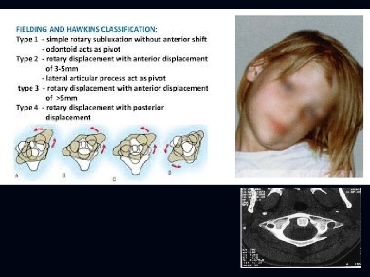
Os Odontoideum n Usually asymptomatic n Pain , mylopathy instability n. Fielding. Os odontoideum. J Bone Joint Surg Am 1980; 62: 376.
Atlanto-Occipital Dislocation n n 2. 5 x more common in children than adults Due to small occipital condyles and horizontal atlanto-occipital joints Suspect if distance between occipital condyles and C 1 is > 5 mm at any point Usually have ++ soft tissue swelling
Dens Fracture n Suspicious for dens fracture: n n n widening of the synchondrosis anterior tilting of the odontoid Believed to have high miss rate – can lead to chronic problems
Hangman’s Fracture n. The hangman's fracture i n. Pseudosubluxation !!
Spinal Cord Injury Without Radiographic Abnormality SCIWORA n n n Defined as traumatic myelopathy in the absence of findings on plain radiographs, flexion-extension radiographs and cervical CT scan mechanism is acceleration-deceleration or rotation injury 30 -50% delayed onset of neurologic deficits from 30 mins-4 days n MRI should be done n require immobilization n Mild SCIWORA : Cervical cord neurapraxia
Thoracic Spine Fractures n Less common spinal fracture in children than in more mobile regions n Child abuse in very young n. Slotkin. Thoracolumbar spinal trauma in children. Neurosurg. Clin. N. Am. 2007; 18: 621.
Thoracolumbar Junction Injuries T 11 -L 2 Classically lap-belt flexion-distraction injuries n Chance fractures and variants n High association with intraabdominal injury (50 -90%) n Neurologic injury infrequent but can occur n n. Arkader. Pediatric chance fractures: a multicenter perspective. J Pediatr Orthop.
Seatbelt Injury Classification n. Rumball. Seat-belt injuries of the spine in young children. J Bone Joint Surg Br. 1992; 74: 571.
4 year Lap Belt Intraabdominal Injuries, Paraplegic
Lumbar Apophyseal Injuries Slipped Apophysis n Compression-shear injuries n Same age group as SCFE n Typically adolescent males, inferior endplates of L 4 or L 5
Thank You