ECG 101 Narrow Complex Tachycardia Scott E Ewing







- Slides: 57
ECG 101 - Narrow Complex Tachycardia Scott E. Ewing DO Lecture #10
Tachyarrhythmias n Atrial n n n n n Sinus tachycardia Inappropriate sinus tachycardia Atrial premature beats Atrial fibrillation Atrial flutter Atrial tachycardia Atrio-ventricular nodal re-entrant tachycardia (AVNRT) Atrioventricular re-entrant tachycardia (AVRT) Ventricular n n n Ventricular tachycardia Ventricular fibrillation Ventricular premature beats
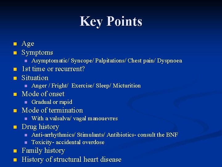
Key Points n n Age Symptoms n n n 1 st time or recurrent? Situation n n With a valsalva/ vagal manouevres Drug history n n Gradual or rapid Mode of termination n n Anger / Fright/ Exercise/ Sleep/ Micturition Mode of onset n n Asymptomatic/ Syncope/ Palpitations/ Chest pain/ Dyspnoea Anti-arrhythmics/ Stimulants/ Antibiotics- consult the BNF Toxicity- accidental overdose Family history History of structural heart disease
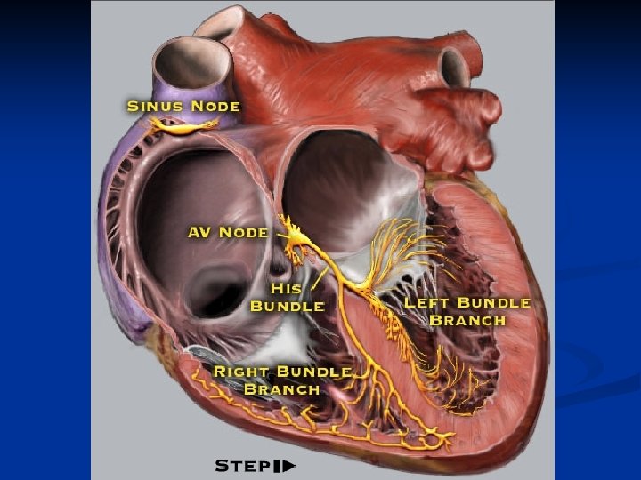
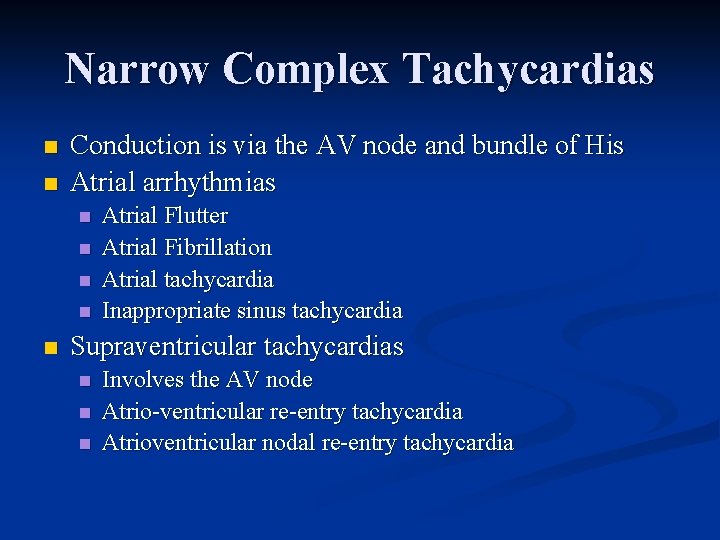
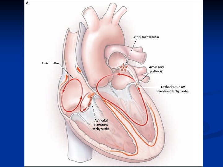
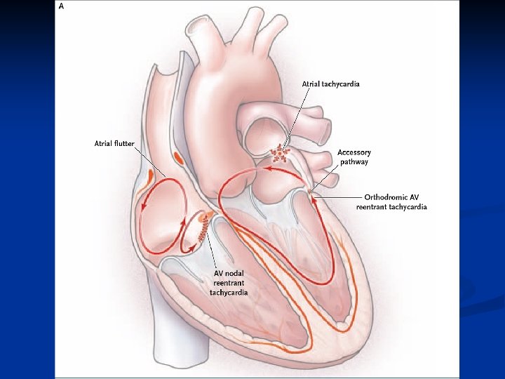
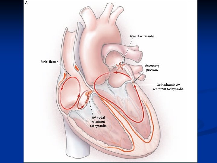
Narrow Complex Tachycardias n n Conduction is via the AV node and bundle of His Atrial arrhythmias n n n Atrial Flutter Atrial Fibrillation Atrial tachycardia Inappropriate sinus tachycardia Supraventricular tachycardias n n n Involves the AV node Atrio-ventricular re-entry tachycardia Atrioventricular nodal re-entry tachycardia
Atrial Fibrillation n Most common arrhythmia 1% patients > 60 yrs n 5% patients > 70 rs n 10% patients >75 yrs n n Presenting features May be asymptomatic n Vagally mediated AF n Alcohol binges n
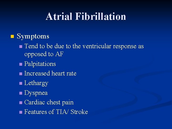
Atrial Fibrillation n Symptoms Tend to be due to the ventricular response as opposed to AF n Palpitations n Increased heart rate n Lethargy n Dyspnea n Cardiac chest pain n Features of TIA/ Stroke n
Atrial Fibrillation n Associated conditions n n n Drugs n n n Thyrotoxicosis Hypertension Heart failure Valve disease Adenosine Digoxin Miscellaneous n n n Chest infection Cardiac surgery Cholecystitis
Atrial Fibrillation n Examination Irregularly irregular pulse n At high ventricular rates there may be a pulse deficit n n i. e. pulse at the apex is higher than the palpated rate at the wrist Hypertension n Absence of A wave in the JVP n Variation of the intensity of S 1 n
Atrial Fibrillation
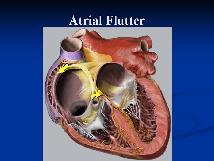
Atrial Flutter
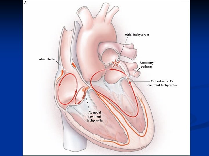
Atrial Flutter n A macro re-entrant arrhythmia n n n Typical atrial flutter n n n Anatomical barrier Zone of slow conduction Contained within the right atrium Constrained anteriorly by the tricuspid valve Constrained posteriorly by the crista terminalis and eustachian ridge Travels in a counterclockwise direction around the atrium Atypical atrial flutters n n n Counterclockwise flutter ASD/ scar related flutter Perimitral flutter
Atrial Flutter n Tends to occur in middle age n n Pulmonary embolism n n Mitral or Tricuspid disease Atrial septal defects Chronic ventricular failure Toxic and Metabolic conditions n n Commonly presents with a sinus tachycardia Associated valve disease n n Probably due to atrial dilatation Alcohol/ thyrotoxicosis/ pericarditis Previous ablation
Atrial Flutter n n n Examination Rarely helpful in establishing the diagnosis Regular pulse (150 bpm- 2: 1, 75 bpm 4: 1 - can be slower) May see rapid, regular flutter waves in the JVP Heart sounds n n Constant intensity of S 1 if relationship of flutter waves to QRS is constant Carotid massage or adenosine n n Allows flutter waves to be seen more easily Ventricular rate will increase when CSM is stopped
Atrial Flutter
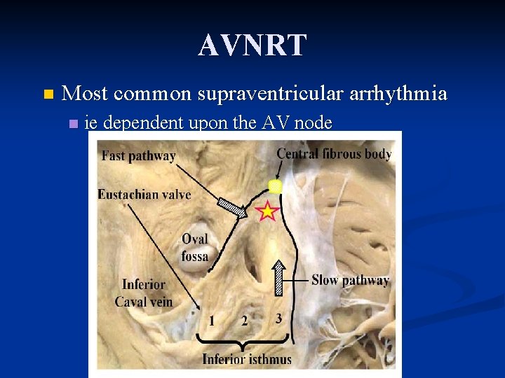
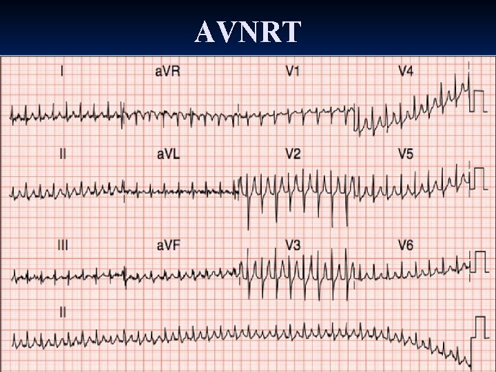
AVNRT n Most common supraventricular arrhythmia n ie dependent upon the AV node
AVNRT Typically 3 rd and 4 th Decade n Recurrent palpitations n RAPID onset and RAPID offset n Patient may feel an ectopic beat to initiate/ terminate the arrhythmia n Vagal maneuvres to terminate the arrhythmia n Anxiety/ breathless/ palpitations n n Syncope (due to high rate or due to transient asystole at termination)
AVNRT
AVNRT
AVNRT
AVRT
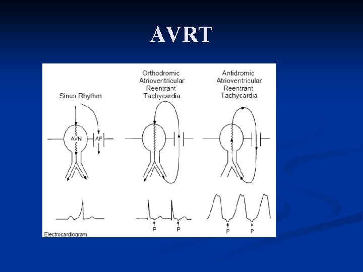
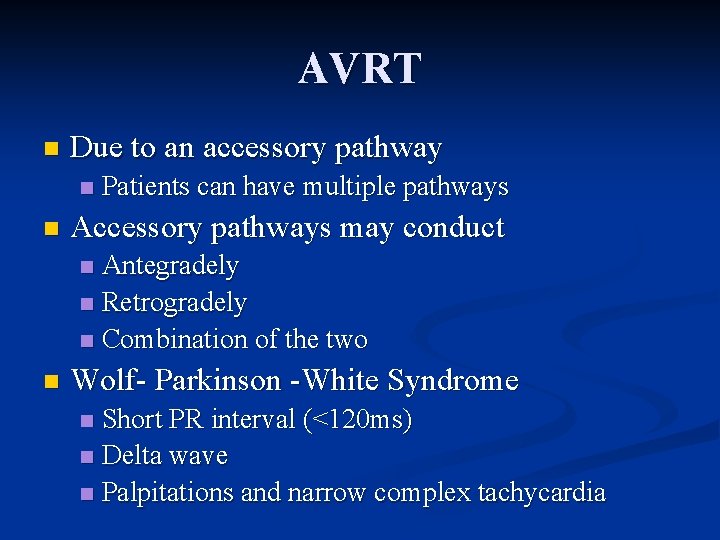
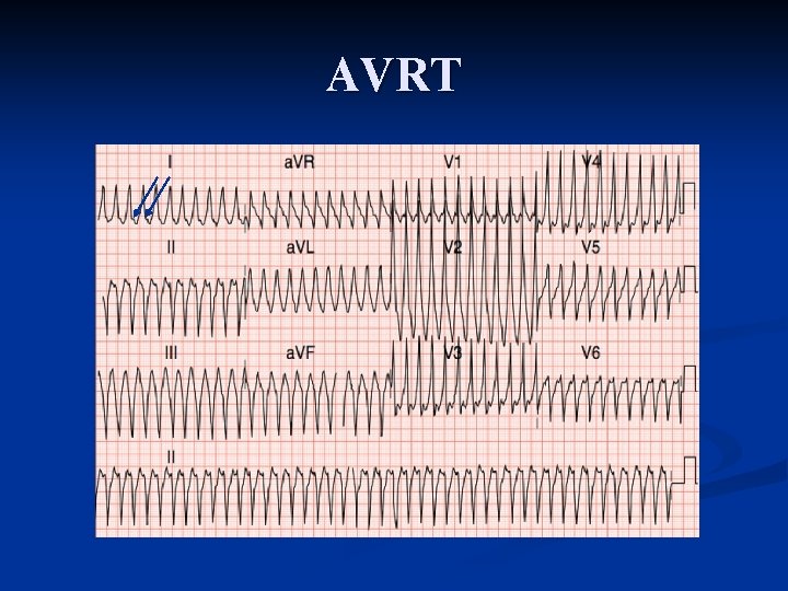
AVRT n Due to an accessory pathway n n Patients can have multiple pathways Accessory pathways may conduct Antegradely n Retrogradely n Combination of the two n n Wolf- Parkinson -White Syndrome Short PR interval (<120 ms) n Delta wave n Palpitations and narrow complex tachycardia n
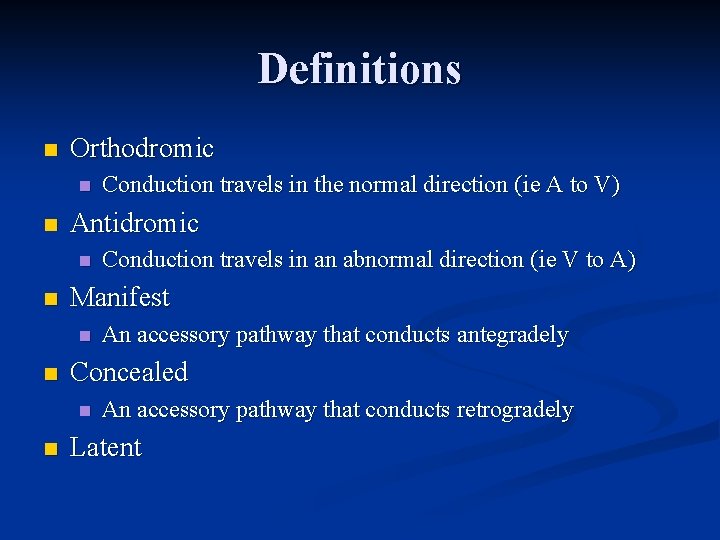
Definitions n Orthodromic n n Antidromic n n An accessory pathway that conducts antegradely Concealed n n Conduction travels in an abnormal direction (ie V to A) Manifest n n Conduction travels in the normal direction (ie A to V) An accessory pathway that conducts retrogradely Latent
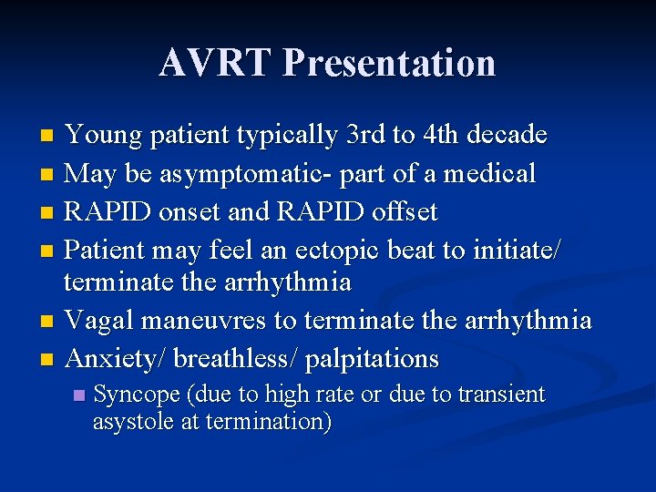
AVRT Presentation Young patient typically 3 rd to 4 th decade n May be asymptomatic- part of a medical n RAPID onset and RAPID offset n Patient may feel an ectopic beat to initiate/ terminate the arrhythmia n Vagal maneuvres to terminate the arrhythmia n Anxiety/ breathless/ palpitations n n Syncope (due to high rate or due to transient asystole at termination)
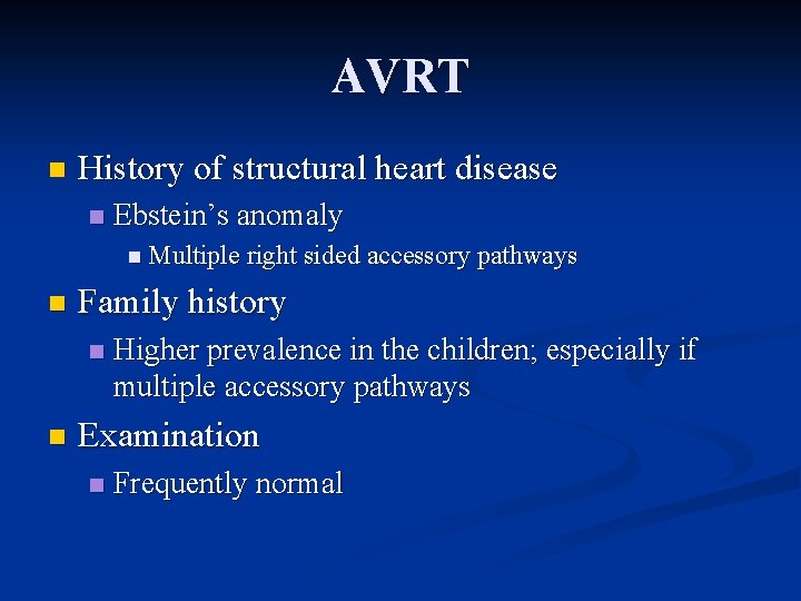
AVRT n History of structural heart disease n Ebstein’s anomaly n Multiple right sided accessory pathways n Family history n n Higher prevalence in the children; especially if multiple accessory pathways Examination n Frequently normal
AVRT
Focal Atrial Tachycardia n n n Typically older patients >6 th decade Frequently have structural heart disease, pulmonary disease Symptoms are related to n n n Rapid initiation n Rate (120 -250 bpm) Underlying heart disease Rate can increase over a few beats as the AV node “warms up” No consistent effect with vagal maneuvres Digoxin / Alcohol/ Lung disease/ Metabolic derangements
Focal Atrial Tachycardia n Regular pulse n Exceptions n If atrial tachycardia is fast the AV node may Wenckebach (Mobitz Type I) n If more than one focus (Multifocal atrial tachycardia) Check for signs of pulmonary disease n Cannon A waves n Variable S 1 n
Examples
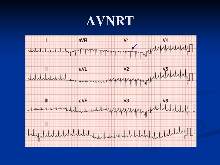
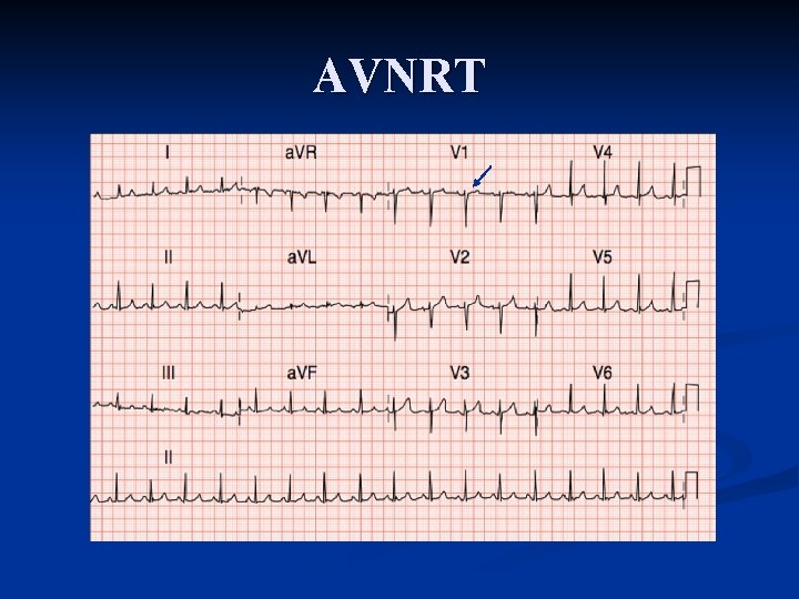
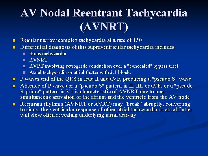
AV Nodal Reentrant Tachycardia (AVNRT) n n Regular narrow complex tachycardia at a rate of 150 Differential diagnosis of this supraventricular tachycardia includes: n n n n Sinus tachycardia AVNRT AVRT involving retrograde conduction over a "concealed" bypass tract Atrial tachycardia or atrial flutter with 2: 1 block. P waves end of the QRS in lead II and a. VF, producing a "pseudo S" wave Absence of P waves or a "pseudo S" pattern in II, III, or a. VF, or a "pseudo R prime" pattern in V 1 is characteristic of AVNRT due to near simultaneous activation of the atrium and the ventricle from the AV node Reentrant rhythms (AVNRT or AVRT) may "break" abruptly, converting to sinus; the ventricular response of other atrial tachcyardia or atrial flutter will slow often revealing underlying atrial activity
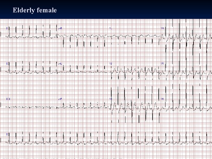
Elderly female
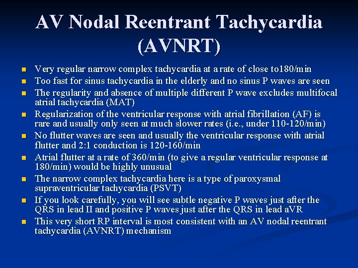
AV Nodal Reentrant Tachycardia (AVNRT) n n n n n Very regular narrow complex tachycardia at a rate of close to 180/min Too fast for sinus tachycardia in the elderly and no sinus P waves are seen The regularity and absence of multiple different P wave excludes multifocal atrial tachycardia (MAT) Regularization of the ventricular response with atrial fibrillation (AF) is rare and usually only seen at much slower rates (i. e. , under 110 -120/min) No flutter waves are seen and usually the ventricular response with atrial flutter and 2: 1 conduction is 120 -160/min Atrial flutter at a rate of 360/min (to give a regular ventricular response at 180/min) would be highly unusual The narrow complex tachycardia here is a type of paroxysmal supraventricular tachycardia (PSVT) If you look carefully, you will see subtle negative P waves just after the QRS in lead II and positive P waves just after the QRS in lead a. VR This very short RP interval is most consistent with an AV nodal reentrant tachycardia (AVNRT) mechanism
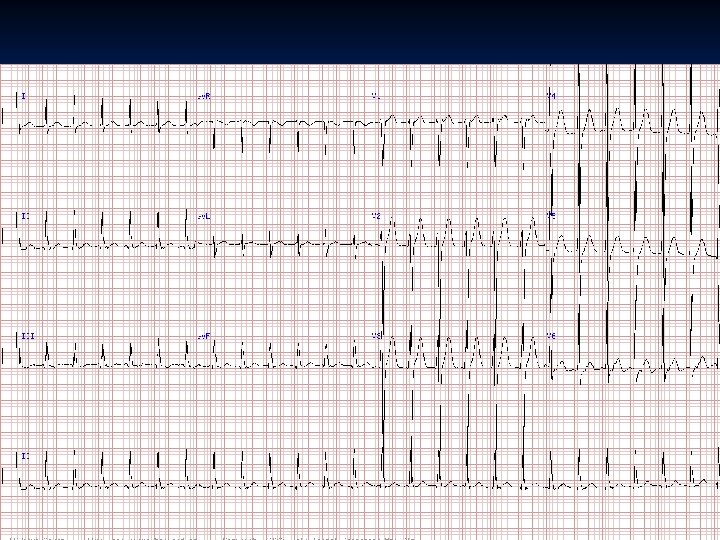
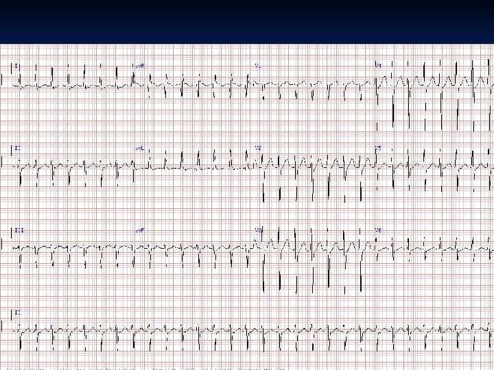
AV Nodal Reentrant Tachycardia (AVNRT) n n n Regular, narrow complex tachycardia at a rate of 150 bpm without preceding P waves However, if you look very closely, there appears to be a negative (retrograde) P wave in the early part of the ST segment of lead V 6 and a subtle P waves just after the terminus of the S waves in leads II, II and a. VF, along with a positive P wave in lead a. VR causing a “pseudo” R wave pattern Most likely diagnosis, therefore, and is AV nodal reentrant tachycardia, a common type of paroxysmal supraventricular tachycardia (PSVT) Resting rate of 150 bpm by itself is strongly against sinus tachycardia in this older gentleman and no sinus P waves are identifiable No atrial flutter waves are seen and the near-metronomically regular and rapid rate is not consistent with atrial fibrillation Ventricular rate here (150/min) also precludes atrial tachycardia with 2: 1 block since this would yield an atrial rate of 300/min, too fast for atrial tachycardia (but typical of atrial flutter)
AV Nodal Reentrant Tachycardia (AVNRT) n n n Narrow complex tachyardia at 180/min with probable subtle retrograde P waves just after the QRS (may need to print this out to see in lead a. VR and II) Therefore, likely diagnosis is AV nodal reentrant tachycardia (AVNRT); although, concealed bypass tract tachycardia or atrial tachycardia not excluded This is, of course, NOT sinus and is also NOT atrial flutter (no flutter waves and ventricular rate is too fast for flutter with 2: 1 conduction)
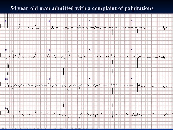
54 year-old man admitted with a complaint of palpitations
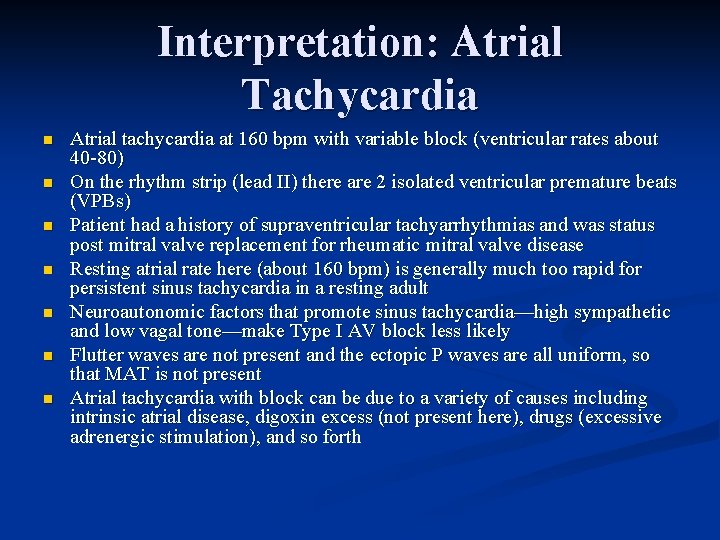
Interpretation: Atrial Tachycardia n n n n Atrial tachycardia at 160 bpm with variable block (ventricular rates about 40 -80) On the rhythm strip (lead II) there are 2 isolated ventricular premature beats (VPBs) Patient had a history of supraventricular tachyarrhythmias and was status post mitral valve replacement for rheumatic mitral valve disease Resting atrial rate here (about 160 bpm) is generally much too rapid for persistent sinus tachycardia in a resting adult Neuroautonomic factors that promote sinus tachycardia—high sympathetic and low vagal tone—make Type I AV block less likely Flutter waves are not present and the ectopic P waves are all uniform, so that MAT is not present Atrial tachycardia with block can be due to a variety of causes including intrinsic atrial disease, digoxin excess (not present here), drugs (excessive adrenergic stimulation), and so forth
AV Reentrant Tachycardia (AVRT)
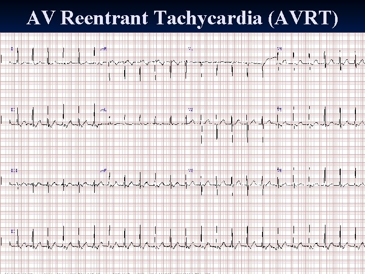
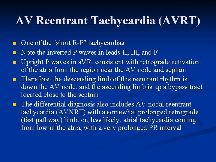
AV Reentrant Tachycardia (AVRT) n n n One of the "short R-P" tachycardias Note the inverted P waves in leads II, III, and F Upright P waves in a. VR, consistent with retrograde activation of the atria from the region near the AV node and septum Therefore, the descending limb of this reentrant rhythm is down the AV node, and the ascending limb is up a bypass tract located close to the septum The differential diagnosis also includes AV nodal reentrant tachycardia (AVNRT) with a somewhat prolonged retrograde (fast pathway) limb, or, less likely, atrial tachycardia coming from low in the atria, with a very prolonged PR interval
Questions?
54 -year-old man with congestive heart failure
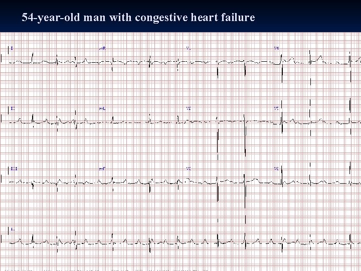
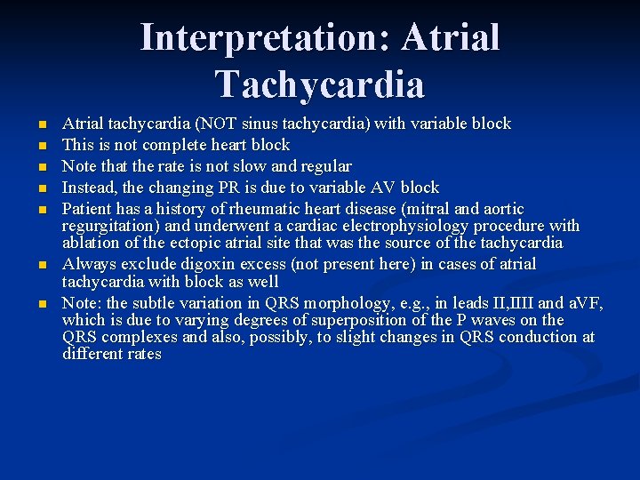
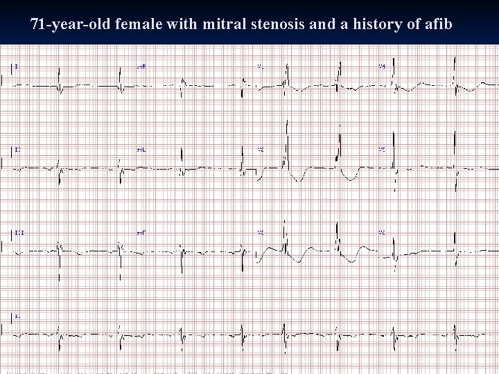
Interpretation: Atrial Tachycardia n n n n Atrial tachycardia (NOT sinus tachycardia) with variable block This is not complete heart block Note that the rate is not slow and regular Instead, the changing PR is due to variable AV block Patient has a history of rheumatic heart disease (mitral and aortic regurgitation) and underwent a cardiac electrophysiology procedure with ablation of the ectopic atrial site that was the source of the tachycardia Always exclude digoxin excess (not present here) in cases of atrial tachycardia with block as well Note: the subtle variation in QRS morphology, e. g. , in leads II, IIII and a. VF, which is due to varying degrees of superposition of the P waves on the QRS complexes and also, possibly, to slight changes in QRS conduction at different rates
71 -year-old female with mitral stenosis and a history of afib
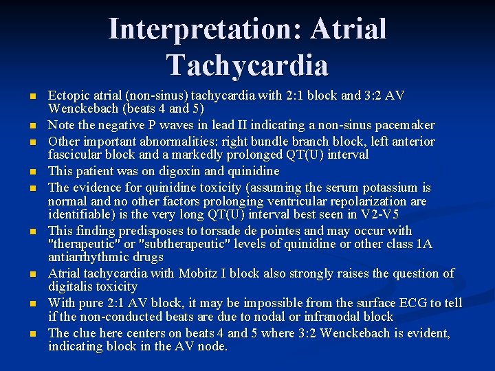
Interpretation: Atrial Tachycardia n n n n n Ectopic atrial (non-sinus) tachycardia with 2: 1 block and 3: 2 AV Wenckebach (beats 4 and 5) Note the negative P waves in lead II indicating a non-sinus pacemaker Other important abnormalities: right bundle branch block, left anterior fascicular block and a markedly prolonged QT(U) interval This patient was on digoxin and quinidine The evidence for quinidine toxicity (assuming the serum potassium is normal and no other factors prolonging ventricular repolarization are identifiable) is the very long QT(U) interval best seen in V 2 -V 5 This finding predisposes to torsade de pointes and may occur with "therapeutic" or "subtherapeutic" levels of quinidine or other class 1 A antiarrhythmic drugs Atrial tachycardia with Mobitz I block also strongly raises the question of digitalis toxicity With pure 2: 1 AV block, it may be impossible from the surface ECG to tell if the non-conducted beats are due to nodal or infranodal block The clue here centers on beats 4 and 5 where 3: 2 Wenckebach is evident, indicating block in the AV node.
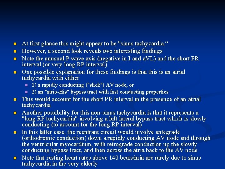
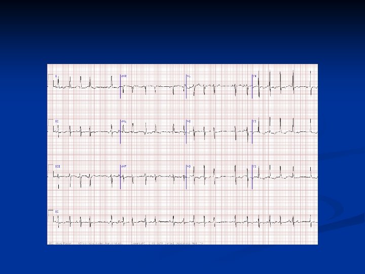
85 -year-old woman
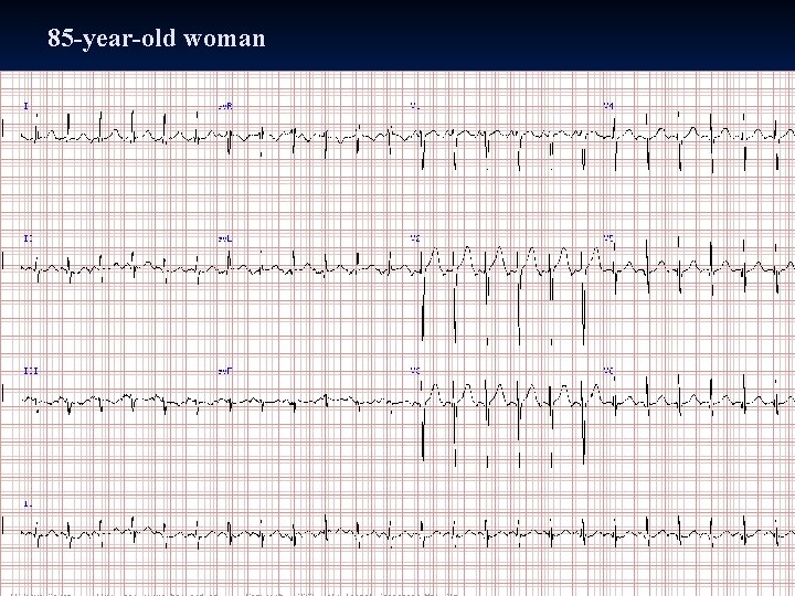
n n At first glance this might appear to be "sinus tachycardia. “ However, a second look reveals two interesting findings Note the unusual P wave axis (negative in I and a. VL) and the short PR interval (or very long RP interval) One possible explanation for these findings is that this is an atrial tachycardia with either n n n 1) a rapidly conducting ("slick") AV node, or 2) an "atrio-His" bypass tract with fast conducting properties This would account for the short PR interval in the presence of an atrial tachycardia Another possibility for this non-sinus tachycardia is that it represents a "long RP tachycardia" involving a left lateral bypass tract which is slowly conducting (to account for the long RP interval) In this latter case, the reentrant circuit would involve antegrade (orthodromic conduction) down a rapidly conducting AV node and through the ventricular myocardium, with retrograde conduction up the slowly conducting bypass tract, and then across the atria back to the AV node Note that resting heart rates above 140 beats/min are rarely due to sinus tachycardia in the very elderly
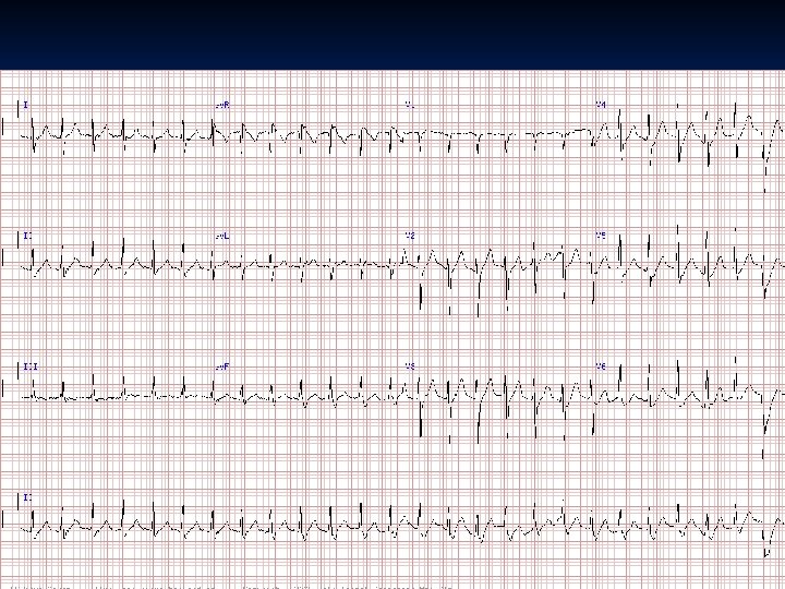
n What is this rhythm? * * * Difficulty rating a) Atrial flutter with variable blockb) Atrial fibrillationc) Wandering atrial pacemakerd) Atrio-ventricular reentrant tachycardiae) Multifocal atrial tachycardia. Answer: e) Three or more consecutive ectopic (non-sinus) P waves are present at a rapid rate. The patient had an exacerbation of severe Chronic obstructive pulmonary disease (COPD), a common substrate in patients with Multifocal atrial tachycardia (MAT). In contrast, Wandering atrial pacemaker (WAP) is a slower rhythm, usually with more gradual change in P waves. (Note that some of the ectopic P waves are non-conducted. )
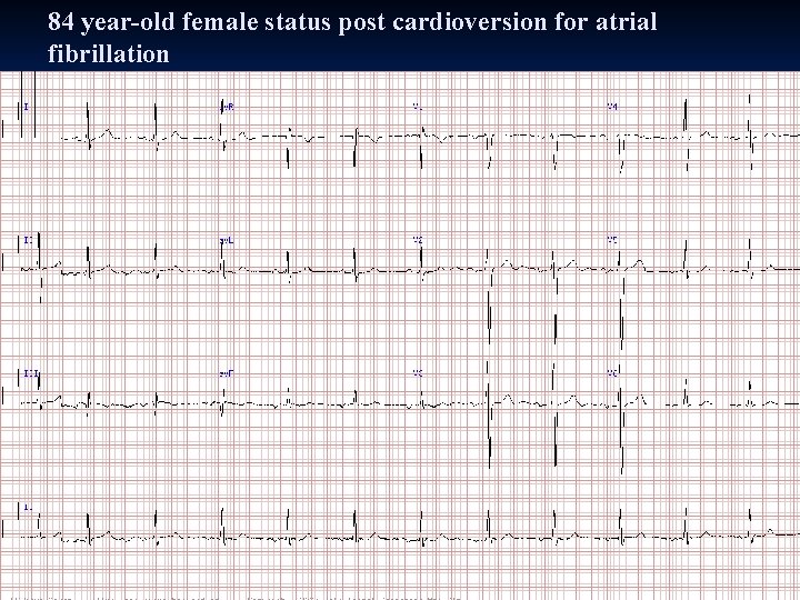
84 year-old female status post cardioversion for atrial fibrillation
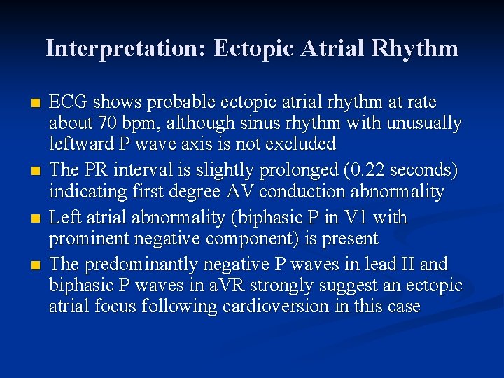
Interpretation: Ectopic Atrial Rhythm n n ECG shows probable ectopic atrial rhythm at rate about 70 bpm, although sinus rhythm with unusually leftward P wave axis is not excluded The PR interval is slightly prolonged (0. 22 seconds) indicating first degree AV conduction abnormality Left atrial abnormality (biphasic P in V 1 with prominent negative component) is present The predominantly negative P waves in lead II and biphasic P waves in a. VR strongly suggest an ectopic atrial focus following cardioversion in this case