DNA RNA and Gel Electrophoresis Central Dogma of

 Transcription Nucleus RNA (copies of")

 � DNA is the genetic material")
. �Three different types of RNA, (messenger (m.")


 ﺩﺍﺧﻞ ﺍﻟﺨﻠﻴﺔ ﺍﻟﺤﻴﺔ DNA ﺍﻟﻨﺴﺦ ﺍﻟﻤﺘﻤﺎﺛﻞ ﻟﻠﺤﻤﺾ ﺍﻟﻨﻮﻭﻱ")


 q PCR is a much quicker tool for duplicating DNA")


, power supply, UV viewing table, camera (or")


- Slides: 36
DNA, RNA and Gel Electrophoresis
Central Dogma of Biology DNA (genetic information in genes) Transcription Nucleus RNA (copies of genes) Translation Cytoplasm proteins (functional molecules)
Structure of DNA
DNA Notes � DNA (DNA = deoxyribonucleic acid) � DNA is the genetic material of all living cells and of many viruses. � DNA is: an alpha double helix of two polynucleotide strands. � The genetic code is the sequence of bases on one of the strands. � A gene is a specific sequence of bases which has the information for a particular protein. � DNA is self-replicating - it can make an identical copy of itself. � Replication allows the genetic information to pass faithfully to the next generation. � The chromosomes contain 90% of the cell’s DNA and 10% is present in mitochondria and chloroplasts. � A single unit in the chain is a nucleotide. This consists of a phosphate group, a pentose sugar (D = DNA; R = RNA) and an organic base (ATGC = DNA; AUGC = RNA) � Adenine (A) and Guanine (G) are purine bases and are long. � Thymine (T) and Cytosine (C) are pyrimidine bases are short. � Need one long and one short nucleotide per pair. � Hydrogen bonds link the complementary base pairs: Two between A and T (A = T) - Three between G and C (G ≡ C)
RNA Notes �RNA (RNA = ribonucleic acid). �Three different types of RNA, (messenger (m. RNA), transfer (t. RNA) and ribosomal (r. RNA). �All are made in the nucleus (transcription). �ribosomes are synthesized in the nucleolus; �m. RNA prepared there too – introns removed �All types of RNA are involved in protein synthesis: �m. RNA: copies the information from the DNA. �t. RNA: carries the specific amino acid to the m. RNA in contact with the ribosome. �r. RNA: makes up 55% of ribosomes (the other 45% = protein).
Differences between DNA and RNA � DNA is double stranded; RNA is a single stranded. � N. B. ATP is also a nucleotide, with ribose as the pentose sugar. � DNA contains the pentose sugar deoxyribose; RNA contains the pentose sugar ribose. � DNA has the base Thymine (T) but not Uracil (U); RNA has U but not T. � DNA is very long (billions of bases); RNA is short (hundreds to thousands of bases) � DNA is self-replicating, RNA is copied from the DNA so it is not self-replicating � The genetic information is held within the base sequence along a DNA strand. � A codon is a sequence of three nucleotides, coding for one amino-acid. � The genetic code is universal, thus all life must have had a common ancestor (i. e. evolution)
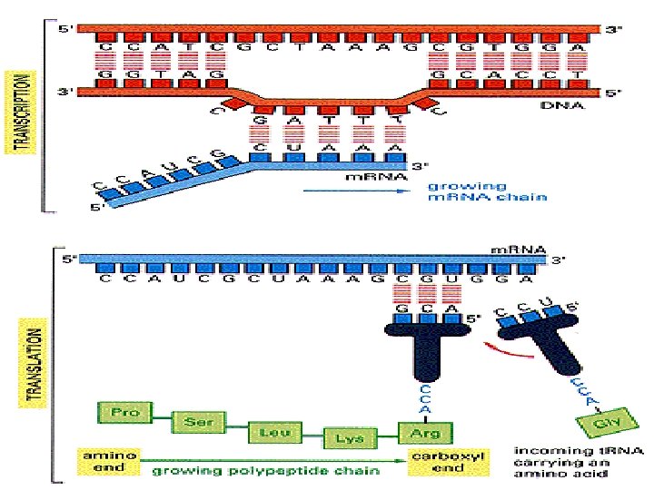
How Does DNA Specify the Sequence of a Protein? �A DNA sequence must be “decoded” to make a protein �This decoding requires creation of an RNA template �Creation of “messenger RNA” is called transcription �Creation of protein from the m. RNA is called translation
Transcription occurs in the nucleus; Translation occurs in the cytoplasm.
Relationship Between Genes and Proteins
DNA Replication in Cells (in vivo) ﺩﺍﺧﻞ ﺍﻟﺨﻠﻴﺔ ﺍﻟﺤﻴﺔ DNA ﺍﻟﻨﺴﺦ ﺍﻟﻤﺘﻤﺎﺛﻞ ﻟﻠﺤﻤﺾ ﺍﻟﻨﻮﻭﻱ q DNA replication is the copying of DNA. q It typically takes a cell just a few hours to copy all of its DNA. q DNA replication is semi-conservative (i. e. one strand of the DNA is used as the template for the growth of a new DNA strand). q This process occurs with very few errors (on average there is one error per 1 billion nucleotides copied). q More than a dozen enzymes and proteins participate in DNA replication.
DNA Replication � This takes place during the S stage of interphase � Nucleotides are synthesized in huge quantities in the cytoplasm. � An enzyme unzips the two complementary strands of DNA. � New complementary nucleotides link to the exposed bases on the separated strands. � The general name for this group of enzymes is DNA polymerase. � A new complementary strand is built along each ‘old’ strand. � Two DNAs, identical to the original and each other, are now present. � Each new DNA molecule is thus ‘half old’ and ‘half new’ ‘semi-conservative replication’.
DNA Key enzymes involved in DNA Replication • • • DNA Polymerase DNA Ligase Primase Helicase Topoisomerase Single strand binding protein (RNA)
DNA Replication enzymes: DNA Polymerase q catalyzes the elongation of DNA by adding nucleoside triphosphates to the 3’ end of the growing strand • A nucleotide triphosphate is a 1 sugar + 1 base + 3 phosphates • When a nucleoside triphosphate joins the DNA strand, two phosphates are removed. q DNA polymerase can only add nucleotides to 3’ end of growing strand
DNA Replication enzymes: DNA Ligase • The two strands of DNA in a double helix are ant parallel (i. e. they are oriented in opposite directions with one strand oriented from 5’ to 3’ and the other strand oriented from 3’ to 5’ • 5’ and 3’ refer to the numbers assigned to the carbons in the 5 carbon sugar • Given the ant parallel nature of DNA and the fact that DNA polymerases can only add nucleotides to the 3’ end, one strand (referred to as the leading strand) of DNA is synthesized continuously and the other strand (referred to as the lagging strand) in synthesized in fragments (called Okazaki fragments) that are joined together by DNA ligase.
Complementary Base-Pairing in DNA �DNA is a double helix, made up of nucleotides, with a sugarphosphate backbone on the outside of the helix. � Note: a nucleotide is a sugar + phosphate + nitrogenous base �The two strands of DNA are held together by pairs of nitrogenous bases that are attached to each other via hydrogen bonds. � The nitrogenous base adenine will only pair with thymine � The nitrogenous base guanine will only pair with cytosine �During replication, once the DNA strands are separated, DNA polymerase uses each strand as a template to synthesize new strands of DNA with the precise, complementary order of nucleotides.
Polymerase Chain Reaction (PCR) q PCR is a much quicker tool for duplicating DNA without cells, developed in 1980 s. q PCR is a means to amplify a particular piece of DNA. Amplify= making many of copies of a segment of DNA. q PCR can make billions of copies of a target sequence of DNA in a few hours. q PCR is a laboratory version of DNA Replication in cells. q The laboratory version is commonly called “in vitro” since it occurs in a test tube while “in vivo” signifies occurring in a living cell.
DNA Extraction 1 - DNA must be purified from cellular material in a manner that prevents degradation. 2 - DNA extraction from plant tissue can vary depending on the material used. 3 - Extraction mean breaking down the cell wall and membranes to allow access to nuclear material, with out its degradation. Materials Needed for Extraction 1 - CTAB buffer. 2 - Microfuge tubs. 3 - Mortar and pestle. 4 - Liquid Nitrogen. 5 - Centrifuge. 6 - Absolute Ethanol (Ice cold). 7 - 70% Ethanol. 8 - 7. 5 M Ammonium Acetate. 9 - 65 o c water bath. 10 - Chloroform. 11 - Sterile water. 12 - Agarose gel. 13 - Loading Buffer.
Steps for Extraction 1 - Liquid nitrogen is employed to break down cell wall material and allow access to DNA. 2 - Once the tissue has been sufficiently ground, it can be re-suspended in a suitable buffer, such as CTAB. 3 - To purify DNA, insoluble particulates are removed through centrifugation. 4 - To remove soluble proteins and other materials, we mixing with chloroform and centrifugation. 5 - DNA must be then be precipitated from the aqueous phase and washed thoroughly to remove contaminating salts. 6 - To purified DNA, then re-suspended and store in TE buffer or sterile distilled water. 7 - To check the quality of the extracted DNA, a sample is run on an agrose gel, stained with Ethidium bromide and visualized under UV light.
PCR requirements: 1 -The DNA. 2 - DNA polymerase. 3 - Buffer. 4. Nucleoside tri-phosphates. 5 - Primers. - All of this are placed in a thin-walled tube and then these tubes are placed in the PCR thermal cycler. PCR Thermo-cycler
RNA Reagents Needed: � DNA sample which you want to amplify. � DNA polymerase. � Taq DNA polymerase – Works at high temps. � Nucleotides , Called (d. NTPs). � Pair of primers: One primer binds to the 5’ end of one of the DNA strands, the other primer binds to the 3’ end of the antiparallel DNA strand. � Water. � Buffer. All of this are placed in a thin-walled tube and then these tubes are placed in the PCR thermal cycler.
The three main steps of PCR � The basis of PCR is temperature changes and the effect that these temperature changes have on the DNA. � In a PCR reaction, the following series of steps is repeated 20 -40 times (note: 25 cycles usually takes about 2 hours and amplifies the DNA fragment of interest 100, 000 fold) Step 1: Denature DNA: At 95 C, the DNA is denatured (i. e. the two strands are separated) Step 2: Primers Annealing: At 40 C- 65 C, the primers anneal (or bind to) their complementary sequences on the single strands of DNA Step 3: DNA polymerase Extends the DNA chain: At 72 C, DNA Polymerase extends the DNA chain by adding nucleotides to the 3’ ends of the primers.
Heat-stable DNA Polymerase �Given that PCR involves very high temperatures, it is imperative that a heat-stable DNA polymerase be used in the reaction. � Most DNA polymerases would denature (and thus not function properly) at the high temperatures of PCR. �Taq DNA polymerase was purified from the hot springs bacterium Thermus aquaticus in 1976 �Taq has maximal enzymatic activity at 75 C to 80 C, and substantially reduced activities at lower temperatures.
1: - Denaturation of DNA This occurs at 95 ºC mimicking the function of helicase in the cell.
2: - Annealing or Primers Binding Reverse Primer Forward Primers bind to the complimentary sequence on the target DNA. Primers are chosen such that one is complimentary to the one strand at one end of the target sequence and that the other is complimentary to the other strand at the other end of the target sequence.
3: - Extension or Primer Extension extension DNA polymerase catalyzes the extension of the strand in the 5 -3 direction, starting at the primers, attaching the appropriate nucleotide (A-T, C-G)
�The next cycle will begin by denaturing the new DNA strands formed in the previous cycle.
The Size of the DNA Fragment Produced in PCR is Dependent on the Primers �The PCR reaction will amplify the DNA section between the two primers. �If the DNA sequence is known, primers can be developed to amplify any piece of an organism’s DNA. Forward primer Reverse primer Size of fragment that is amplified
How PCR Works
Gel Electrophoresis of DNA • Gel electrophoresis detects the presence of DNA in a sample • Gel electrophoresis detects the number of nucleotides in a fragment of DNA – e. g. , the number of nucleotides in a DNA region which was amplified by PCR – Is a rough estimate, is not exact, need more sophisticated sequencing techniques to get an exact number of nucleotides – Can be used to tentatively identify a gene because we know the number of nucleotides in many genes
Equipment Needed � Box to hold the gel � Comb to create small wells in the agarose gel to put the DNA sample into at the beginning of the gel � Positive and negative electrodes to create the electrical current � Power supply � Gel photo imaging system � Buffer.
Materials �Gel box/tank, gel tray, gel comb(s), power supply, UV viewing table, camera (or more sophisticated gel. �viewing and image producing equipment), pipettes, pipette tips, Kim Wipes, gloves, goggles, 250 ml. �Erlenmeyer flask, graduated cylinder. �Ethidium Bromide (10 mg/ml). �Buffer.
How Gel Electrophoresis of DNA Works • A sample which contains fragments of DNA is forced by an electrical current through a firm gel which is really a sieve with small holes of a fixed size – Phosphate group in DNA is negatively charged so it is moved towards a positive electrode by the current – Longer fragments have more nucleotides • So have a larger molecular weight • So are bigger in size • So aren’t able to pass through the small holes in the gel and get hung up at the beginning of the gel – Shorter fragments are able to pass through and move farther along the gel – Fragments of intermediate length travel to about the middle of the gel • DNA fragments are then visualized in the gel with a special dye • The number of nucleotides are then estimated by comparing it to a known sample of DNA fragments which is run through the gel at the same time
How Gel Electrophoresis of DNA Works
The results from a DNA gel • Many samples can be run on one gel- but it is important to keep track. • Most gels have one lane as a ‘DNA ladder’ - DNA fragments of known size are used for comparison.