Cutaneous Larva Migrans Cutaneous Leishmaniasis Larva migrans Definition


 Mode of infection 1. Human")


 Cutaneous leishmaniasis Caused by §L. Donovani §L.")

. 2")

 L. major (Wet or rural CL) -Has chronic")


 New World Cutaneous Leishmaniasis Leishmania mexicana Chiclero ulcer")


 Local ØCryosurgery, 2) Chemotherapy curettage or local application of heat to raise")


- Slides: 38
Cutaneous Larva Migrans & Cutaneous Leishmaniasis
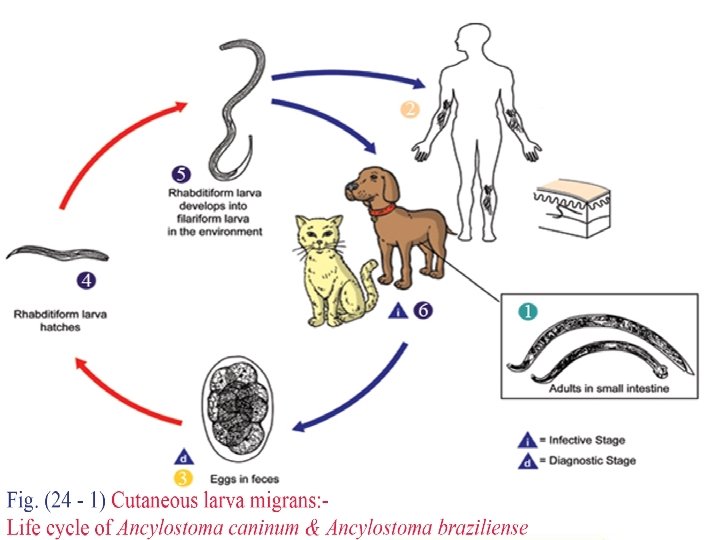
Larva migrans Definition : It is an infection of human tissue by migrating larvae of non human nematodes. Rarely caused by larvae of human nematodes. Larva migrans in man includes: ØCutaneous larva migrans ØVisceral larva migrans.
Cutaneous larva migrans (Creeping eruption, Plumber's itch, Sand worm) Mode of infection 1. Human infection is caused by penetration of the skin by animal hookworm’s filariform larvae which are not adapted to man. 2. Infection occurs due to contact with contaminated soil (moist or sandy) with dog & cat excreta. 3. The larvae migrate in the superficial layers of the skin and not go beyond the basal layer of the skin and keep migrating in the epidermis without development and rarely reaching the circulation. 4. Hookaorms braziliense are Ancylostoma caninum & Ancylostoma
Pathogenesis and symptomatology ØAt the site of entry red itchy papule erythematous zigzag tunnel (1 -2 mm) vesicles 2 ry bacterial infection sever irritation and pruritis. ØLarvae remain active, move very slowly in the epidermis layer only for several weeks or months till die. Commonly affect the skin of feet, hands or buttocks and may advances to 12 cm / day. ØThe skin lesion heals leaving linear white scars at the affected sites. ØRarely larvae reach the lung pulmonary symptoms, eosinophilia and pneumonitis.
CUTANEOUS LARVA MIGRANS It penetrates the skin of man especially feet and legs It fails to penetrate the skin fully (not go beyond the epidermis) Produce Zigzag tunnel
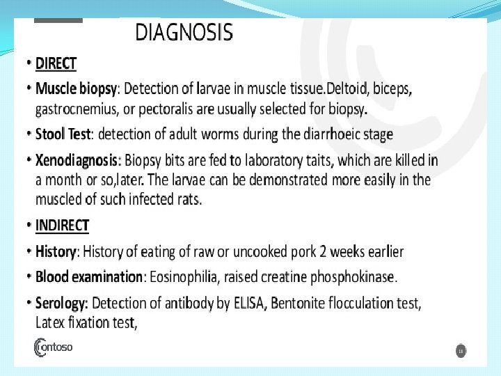
Laboratory diagnosis ØSkin biopsy for larval identification. ØNo eosinophilia. Treatment ØAntihistaminics. ØAntibiotics for 2 rybacterial infection Local freezing: Spray of skin by ethyl chloride (local freezing) or carbon dioxide snow which produce freezing of larvae till death skin bleb larvae are lost with epidermal sloughs. ØThiabendasole ØIvermectin
Leishmaniasis classified into Visceral Leishmaniasis (Kala azar) Cutaneous leishmaniasis Caused by §L. Donovani §L. Infantum v. L. chagasi § Old §L. tropica. Mucocutaneous leishmaniasis Caused by v. L. braziliensis. §L. major. §L. aethiopica. v. L. mexicana. World Leishmaniais v New World Leishmaniasis
Old World Cutaneous Leishmaniasis Morphological characters: 1 - Amastigote Shape: Oval 2 - Promastigote Fusiform or spindle Kinetoplast: Beside the nucleus Flagellum: Absent Nucleus: -Eccentric with central At the anterior end Present -Central with central Karyosome Habitat: -Intracellular (macrophage) -Tissue culture Karyosome -Midgut of the insect -Culture media
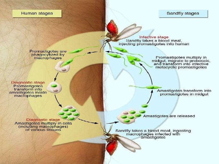
v. Mode of transmission 1 - Bite of female sand fly (Phlebotomus species). 2 - Direct contact with infected lesions. 3 - Mechanical transmission by blood sucking fly as Stomoxys. D. H: Man R. H: Dogs in L. tropica. Rodents in L. major & L. aethiopica. I. S: Metacyclic promastigotes in the mouth part of the female sand fly Vector: Female sand fly (Phlebotomus)
Pathogenesis & Symptomatology 1 - Old World Cutaneous Leishmaniasis L. tropica (Dry or urban CL) G. D: L. major (Wet or rural CL) Middle East, Asia , Africa Middle East, Asia, Africa in big cites (urban areas) in villages (rural areas) L. aethiopica (lepromatous lesions) East Africa (Ethiopia & Kenya) Pathog. & Oriental sore (Baghdad The same as L. tropica Diffuse Cutaneous CL/P Except the followings: Leishmaniasis boil or Delhi boil) -Localized nodules at the bite site necrosis (in the next slide) -Thickening of skin, papules & multiple painless ulcer é sharp nodules like edges & raised indurated lepromatous leprosy margin. -2 nd bacterial infection is common.
L. tropica (Dry or urban CL) L. major (Wet or rural CL) -Has chronic course if - Has an acute course. untreated. L. aethiopica (lepromatous lesion) -Usually affects immunocompromised patients. -Has long I. P(2 -12 ms). -Short I. P(2 -6 ws). -Scanty exudate & slow -Serous exudate & rapid healing (12 ms). healing (3 -6 ms). -The ulcer mainly on -Usually on the lower the face & limbs. -Single or multiple ulcers. - Multiple ulcers. -No mucosal infection or ulceration -Mainly on face & limbs -Usually multiple nodules -Heals spontaneously after 1 -2 years giving large disfiguring depigmented disfiguring scars. -No spontaneous healing & can be relapse. scars. -Gives solid immunity to -Gives immunity against -No solid immunity
Oriental sore
Leishmania aethiopica
New world cutaneous leishmaniasis (American leishmaniasis) New World Cutaneous Leishmaniasis Leishmania mexicana Chiclero ulcer or bay sore New World Mucocutaneous Leishmaniasis Leishmania braziliensis Mucocutaneous Leishmaniasis (Espundia)
Chiclero ulcer Espundia Caused by L. mexicana Caused by L. braziliensis -A small single nodule at the site of -Primary skin lesion: Nodule in sand fly bite ulcerates. exposed regions ulcerates. -Usually on the face & ear pinna - The ulcer with raised indurated heals within 6 months. margin heals in scar in months. -Ear lesion causes destruction of the -Secondary metastatic lesion: catilage of the ear pinna. The parasite migrates from the -Seen in chicleros who live in forests primary site to blood & lymph & collect gum from chicle trees. to mucocutaneous junctions. -Sites: nasal septum, lips, palate nasopharynx & larynx. -Deformity & 2 nd bacterial infection. - Death from septicaemia and bronchopneumonia.
Chiclero ulcer Espundia
Laboratory diagnosis Direct Indirect 1 -Scraping the edge of the ulcer or aspiration by a needle (not the base as contains pus and necrotic tissues) and examined by : • Direct smear stained by Giemsa or leishman. • Culture on NNN medium (amastigotes changes into promastigotes). 1 -Immunodiagnosis: §Leishmanin I. D test (Montenegro test): Not a specific test. It is +ve with cutaneous and mucocutaneous leishmaniasis but negative in diffuse cutaneous leishmaniasis. §Serological tests: Not useful for the diagnosis. 2 - Biopsy from the edge of the ulcer and examined by direct smear. 2 - PCR: A reliable diagnostic test. than routine smear and culture and it used also for species differentiation.
Treatment 1) Local ØCryosurgery, 2) Chemotherapy curettage or local application of heat to raise the intra- lesional temperature to 37 - 43 ° C for 12 ØNon-ulcerated lesion: Intra- lesional injection of pentavalent antimony compounds. ØUlcerated lesion: Should be treated with systemic pentavalent antimony hours as amastigote do not compounds (ex. Pentostam). grow above 33 ° C. ØAlternative drugs to pentostam: ØSurgical lesion. excision of the Amphotericin B, imidazoles or Allopurinol. ØAntibiotics for secondary bacterial infection of lesions.
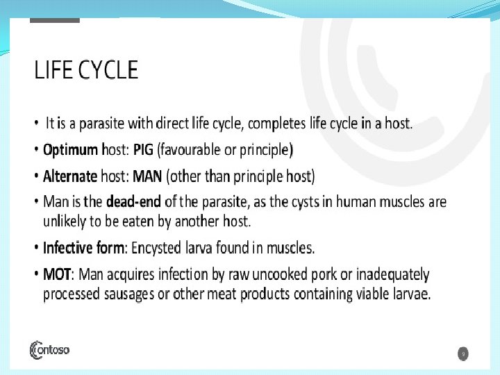
TRICHINELLA SPIRALIS A NEMATODE