
Protoz Oal Infecion Dr Fariborzi Visceral leishmaniasis The
Protoz. Oal Infecion Dr. Fariborzi
Visceral leishmaniasis �The most important clinical manifestation of VL is the syndrome known as kala-azar (Hindi for "black fever"). � The incubation period is usually 2 to 6 months but can range from a few weeks to several years. �Onset of symptoms is usually insidious or subacute, with slow progression of malaise, fever, weight loss, and splenomegaly (with or without hepatomegaly) over a period of months �In rare cases, acute febrile illness can occur with rapidly progressive symptoms
�Kala-azar ("black fever") refers to darkening of the skin, which is a common symptom in South Asia. � Symptoms included fever, malaise, cough, intermittent diarrhea, abdominal pain, adenopathy and mild hepatosplenomegaly.
Laboratory findings � Nonspecific laboratory findings of VL include anemia, neutropenia, eosinopenia, and thrombocytopenia. �The anemia is normocytic and normochromic, unless iron deficiency is also present. � The etiology of anemia is thought to be multifactorial, including bone marrow suppression, hemolysis, and hypersplenism. � An elevated neutrophil count is uncharacteristic of VL and should prompt a search for secondary bacterial infection. � Elevated liver enzymes and bilirubin are also seen. �Hypergammaglobulinemia results from polyclonal B cell activation. �Severe anemia and frank jaundice are associated with a poor prognosis
DIAGNOSIS �Specific laboratory testing is needed to establish the diagnosis of visceral leishmaniasis. � Definitive diagnosis requires the demonstration of parasite by smear or culture in tissue (usually bone marrow or spleen). � The utility of less invasive diagnostic tools (such as demonstration of specific antibodies, antigens, or parasite DNA in peripheral blood specimens) depends on the clinical status of the patient, the geographic origin of the parasite, the methods employed, and laboratory experience
Treatment � amphotericin B � pentavalent antimonial drug � paromomycin (a parenteral aminoglycoside), � miltefosine (the first oral drug for treatment of VL)
Giardia INTRODUCTION : �Giardia lamblia (also known as G. duodenalis or G. intestinalis) is a protozoan parasite capable of causing epidemic or sporadic diarrheal illness. � It is an important cause of water-borne and foodborne disease, day care center outbreaks, and illness in international travelers
EPIDEMIOLOGY � G. lamblia infection occurs worldwide. �High risk groups include infants, young children, international adoptees, travelers, immunocompromised individuals, and patients with hypochlorhydria or cystic fibrosis � Giardiasis is especially common in areas with poor sanitary conditions and insufficient water treatment facilities. �Giardia also infects mammalian animals including beavers, dogs, cattle, and sheep. �Different genotypes of Giardia are almost always found in dogs and humans; therefore zoonotic transmission between dogs and humans is very uncommon
Routes of transmission � Giardia cysts may be transmitted via water, food, or fecal-oral transmission between infected individuals
CLINICAL MANIFESTATIONS �The severity of clinical manifestations associated with giardiasis is variable. � In general, about half of exposed individuals clear the infection in the absence of clinical symptoms, and approximately 5 to 15 percent of individuals shed cysts asymptomatically. � The remaining 35 to 45 percent of individuals have symptomatic infection. � The nature of clinical manifestations in an individual patient likely depends on a number of factors, including the virulence of the isolate, the parasite load, and the host immune response
Asymptomatic infection � Asymptomatic infection occurs in both children and adults, and asymptomatic cyst shedding can last six months or more.
�Symptoms usually develop after an incubation period of 7 to 14 days (range 1 to 45 days). �Symptoms may last two to four weeks. Significant weight loss (10 percent of body weight) occurs in about 50 percent of symptomatic patients
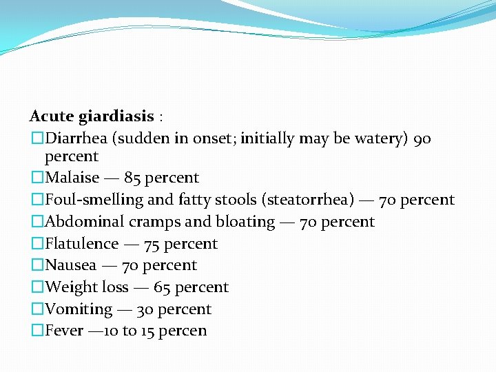
Acute giardiasis : �Diarrhea (sudden in onset; initially may be watery) 90 percent �Malaise — 85 percent �Foul-smelling and fatty stools (steatorrhea) — 70 percent �Abdominal cramps and bloating — 70 percent �Flatulence — 75 percent �Nausea — 70 percent �Weight loss — 65 percent �Vomiting — 30 percent �Fever — 10 to 15 percen
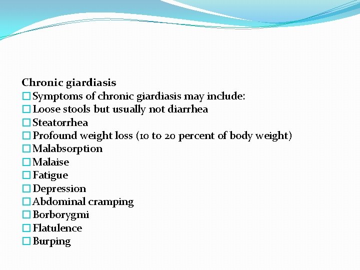
Chronic giardiasis �Symptoms of chronic giardiasis may include: �Loose stools but usually not diarrhea �Steatorrhea �Profound weight loss (10 to 20 percent of body weight) �Malabsorption �Malaise �Fatigue �Depression �Abdominal cramping �Borborygmi �Flatulence �Burping
DIAGNOSIS �Stool microscopy �Immunoassays �Duodenal biopsy
TREATMENT � tinidazole � metronidazole � nitazoxanide. �Alternatives include � albendazole � paromomycin �furazolidone � quinacrine
Amebiasis INTRODUCTION � Intestinal amebiasis is caused by the protozoan Entamoeba histolytica. � Most infection is asymptomatic � clinical manifestations include amebic dysentery and extraintestinal disease. � Extraintestinal manifestations include amebic liver abscess and other more rare manifestations such as pulmonary, cardiac, or brain involvement
EPIDEMIOLOGY � Amebiasis occurs worldwide; the prevalence is disproportionately increased in developing countries because of poor socioeconomic conditions and sanitation levels. �Areas with high rates of amebic infection include India, Africa, Mexico, and parts of Central and South America.
Transmission � The parasite exists in two forms, a cyst stage (the infective form), and a trophozoite stage (the form that causes invasive disease). �Infection occurs following ingestion of amebic cysts; this is usually via contaminated food or water but can be associated with venereal transmission through fecal-oral contact
CLINICAL MANIFESTATIONS � The majority of entamoeba infections are asymptomatic; this includes 90 percent of E. histolytica infections and all E. dispar infection and E. moshkovskii infections. � Factors that influence whether infection leads to asymptomatic or invasive disease include the E. histolytica strain and host factors such as genetic susceptibility, age, and immune status �Risk factors for severe disease and increased mortality include young age, pregnancy, corticosteroid treatment, malignancy, malnutrition, and alcoholism
� Clinical amebiasis generally has a subacute onset, usually over one to three weeks. � Symptoms range from mild diarrhea to severe dysentery-producing abdominal pain (12 to 80 percent), diarrhea (94 to 100 percent), and bloody stools (94 to 100 percent) to fulminant amoebic colitis. � Weight loss occurs in about half of patients � Fever occurs in 8 to 38 percent. � Amebic dysentery is diarrhea with visible blood and mucus in stools and the presence of hematophagous trophozoites (trophozoites with ingested red blood cells) in stools or tissues � Fulminant colitis with bowel necrosis leading to perforation and peritonitis has been observed in approximately 0. 5 percent of cases; associated mortality rate is more than 40 percent � Toxic megacolon can also develop
DIAGNOSIS � Diagnostic techniques include microscopy, antigen detection, serology, molecular techniques, and colonoscopy with histological examination. � Culture techniques are limited to research settings
TREATMENT �All E. histolytica infections should be treated, even in the absence of symptoms, given the potential risk of developing invasive disease and the risk of spread to family members � The goals of antibiotic therapy of intestinal amebiasis are to eliminate the invading trophozoites and to eradicate intestinal carriage of the organism.
�Invasive colitis is generally managed with metronidazole (alternative therapies include tinidazole, ornidazole, and nitazoxanide), followed by a luminal agent (such as paromomycin, diiodohydroxyquin or diloxanide furoate) to eliminate intraluminal cysts �A ten day course of metronidazole eliminates intraluminal infection in many cases, but a second agent is still warranted � Asymptomatic patients with E. histolytica (and not E. dispar/E. moshkovskii) should be treated with an intraluminal agent alone.
Malaria Etiology � Malaria is caused by obligate intracellular protozoa of the genus Plasmodium, including P. falciparum, P. malariae, P. ovale, and P. vivax. � Plasmodium has a complex life cycle that enables survival in different cellular environments in the human host and in the mosquito vector. There are two major phases in the life cycle, an asexual phase (schizogony) in humans and a sexual phase (sporogony) in mosquitoes. � The erythrocytic phase of Plasmodium asexual development begins when the merozoites released from exoerythrocytic schizonts in the liver penetrate erythrocytes. � When inside the erythrocyte, the parasite transforms into the ring form, which enlarges to become a trophozoite. � These latter two forms can be identified with Giemsa stain on blood smear, which is the primary means of confirming the diagnosis of malaria
�The parasites usually are transmitted to humans by female Anopheles mosquitoes. �Malaria also can be transmitted through blood transfusion, via contaminated needles, and transplacentally to a fetus
Clinical Manifestations �The clinical manifestations of malaria range from asymptomatic infection to fulminant illness and death depending on the virulence of the infecting malaria species and the host immune response. � The incubation period ranges from 6 to 30 days depending on the Plasmodium species. � The most characteristic clinical feature of malaria, which is seldom noted with other infectious diseases, is febrile paroxysms alternating with periods of fatigue but otherwise relative wellness. �The classic symptoms of the febrile paroxysms of malaria include high fever, rigors, sweats, and headache. �Paroxysms coincide with the rupture of schizonts that occur every 48 hours with P. vivax and P. ovale (tertian periodicity) and every 72 hours with P. malariae (quartan periodicity).
Laboratory and Imaging Studies � The diagnosis of malaria is established by identification of organisms on stained smears of peripheral blood. � In nonimmune persons, symptoms typically occur 1 to 2 days before parasites are detectable on blood smear. � Although P. falciparum is most likely to be identified from blood during a febrile paroxysm, the timing of the smears is less important than obtaining smears several times each day over 3 successive days. � Both thick and thin blood smears should be examined. � The concentration of erythrocytes on a thick smear is approximately 20 to 40 times greater than on a thin smear. � Thick smears are used to scan large numbers of erythrocytes quickly. � Thin smears allow for positive identification of the malaria species and determination of the percentage of infected erythrocytes, which also is useful in following the response to therapy
Differential Diagnosis �The most important aspect of diagnosis of malaria in children is to consider the possibility of malaria in any child who has fever, chills, splenomegaly, anemia, or decreased level of consciousness with a history of recent travel or residence in an endemic area, regardless of the use of chemoprophylaxis. � The differential diagnosis is broad and includes many infectious diseases, such as tuberculosis, typhoid fever, brucellosis, relapsing fever, infective endocarditis, influenza, poliomyelitis, yellow fever, trypanosomiasis, kala-azar, and amebic liver abscess
Treatment �Oral chloroquine is the recommended treatment except for chloroquine-resistant P. falciparum. � Quinine sulfate plus tetracycline, quinine plus pyrimethamine-sulfadoxine, or mefloquine are used for chloroquine-resistant malaria. � Patients with malaria usually require hospitalization and may require intensive care unit (ICU) admission. �Quinidine gluconate is used for parenteral treatment
Complications and Prognosis �Cerebral malaria is a complication of P. falciparum infection and a frequent cause of death (20 -40% of cases), especially among children and nonimmune adults. � Similar to other complications, cerebral malaria is more likely to occur among patients with intense parasitemia (≥ 5%). � Other complications include splenic rupture, renal failure, severe hemolysis (blackwater fever), pulmonary edema, hypoglycemia, thrombocytopenia, and algid malaria (sepsis syndrome with vascular collapse).
Prevention �There are two components of malaria prevention: 1 - reduction of exposure to infected mosquitoes 2 -chemoprophylaxis. Mosquito protection is necessary because no prophylactic regimen can guarantee protection in every instance because of the widespread development of resistant organisms
Toxoplasmosis �Toxoplasmosis is a zoonosis caused by Toxoplasma gondii, an intracellular protozoan parasite. � Infection is acquired by infectious oocysts, such as those excreted by newly infected cats, which play an important role in amplifying the organism in nature, or from ingesting cysts in contaminated, undercooked meat. � Less commonly, transmission occurs transplacentally during acute infection of pregnant women.
� Acquired toxoplasmosis is usually asymptomatic. � Symptomatic infection is typically a heterophile-negative mononucleosis syndrome that includes lymphadenopathy, fever, and hepatosplenomegaly. � Disseminated infection, including myocarditis, pneumonia, and central nervous system (CNS) toxoplasmosis, is more common among immunocompromised persons, especially persons with acquired immunodeficiency syndrome (AIDS). � Among women infected during pregnancy, 40% to 60% give birth to an infected infant. � The later in pregnancy that infection occurs, the more likely it is that the fetus will be infected, but the less severe the illness. � Serologic diagnosis can be established by a fourfold increase in antibody titer or seroconversion, a positive Ig. M antibody titer, or positive polymerase chain reaction (PCR) for T. gondii in peripheral white blood cells (WBCs), cerebrospinal fluid (CSF), serum, or amniotic fluid.
�Treatment includes pyrimethamine and sulfadiazine, which act synergistically against Toxoplasma. � Because these compounds are folic acid inhibitors, they are used in conjunction with folinic acid. �Spiramycin, also is used in therapy of pregnant women with toxoplasmosis. �Corticosteroids are reserved for patients with acute CNS or ocular infection.
�Ingesting only well-cooked meat and avoiding cats or soil in areas where cats defecate are prudent measures for pregnant or immunocompromised persons. �Administration of spiramycin to infected pregnant women has been associated with lower risks of congenital infection in their offspring.
- Slides: 36