Complications of Heart Failure 1 Renal failure 2


- Slides: 23
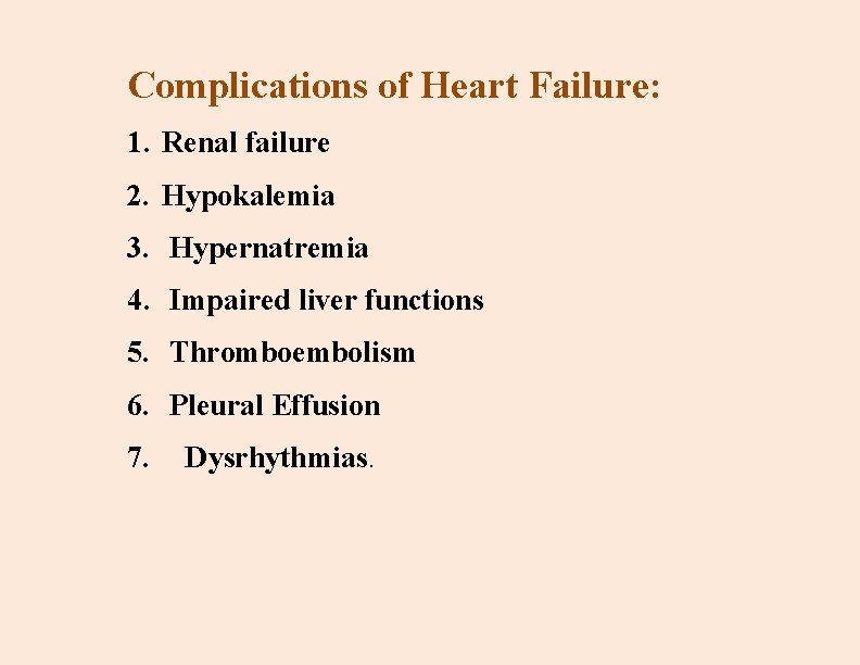
Complications of Heart Failure: 1. Renal failure 2. Hypokalemia 3. Hypernatremia 4. Impaired liver functions 5. Thromboembolism 6. Pleural Effusion 7. Dysrhythmias.
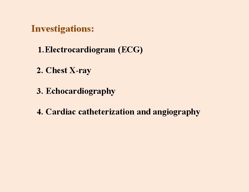
Investigations: 1. Electrocardiogram (ECG) 2. Chest X-ray 3. Echocardiography 4. Cardiac catheterization and angiography
5 -Laboratory studies usually performed during the initial workup include serum electrolytes, blood urea nitrogen (BUN), and creatinine, thyroidcomplete blood cell count, stimulating hormone, serum B-type natriuretic peptide (BNP), and routine urinalysis. The BNP level is a key diagnostic indicator of HF.
Medical management: - 1 Nutritional Therapy: 1 A low-sodium (2 to 3 g/day) diet. 2 Avoidance of drinking excessive amounts 3 Examine labels todetermine sodium content. 4 Examine the labels of over-the-counter drugs such as laxatives, coughmedicines, and antacids for sodium content 5. Avoid using salt when preparing foods or adding salt to foods. 6. Eat smaller, more frequent meals.
2 -Pharmacologic Therapy: a. Angiotensin-Converting Enzyme(ACE) Inhibitors b. Angiotensin II Receptor Blockers c. Beta-Blockers d. Diuretics: - Loop diuretics - Thiazide diuretics -Potassium-sparing diuretics.
e. Positive Inotropes e. g. , Digitalis: - Digoxin Toxicity: - signs and symptoms • Anorexia, nausea, vomiting, fatigue, depression, and malaise )early effects of digitalis toxicity) • Changes in heart rate or rhythm; onset of irregular rhythm f. . Vasodilators e. g. , nitroglycerin. g. Morphine. . h. Other Medications: - Anticoagulants -hyperlipidemia (e. g, statins) - antidysrhythmic drugs
3 Additional Therapy: 1 -Supplemental Oxygen therapy may become necessary as HF progresses. The need is based on the degree of pulmonary congestion and resulting hypoxia. Some patients require supplemental oxygen only during periods of activity.
2 -Other Interventions: - Percutaneous coronary intervention or coronary artery bypass surgery. - implantable cardio verter defibrillator (ICD) - Cardiac Resynchronization Therapy - Intra-Aortic Balloon Pump - Cardiac transplantation is the only option for long-term survival.
Nursing management: Assessment: A- Subjective Data: *Respiratory system: - Lung disease? - How many flights of stairs can be climbed without dyspnea? - How many pillows used for sleeping? - Dyspnea at rest or that awakens from sleeping?
*Cardiovascular: - Any cardiac disease history: hypertension, myocardial infarction, valvular problem, anemia, dysrhythmias, palpitations? - Chest pain: precipitating factors, severity, relieving factors? -Can activities of daily living be performed?
- Can activities performed 6 months, 4 months, 2 weeks ago still be done? - Any dizziness )vertigo) or fainting (syncope ) ? *Fluid retention: -Daily sodium intake? -Are shoes tight? Do ankles swell?
*GIT: - Is appetite good? - Any nausea, vomiting, or abdominal pain ? *Neurological: -Any change in behavior ? *Medications, Knowledge of Condition, Coping Skills.
B- Objective Data: Integumentary: Cool, diaphoretic skin; cyanosis or pallor; peripheral edema )right-sided heart Failure). Respiratory: Tachypnea, crackles, wheezes; frothy, bloodtinged sputum. Cardiovascular: Tachycardia, S 3, S 4, murmurs; pulsus alternans, jugular venous distention.
Gastrointestinal: Abdominal distention, hepatosplenomegaly, ascites. Neurologic: Restlessness, confusion, decreased attention or memory. Possible Diagnostic Findings:
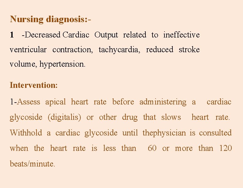
Nursing diagnosis: 1 -Decreased Cardiac Output related to ineffective ventricular contraction, tachycardia, reduced stroke volume, hypertension. Intervention: 1 -Assess apical heart rate before administering a glycoside (digitalis) or other drug that slows cardiac heart rate. Withhold a cardiac glycoside until thephysician is consulted when the heart rate is less than beats/minute. 60 or more than 120
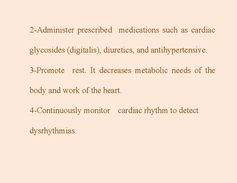
2 -Administer prescribed medications such as cardiac glycosides (digitalis), diuretics, and antihypertensive. 3 -Promote rest. It decreases metabolic needs of the body and work of the heart. 4 -Continuously monitor cardiac rhythm to detect dysrhythmias.
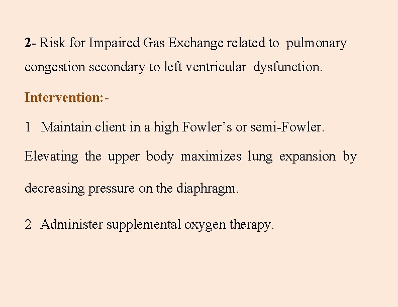
2 - Risk for Impaired Gas Exchange related to pulmonary congestion secondary to left ventricular dysfunction. Intervention: - 1 Maintain client in a high Fowler’s or semi-Fowler. Elevating the upper body maximizes lung expansion by decreasing pressure on the diaphragm. 2 Administer supplemental oxygen therapy.
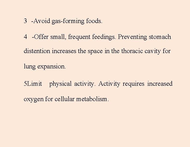
3 -Avoid gas-forming foods. 4 -Offer small, frequent feedings. Preventing stomach distention increases the space in the thoracic cavity for lung expansion. 5 Limit physical activity. Activity requires increased oxygen for cellular metabolism.
3 --Excess Fluid Volume related to reduced renal function secondary to increased antidiuretic hormone aldosterone production and reduced cardiac output. Interventions: 1 -Monitor for edema, weight gain, jugular venous distention (JVD(, and lung crackles. 2 - Monitor intake and output (I&O). and
3 -Administer prescribed diuretic. Diuretics promote the excretion of sodium and water. 4 - Provide sodium-restricted diet as prescribed. 5 - Maintain fluid restriction as ordered
4 --Activity Intolerance related to hypoxemia secondary to decreased cardiac output. Interventions: - 1 -Provide rest and space activities. 2 -Keep personal items within easy reach. Reduced exertion decreases oxygen expenditure. 3 -Teach use of assistive devices and lifestyle changes. 4 - Assist as needed with activities of daily living (ADLs).
23