CHAPTER 5 TISSUES Introduction Cells are arranged in


 Simple Squamous 2) Simple Cuboidal")


 � Varying �")


 � White blood cells")


 � Made of: Collagen Add")

 Tissue � Forms: � Delicate, � Binds body parts together �")


 Densly packed collagenous fibers Very Strong!")


 Skeletal 2) Smooth 3)")


- Slides: 57
CHAPTER 5 TISSUES
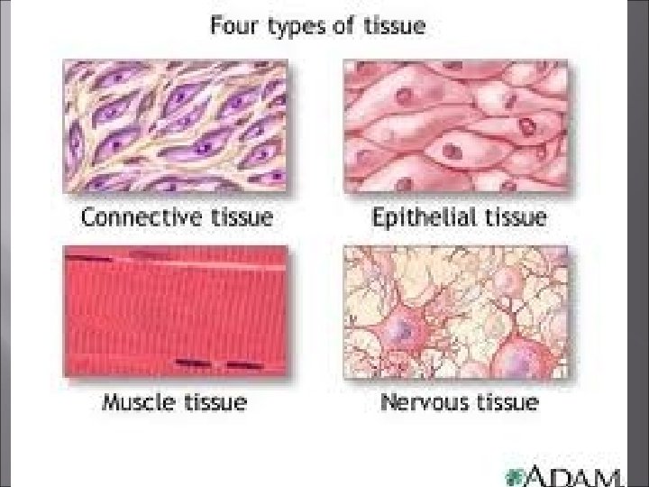
Introduction � � � Cells are arranged in tissues that provide specific functions for the body. Cells of different tissues are structured differently, which leads to their differences in function. The tissues of the human body include four major types. � Epithelial � Connective � Muscle � Nervous
� Function Epithelial � Protection, secretion, absorption, excretion � Sensory reception � Location � Covers your body surfaces, covers and lines internal organs, compose glands � Characteristics � Lack blood vessels, readily divide, cells are tightly packed � Anchored to a basement membrane � Replaced frequently
Epithelial � � � Epithelium- tissue that constitutes the outermost layer of the skin. Carcinoma is a cancer originating from epithelium tissue. About 90% of all cancers originate from epithelium tissue.
Types of Epithelium � � � � � 1) Simple Squamous 2) Simple Cuboidal 3) Simple Columnar 4) Pseudostratified Columnar 5) Stratified Squamous 6) Stratified Cuboidal 7) Stratified Columnar 8) Transitional 9) Glandular
Simple Squamous Epithelium � � Made up of a single layer of thin, flattened cells. Suited for diffusion � Exchanges � gases in the lungs Lines: � Blood and lymph vessels � Body cavities � A basement membrane forms between epithelial and connective tissues
Simple Squamous Epithelium
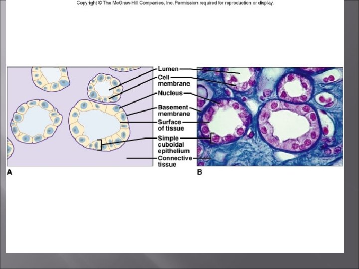
Simple Cuboidal Epithelium � � Consists of a single layer of cube-shaped cells with a centrally located nuclei. It functions in secretion and absorption in the kidneys, and in secretion in glands.
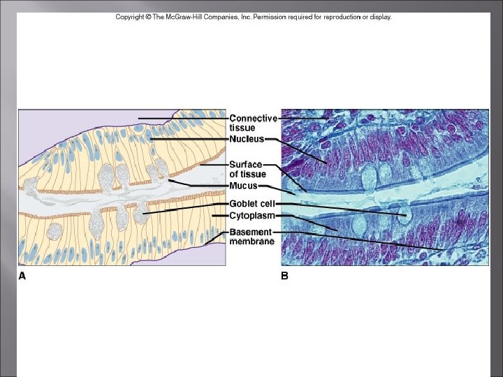
Simple Columnar Epithelium � � Cells are longer than they are wide Single layer of cells nuclei are located near the basement membrane � May be ciliated � � It lines: � The � uterus, stomach, and intestines It also protects underlying tissues, secretes digestive fluids, absorbs nutrients
Simple Columnar Epithelium � � Inner lining organs of the digestive system contain this type. Goblet Cells: � Secrete Mucus � Embedded within the tissues � Unicellular gland. � Have microvilli Increases the surface area for available for absorption.
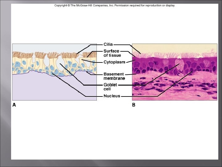
Pseudostratified Columnar Epithelium � Appear Layered (but are not truly layered) � Varying � positions of the nuclei Cilia may be present � Also, Goblet Cells Function to line and sweep debris from respiratory tubes. � Commonly found forming the inner lining of the respiratory passages.
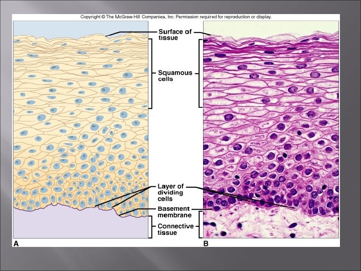
Stratified Squamous Epithelium � Made up of layers of flattened cells � Designed � to protect underlying layers. Where is it found? � It makes up the outer layer of skin. � It lines the mouth, throat � Outer layers of cells undergo keratinization � Does not occur where tissues remain moist throat
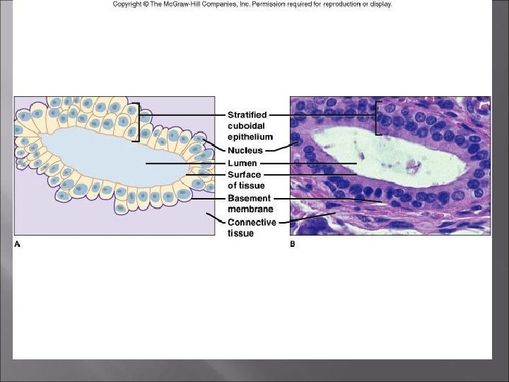
Stratified Cuboidal Epithelium � � � 2 -3 layers of Cuboidal Cells Several layers of cells provides more protection than one single layer. Lines: � Sweat glands � Salivary glands � Pancreas � Mammary glands
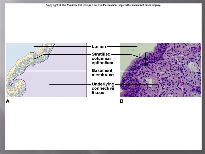
Stratified Columnar Epithelium � � Several layers of cells- stratified Where is it found? � Vas deferens � Parts of the male urethra � Parts of the pharynx
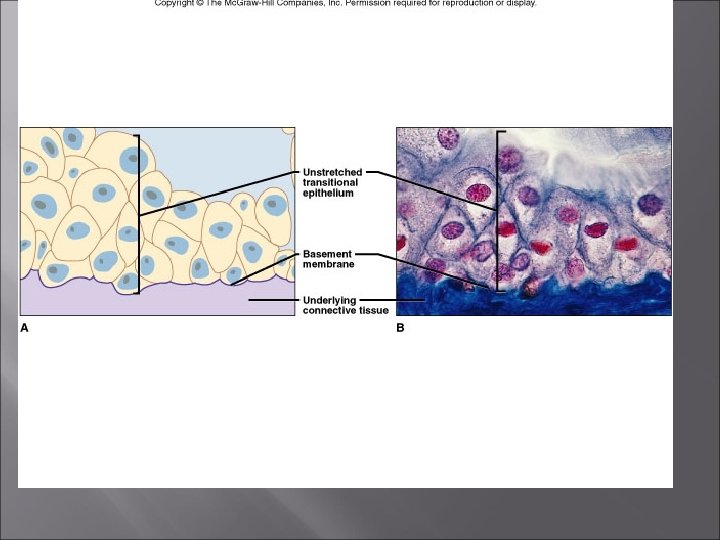
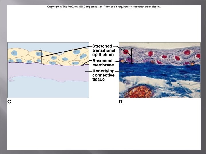
Transitional Epithelium � Designed to distend and return to its normal size � Lining of the urinary bladder � Provides distensibility � Keeps urine from diffusing back into the internal cavity
Glandular Epithelium � � � Produces and secretes substances into ducts or into body fluids Exocrine- glands that secret products into ducts Endocrine (ductless) - glands that secret products into body fluids and blood
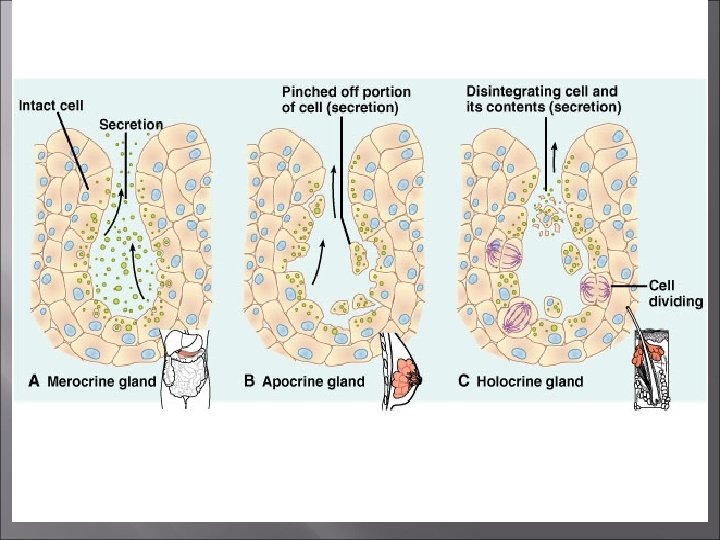
Glandular Epithelium � Gland classification = way product is secreted � Merocrine Glands Release fluid products – exocytosis � Apocrine Glands Lose portions of their cell bodies during secretion �Ex. Mammary glands � Holocrine Glands Release entire cells �Ex. Sebaceous glands
� Function Connective � Bind, support, protect, fill spaces, store fat, produce blood cells, serves as framework, protect against infection, repairs tissue damage. � Has an abundant matrix (unlike epithelial) Intercellular material Has good blood supply (except cartilage) � Location � Widely � distributed throughout the body Characteristics � Mostly have good blood supply; cells are farther apart then epithelial cells with extracellular matrix between
2 Major Types � � 1. Liquid or Vascular Connective Tissue 2. Solid Connective Tissue
Vascular Tissue � Blood: � Red blood cells (carry oxygen) � White blood cells (fight infection) � Platelets (cell fragments used for agglutination) � Plasma (fluid intracellular matrix)
Major Cell Types � Fibroblast Cell � Most common � Fixed, star-shaped cell � Large � Secretes Fibers � Macrophages (wandering) � Function Scavenger cells Defend against infection
Major Cell Types � Mast Cells � Large � Located near blood vessels � Release Heparin (anticoagulant) Histamine (promotes inflammation)
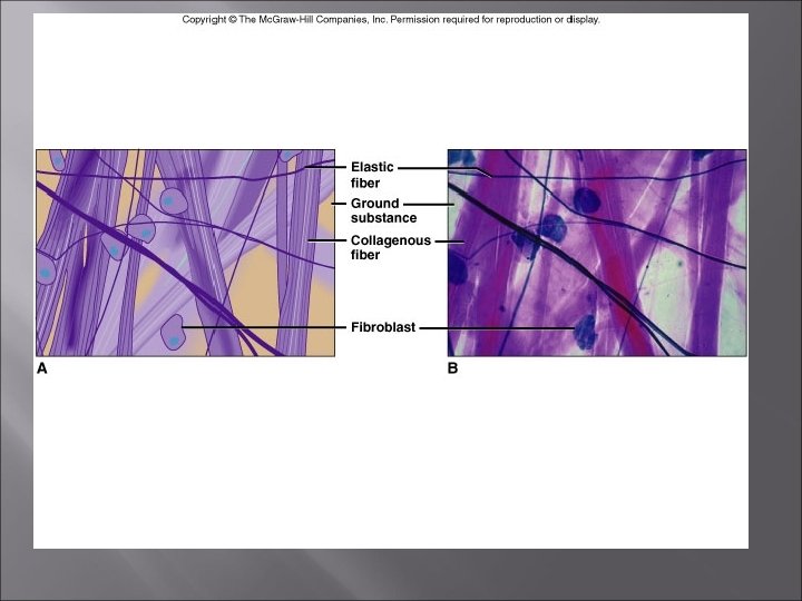
Connective Tissue Fibers � Collagenous Fibers � (white fibers) � Made of: Collagen Add Strength (for holding body parts together) � Elastic Fibers (yellow fibers) � Made of: Elastin Stretchy Add Flexibility to certain types of connective tissues
Connective Tissue Fibers � Reticular � Thin collagenous � Form supportive networks � Osteocytes (bone cells) � Arranged in concentric circles around the osteonic or Haversian Canals
Loose Connective (areolar) Tissue � Forms: � Delicate, � Binds body parts together � Skin � thin membranes and underlying organs The majority of the cells are fibroblasts that are separated by a gel-like intracellular material that contains collagenous and elastic fibers.
Adipose Tissue � Loose connective tissue � Stores � fat Where is it found? � Beneath the skin � Around joints � Padding the kidneys, and internal organs � Abdominal membranes
Adipose Tissue
Dense Connective Tissue � � (Fibrous Connective Tissue) Densly packed collagenous fibers Very Strong! � Lacks � a good blood supply Where is it found? � Tendons and Ligaments
Cartilage � Rigid connective tissue � Provides � � Enclosed within Perichondrium Lacks a vascular system � Healing � a supportive framework takes longer Chondrocytes- cartilage cells � Lie within the lacunae Small chamber within a gel-like fluid matrix
Cartilage � Hyaline Cartilage � Most common � White � Abundant fine collagen fibers � Where is it found? � Ends � of bones Function? � Supports respiratory passages Trachea is composed of Hyaline Cartilage
Cartilage � Elastic Fibers � Forms Framework Ex. External Ears & Parts of Larynx � Fibrocartilage � Collagenous Fibers � Tough- Tissue Shock- absorbing �Ex. Intervertebral Disks of Backbone �Disks separate individual parts of backbone �Ex. Knees, Pelvic Girdle
Bone � � Most Rigid Connective Tissue Matrix Contains: � Deposits of Mineral Salts � Collagen � Functions: � Internally Supports the Body � Forms Muscle Attachments � Site for Blood Cell Formation � Osteocytes- Bone Cells � Found in Lacunae � Arranged in Osteons (concentric circles) Around osteonic canals interconnected by canaliculi
Bone � Bone has a good blood supply, enabling rapid recovery after an injury.
Bone
Blood � Composed of red and white cells � Suspended in plasma Liquid matrix � Function: � Transport substances throughout body
� Function Muscle � Movement � Location � Attached to bones, in the walls of hollow internal organs, heart � Characteristics � Able to contract in response to specific stimuli
3 Major Types of Muscle Tissue � � � 1) Skeletal 2) Smooth 3) Cardiac
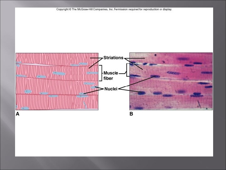
Skeletal Muscle Tissue � Attached to bone � Controlled � Muscle Fibers � Long, � striated, cylindrical, many nuclei Major Characteristic � Ability � voluntarily to contract from nervous impulse Least likely cell type to reprodue
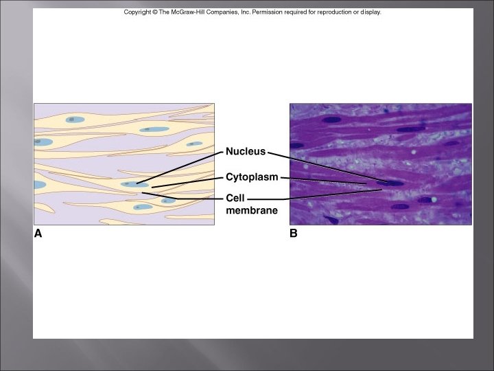
Smooth Muscle Tissue � � Lacks Striations Uninucleate Spindle-shaped cells Involuntary Muscle � Found in the walls of internal organs Ex. Stomach, Digestive Tract, blood vessels, urinary bladder
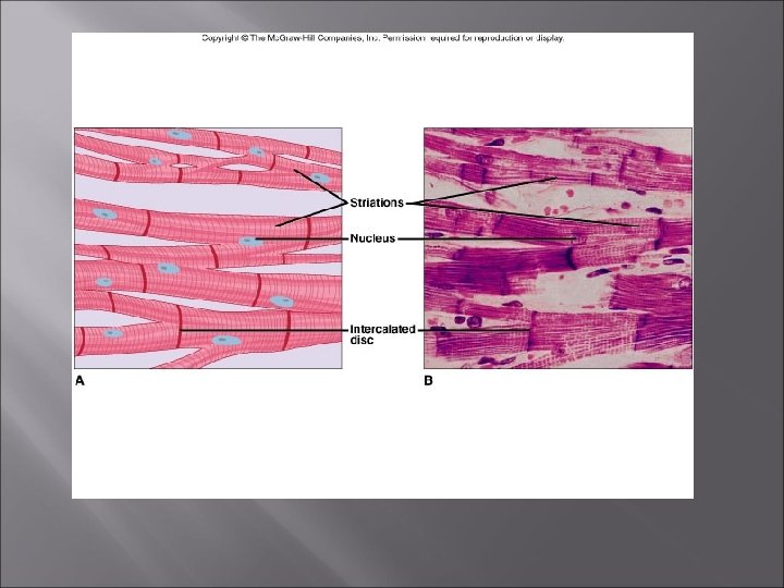
Cardiac Muscle Tissue 1. Cardiac muscle tissue is found only in the heart and consists of branching fibers that are connected to each other with intercalated disks. This is the band that occurs where two cardiac muscle cells join. 2. This involuntary muscle has a single nucleus in each cell but appears striated. 3. When cardiac muscle cells are damaged by a heart attack, they are usually replaced by “connective tissue cells”
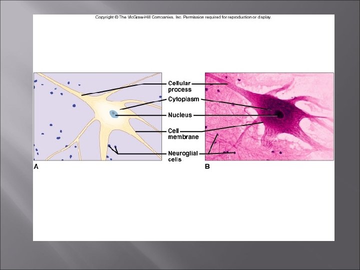
Nervous Tissues: A. Nervous tissues are found in the brain, spinal cord, and nerves. B. Neurons, or nerve cells, conduct nervous impulses while helper cells, or neuroglia, support and nourish the neurons. C. Generally nervous tissue conducts nerve impulses from one neuron to another and it coordinates body activities.
Nervous � Function � Transmit impulses for coordination, regulation, integration, and sensory reception � Location � Brain, � spinal cord, nerves Characteristics � Cells connect to each other and to other body parts