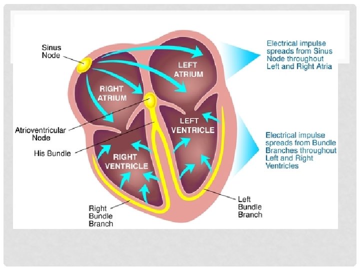
ARRHYTHMIAS DANNY HAYWOOD FY 1 INTRO Conduction system


 • If symptomatic/clinical deterioration • IV atropine")
 vs Broad complex (Ventricular) • Narrow • QRS <0.")
 Atrial Flutter Atrioventricular nodal")


 • Medical")
")
 1 CHADSVASC H Hypertension:")


")

 short refractory period + slow")

 • Pre-excitation (delta wave) on")


- Slides: 42
ARRHYTHMIAS DANNY HAYWOOD FY 1
INTRO • • • Conduction system of heart Symptoms/signs Investigations Tachy vs Bradyarrhythmias • Different types • Management • Tachyarrhythmias • Broad vs narrow • Types of each • Management of each • Summary • Some example ECGs
SYMPTOMS/SIGNS • • Syncope Dizziness Palpitations Heart Failure Chest pain Sudden death No symptoms
INVESTIGATIONS • Bedside • ECG • Bloods • TFTs, U+E, FBC, Troponins • Imaging • Echo, CXR • Special tests • Holter monitor
ARRHYTHMIAS • Bradyarrhythmias vs Tachyarrhythmias • Brady • HR < 60 bpm • Tachy • HR > 100 bpm
BRADYARRHYTHMIAS • Type I heart block • 1 st degree heart block • Prolonged PR interval > 0. 2 seconds • Type II heart block • Mobitz type 1 – Wenckebach • Gradually increased PR intervals until missed QRS • Mobitz type 2 • Intermittently P wave not followed by QRS • May be pattern eg 2: 1, 3: 1 ratio of P waves to QRS complexes – no increase in PR interval • Type III heart block • Complete heart block • No correlation between P waves and QRS complexes
MANAGEMENT • Acute (eg. Secondary to MI) • If symptomatic/clinical deterioration • IV atropine • External (transcutaneous) pacing • Chronic • Mobitz type II or complete AV block • Permanent pacemaker
TACHYARRHYTHMIAS • Narrow complex (Supraventricular) vs Broad complex (Ventricular) • Narrow • QRS <0. 12 seconds • Broad • QRS >0. 12 seconds
NARROW COMPLEX • • • Sinus tachycardia Atrial Fibrillation (AF) Atrial Flutter Atrioventricular nodal re-entry tachycardia (AVNRT) Atrioventricular reciprocating tachycardia (AVRT)
AF • Continuous, rapid activation of atria – due to rapidly depolarising foci within the atria • Often located by pulmonary veins • No coordinated mechanical action
AF – CAUSES • ATRIAL Ph. IB • • • A – Alcohol T – Thyroid disease R – Rheumatic heart disease I – Ischaemic heart disease A – Atrial myxoma L – Lung pathology (pneumonia, PE) • Ph – Pheochromocytoma • I – Idiopathic • B – Blood pressure (hypertension)
AF - MANAGEMENT • Conservative • Alcohol cessation • Lifestyle factors (diet/exercise/smoking) • Medical • Treat underlying cause • Rate control vs rhythm control • Interventional • Catheter ablation
RATE CONTROL • Older age, permanent AF • Bisoprolol/verapamil and Warfarin (CHADSVASc)
C Congestive heart failure (or Left ventricular systolic dysfunction) 1 CHADSVASC H Hypertension: blood pressure consistently above 140/90 mm. Hg (or treated hypertension on medication) 1 A 2 Age ≥ 75 years 2 D Diabetes Mellitus 1 S 2 Prior Stroke or TIA or thromboembolism 2 V Vascular disease (e. g. peripheral artery disease, myocardial infarction, aortic plaque) 1 A Age 65– 74 years Sc Sex category (i. e. female gender) Score Risk Anticoagulation Therapy 0 Low No antithrombotic therapy (or Aspirin) 1 Moderate Oral anticoagulant (or Aspirin) 2 or greater High Oral anticoagulant 1 1
RHYTHM CONTROL • Cardioversion • • • Pharmacological vs DC younger, symptomatic, physically active patients Congestive heart failure Paroxysmal AF failure of rate control • < 48 hours • Cardioversion + heparin • > 48 hrs – TOE/anti-coagulation (3 weeks) • risk of failure? • High – 4 weeks sotalol/amiodarone then electrical. • Low - electrical
RHYTHM CONTROL • Pharmacological • No structural heart disease • 1 st - Flecainide • 2 nd – Sotalol • 3 rd – Amiodarone • Structural heart disease • Amiodarone • Interventional • Pulmonary vein isolation - catheter ablation
ATRIAL FLUTTER • Organised atrial rhythm, coming from ectopic focus in atria (usually left) • Usually 300 bpm • Ventricular rate depends on degree of AV block eg 2: 1 = 150 bpm • Saw tooth pattern
ATRIAL FLUTTER • Management • Conservative • Vagal manoeuvres • Medical – similar to AF • Acute • DC cardioversion or IV adenosine (<48 hours) • > 48 hours - 3 weeks anticoag then cardiovert • Chronic • Pill in pocket • Regular anti-arrhythmics • Interventional • Radiofrequency catheter ablation
AVNRT • 2 pathways within the AV node 1) short refractory period + slow conduction 2) long refractory period + fast conduction • Normally conducts through fast pathway • If premature atrial beat, fast pathway still refractory (long refractory period) therefore travels down slow pathway and back up the fast pathway.
AVNRT
AVRT • Accessory pathway (Bundle of Kent most common) • Pre-excitation (delta wave) on ECG • Wolff-Parkinson-White syndrome
MANAGEMENT OF SVTS • Haemodynaically unstable • Electrical cardioversion • Conservative • Vagal manoeuvres • Valsalva, carotid massage, cold water • Medical • Adenosine (acute) • Anti-arrhythmics (regular and pill-in-pocket) • Interventional • Catheter ablation
BROAD COMPLEX TACHYS VT VS VF • VT • Unstable • electrical cardioversion • Stable • 1 st – Class I Anti-arrhythmics (lidocaine) • 2 nd – Amiodarone • 3 rd – DC cardioversion
BROAD COMPLEX TACHYS VT VS VF • • Cardiac arrest Rapid, irregular activity – no cardiac output Usually provoked by ventricular ectopic beat Management • Electrical defibrillation
BROAD COMPLEX TACHYS • Something to be aware of • SVT with concomitant bundle branch block = broad complex tachy
SUMMARY • Brady vs tachy • Brady • • Sinus Brady 1 st degree heart block Mobitz I & II Complete • Tachy • Narrow • Sinus tachy, AF, Flutter, AVNRT, AVRT • Broad • VT, VF, • Remember causes of AF
ECGS – TEST YOURSELF
A
B
C
D
E
F
G
H
I
J
K
L
ANSWERS A. B. C. D. E. F. G. H. I. J. K. L. Sinus rhythm AF Atrial Flutter VT VF 1 st degree heart block Complete heart block Mobitz type II AVRT Mobitz type I AVNRT Right bundle branch block
REFERENCES • All images and ECGs borrowed gratefully from google images • Kumar & Clarke: Clinical Medicine 7 th Ed • NICE guidelines: AF (CG 36)