CONDUCTION SYSTEM ANATOMY Haitham Taani Conduction system SA


")


- Slides: 18
CONDUCTION SYSTEM ANATOMY Haitham Taani
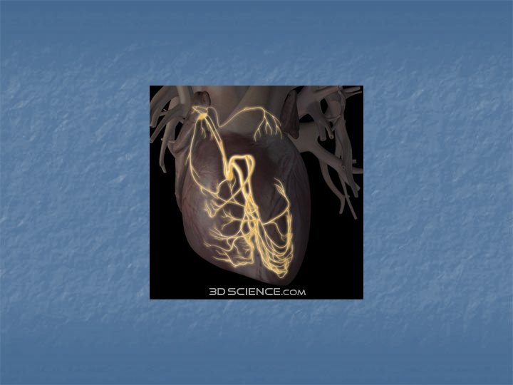
Conduction system *SA node *internodal pathways *atrioventricular Node *atrioventricular bundle *Lt bundle branch *Rt bundle branch *purkinje fibers *ventricular myocardial fibers
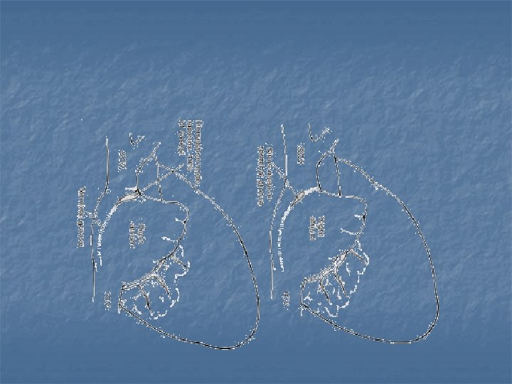
SA node *15 x 5 x 1. 5 mm *on the lateral surface of SVC and RA near the crista terminalis *lies superficially at the anteriolateral aspect of junction between SVC and RA appandage * In rare cases : medially along the ridge of atriocaval junction
SA node…. . Arterial supply * Branch of RCA in 55% *branch of Cx in 45% * When arise from the RCA : it courses posteriorly and superiorly over the anterior wall of RA beneath the RA appandage to the base of SVC the it encircle the SVC either clockwise, counterclockwise or bifurcates
SA node …. arterial supply *when arises from Cx it courses over the LA appandage
Internodal pathways *the ends of SA node fibers fuse with surrounding atrial muscle fibers * The A. P originating in SA node travels through the entire atrial mass to reach the AV node *classically divided into ant. and medial: ant and post. To F. O post. Pathway: caudal to F. O
AV node * On the Rt. Atrial side of central fibrous body (Rt. Trigone) in the muscular portion of atrioventricular septum, *just anteriosuperior to ostium of C. S *Avarage dimentions in adult: 1 x 3 x 6 mm * The Lt. surface lies against mitral annulus *it is flat and oval in shape
AV node *within the koch triangle which formed by septal leaflt of TV Tendon of Todaro coronary sinus
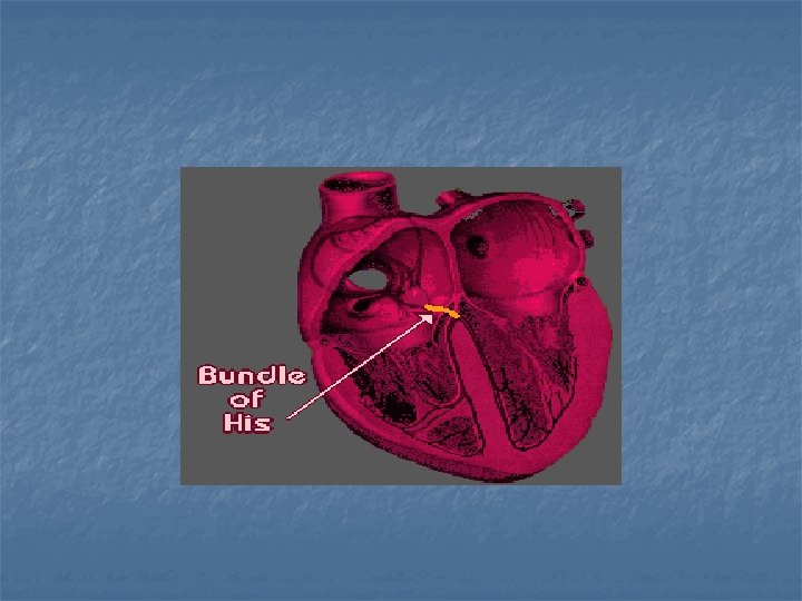
Bundle of HIS *Direct continuation of AV node *passes through the Rt. Trigone of central fibrous body to reach the posterioinferior surface of membranous septum *membranous septum: localized from the atrial side by identifying the commissure of septal and ant. Leaflet of TV (just inferior)
. . …Bundle of HIS *Lies on the Lt side 75 -80 % Rt. Side 20 -25% * Branching occurs neneathe commissure between Rt. And NC cusp of AO. V
*AV node directly in front of ostium of C. S *Line between C. S and commissure of ant. and septal leaflet of TV : Area of AV node &HIS bundle *membranous septum: just inf. To that commissure
*relation to AO. V: AV node: in atrioventricular septum on the Rt. Atrial side of trigone post to noncoronary cusp HIS BUNDLE: branching occur beneath the commissure of Rt. And N. C cusp
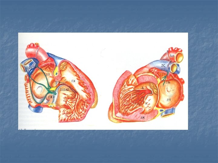
LEFT BUNDLE BRANCH *connected to HIS bundle by narrow stem *subdivided to 2 or 3 main radiation anterior : towards the ant. lat. Pappilary muscle of LV Posterior : towards theposteriomedial pappilary muscle (wider)
Right bundle branch *originated from HIS in the region of ant. Inf. Margin of membranous septum * Then along the moderator band passing toward the base of anterior pappilary muscle