
Complications of Acute M I Douglas Burtt M
Complications of Acute M. I. Douglas Burtt, M. D. Bio-Med 350
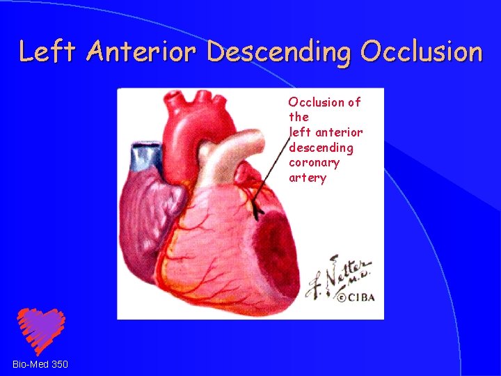
Left Anterior Descending Occlusion of the left anterior descending coronary artery Bio-Med 350
Experimental Data Canine studies – transient artery clamping or ligation © Balloon angioplasty studies © © Time dependent series of events © Chest Pain as a late event Bio-Med 350
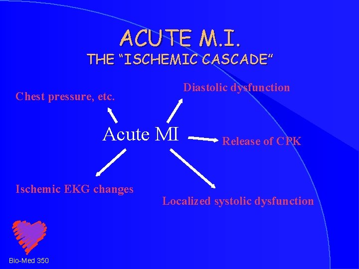
ACUTE M. I. THE “ISCHEMIC CASCADE” Diastolic dysfunction Chest pressure, etc. Acute MI Ischemic EKG changes Bio-Med 350 Release of CPK Localized systolic dysfunction
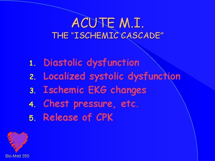
ACUTE M. I. THE “ISCHEMIC CASCADE” 1. 2. 3. 4. 5. Bio-Med 350 Diastolic dysfunction Localized systolic dysfunction Ischemic EKG changes Chest pressure, etc. Release of CPK
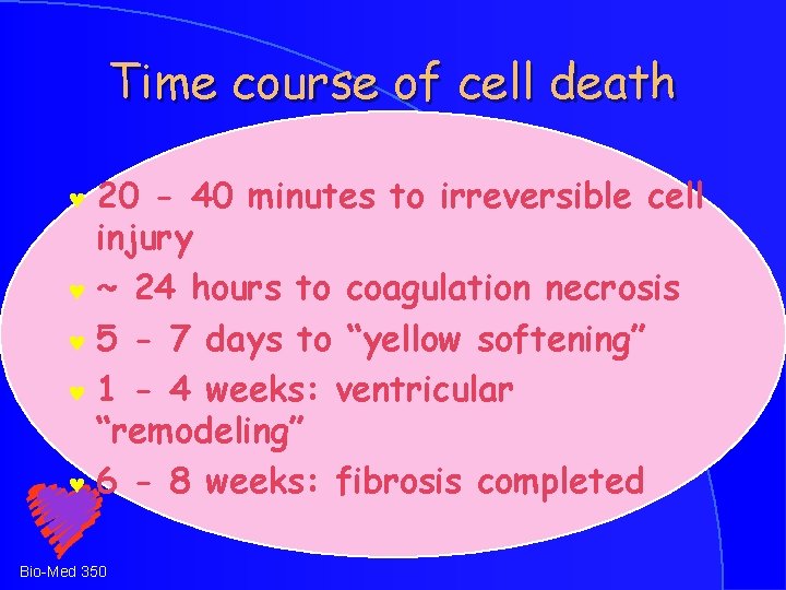
Time course of cell death 20 - 40 minutes to irreversible cell injury © ~ 24 hours to coagulation necrosis © 5 - 7 days to “yellow softening” © 1 - 4 weeks: ventricular “remodeling” © 6 - 8 weeks: fibrosis completed © Bio-Med 350
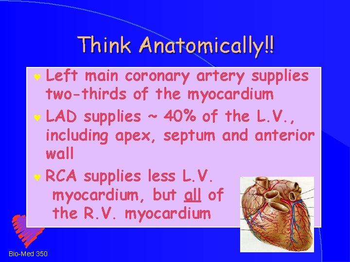
Think Anatomically!! Left main coronary artery supplies two-thirds of the myocardium © LAD supplies ~ 40% of the L. V. , including apex, septum and anterior wall © RCA supplies less L. V. myocardium, but all of the R. V. myocardium © Bio-Med 350
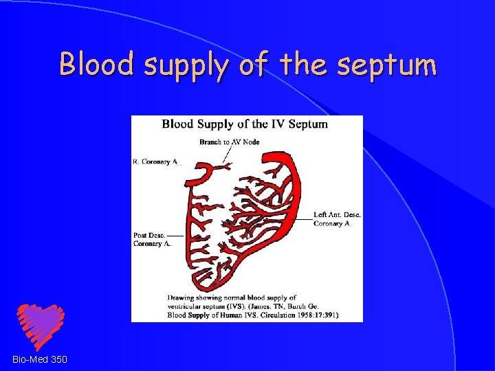
Blood supply of the septum Bio-Med 350
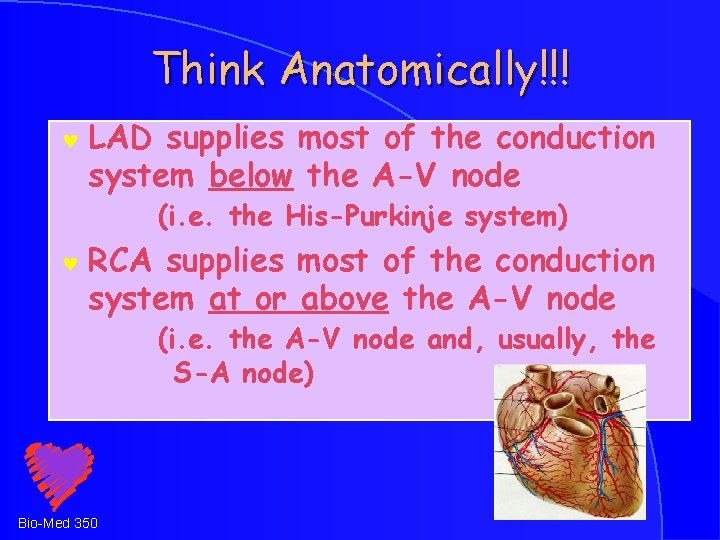
Think Anatomically!!! © LAD supplies most of the conduction system below the A-V node (i. e. the His-Purkinje system) © RCA supplies most of the conduction system at or above the A-V node (i. e. the A-V node and, usually, the S-A node) Bio-Med 350
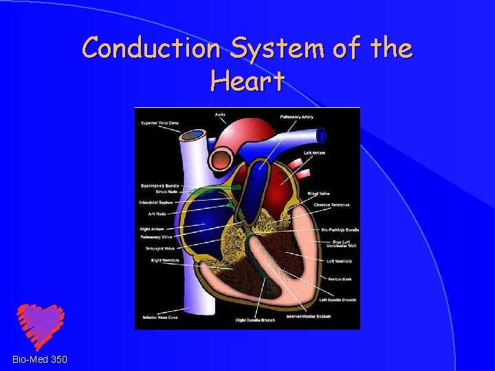
Conduction System of the Heart Bio-Med 350
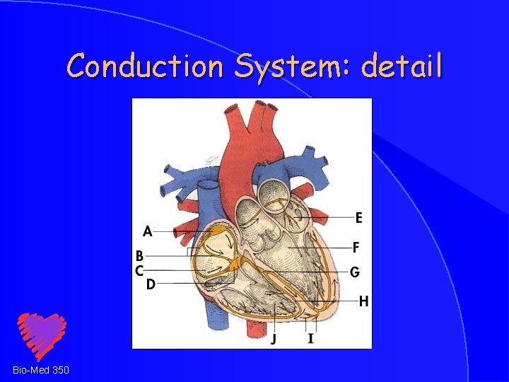
Conduction System: detail Bio-Med 350
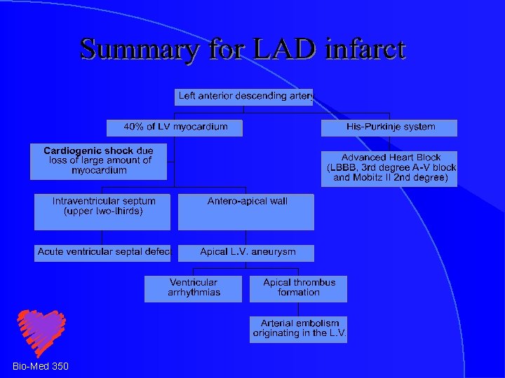
ACUTE M. I. Anatomical correlates LAD occlusion causes extensive infarction associated with: © LV failure High grade heart block © Apical aneurysm formation Thrombo-embolic complications © © Bio-Med 350
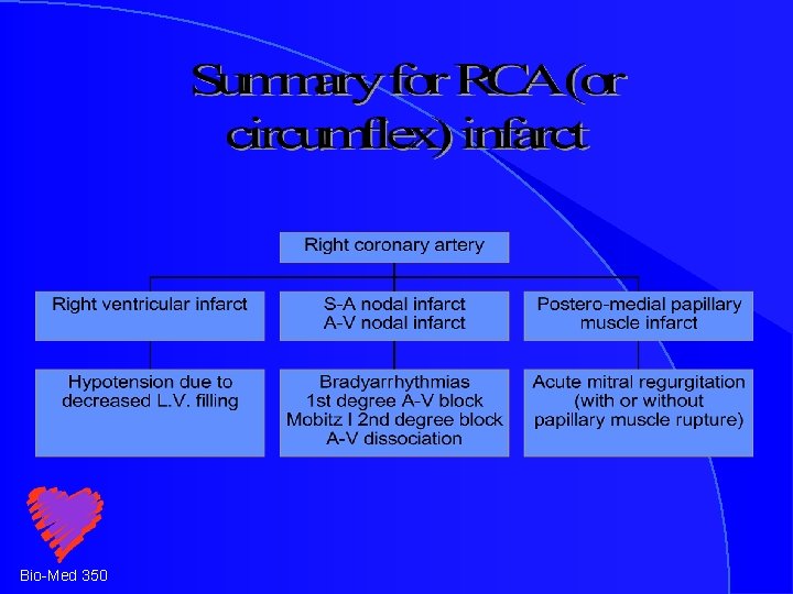
ACUTE M. I. Anatomical correlates RCA occlusion causes moderate infarction associated with: © RV failure Bradyarrhythmias Occasional mechanical complications © © Bio-Med 350
ACUTE M. I. Arrhythmias Sinus bradycardia © Sinus tachycardia © Atrial fibrillation © PVCs / ventricular tachycardia /ventricular fibrillation © Heart block © Bio-Med 350
Arrhythmias: Inferior M. I. © © Sinus bradycardia -- S. A. nodal artery and increased vagal tone Heart block -- A-V nodal artery 1 st degree A-V block Wenckebach 2 nd degree A-V block A-V dissociation Atrial fibrillation -- L. A. stretch Ventricular tachycardia / fibrillation -via “re-entry” or increased automaticity Bio-Med 350
Arrhythmias: Anterior M. I. Sinus tachycardia -- low stroke volume © Heart block -- His-Purkinje system © Left or Right Bundle branch block Complete Heart Block © Ventricular tachycardia / fibrillation due to “re-entry” or increased automaticity Bio-Med 350
ACUTE M. I. Hypotension Identify hemodynamic subset © Distinguish decreased preload from decreased cardiac output © Think about hemodynamic monitoring © Bio-Med 350
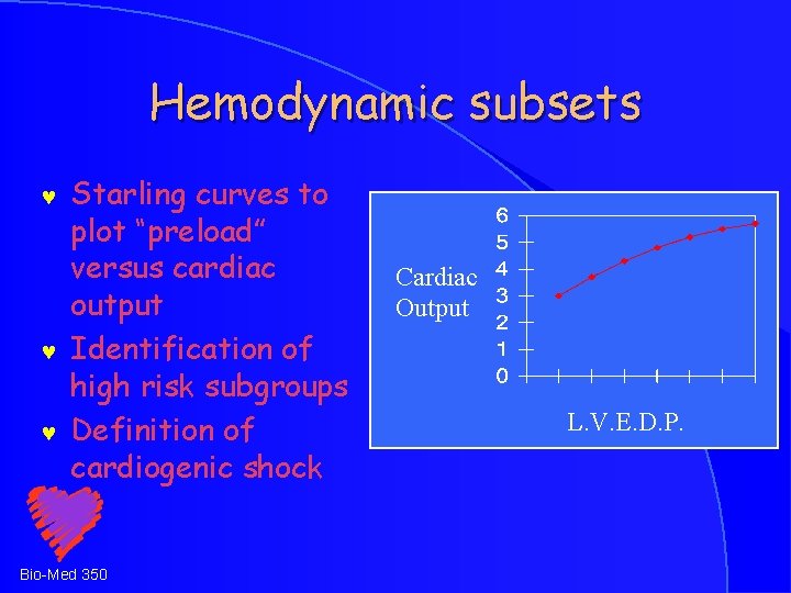
Hemodynamic subsets © © © Starling curves to plot “preload” versus cardiac output Identification of high risk subgroups Definition of cardiogenic shock Bio-Med 350 Cardiac Output L. V. E. D. P.
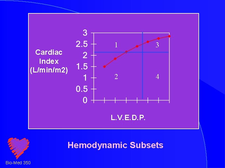
Cardiac Index (L/min/m 2) 1 3 2 4 L. V. E. D. P. Hemodynamic Subsets Bio-Med 350
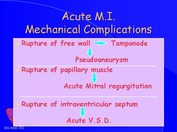
Acute M. I. Mechanical Complications ª Rupture of free wall Tamponade Pseudoaneurysm ª Rupture of papillary muscle Acute Mitral regurgitation ª Rupture of intraventricular septum Acute V. S. D. Bio-Med 350
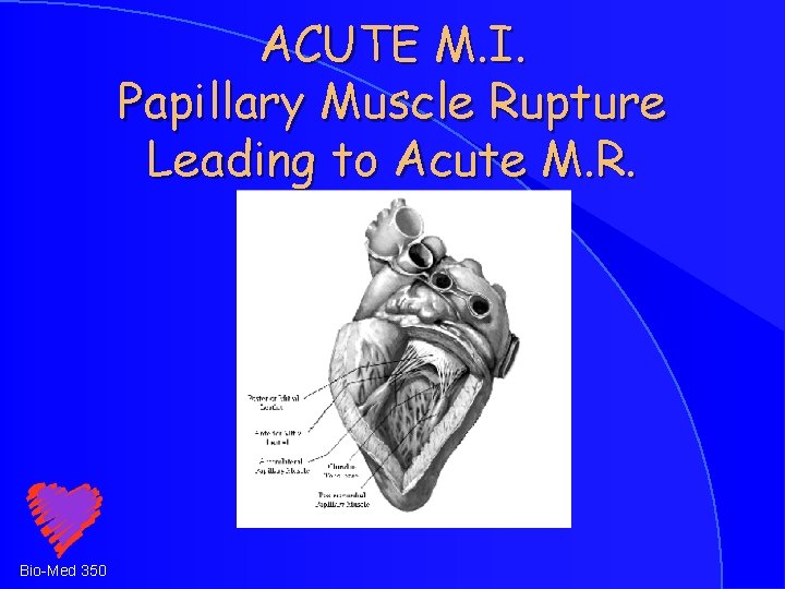
ACUTE M. I. Papillary Muscle Rupture Leading to Acute M. R. Bio-Med 350
ACUTE M. I. Papillary Muscle Rupture Leading to Acute M. R. Systolic murmur © Giant V - waves on PC Wedge tracing © Echo/Doppler confirmation © RX with Afterload reduction © Intra-aortic balloon pump © Bio-Med 350
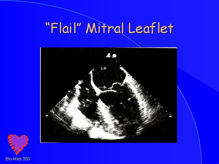
“Flail” Mitral Leaflet Bio-Med 350
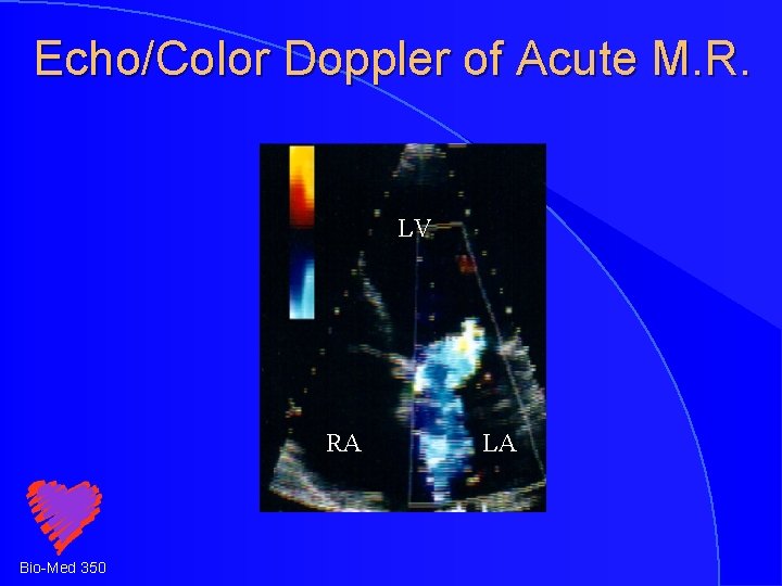
Echo/Color Doppler of Acute M. R. LV RA Bio-Med 350 LA
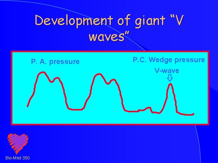
Development of giant “V waves” P. A. pressure Bio-Med 350 P. C. Wedge pressure V-wave
Acute Mitral Regurgitation: Treatment Rapid diagnosis © Afterload reduction © Inotropic support © Intra-aortic balloon pump © Surgical valve replacement © Bio-Med 350
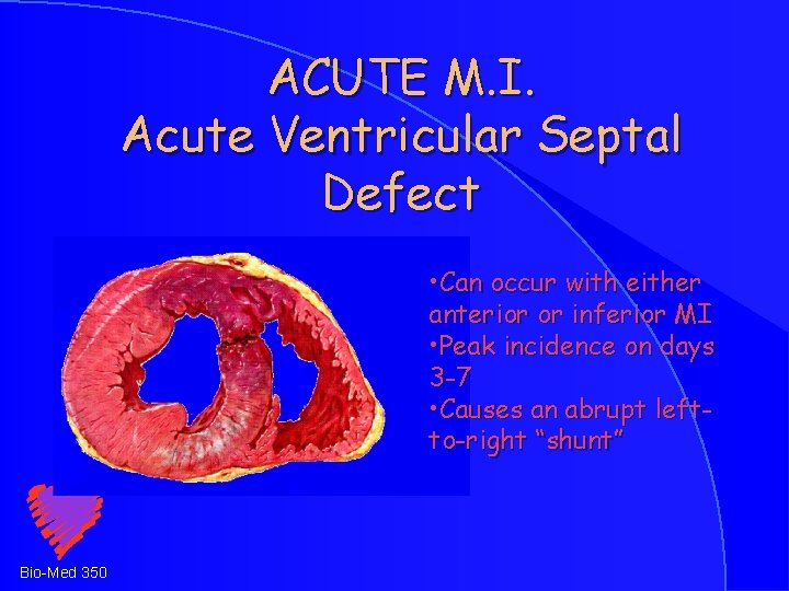
ACUTE M. I. Acute Ventricular Septal Defect • Can occur with either anterior or inferior MI • Peak incidence on days 3 -7 • Causes an abrupt leftto-right “shunt” Bio-Med 350
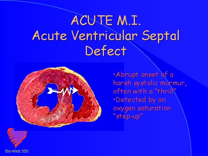
ACUTE M. I. Acute Ventricular Septal Defect • Abrupt onset of a harsh systolic murmur, often with a “thrill” • Detected by an oxygen saturation “step-up” Bio-Med 350
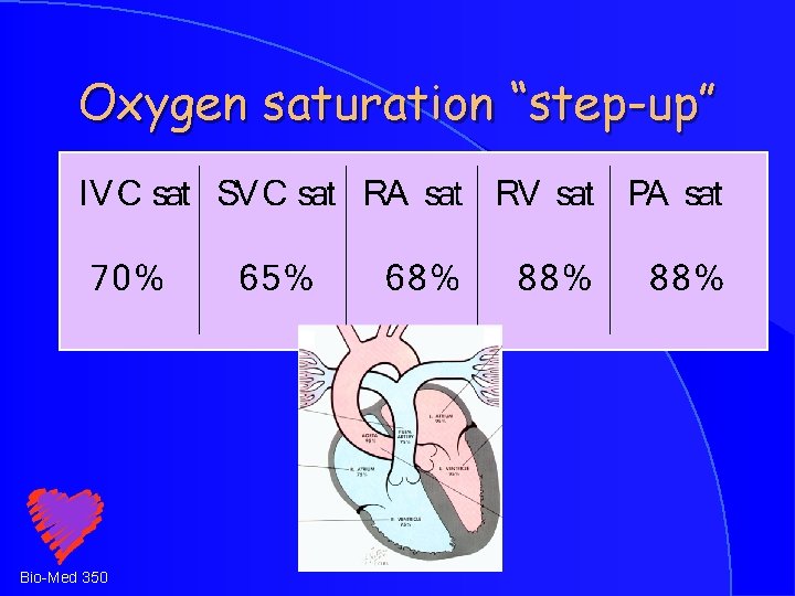
Oxygen saturation “step-up” Bio-Med 350
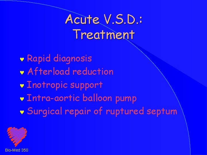
Acute V. S. D. : Treatment Rapid diagnosis © Afterload reduction © Inotropic support © Intra-aortic balloon pump © Surgical repair of ruptured septum © Bio-Med 350
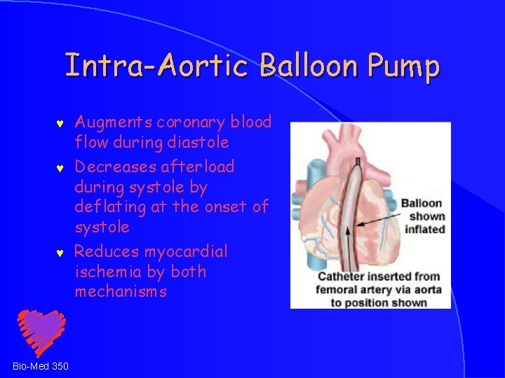
Intra-Aortic Balloon Pump © © © Bio-Med 350 Augments coronary blood flow during diastole Decreases afterload during systole by deflating at the onset of systole Reduces myocardial ischemia by both mechanisms
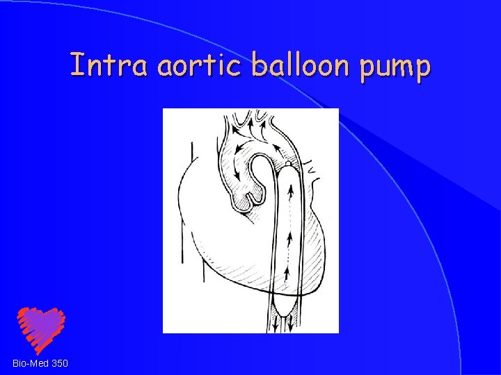
Intra aortic balloon pump Bio-Med 350
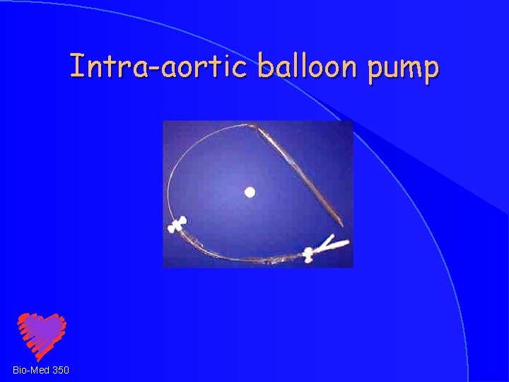
Intra-aortic balloon pump Bio-Med 350
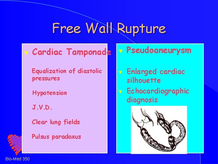
Free Wall Rupture © Cardiac Tamponade © Pseudoaneurysm Equalization of diastolic pressures © Hypotension © Enlarged cardiac silhouette Echocardiographic diagnosis J. V. D. Clear lung fields Pulsus paradoxus Bio-Med 350
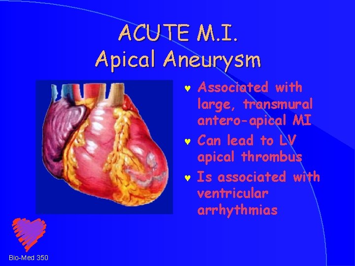
ACUTE M. I. Apical Aneurysm © © © Bio-Med 350 Associated with large, transmural antero-apical MI Can lead to LV apical thrombus Is associated with ventricular arrhythmias
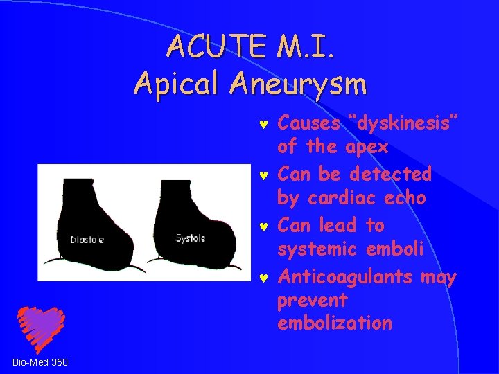
ACUTE M. I. Apical Aneurysm © © Bio-Med 350 Causes “dyskinesis” of the apex Can be detected by cardiac echo Can lead to systemic emboli Anticoagulants may prevent embolization
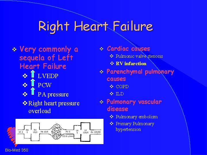
Right Heart Failure v Very commonly a sequela of Left Heart Failure v LVEDP v PCW v PA pressure v Right heart pressure overload Bio-Med 350 v Cardiac causes v Pulmonic valve stenosis v RV infarction v Parenchymal pulmonary causes v COPD v ILD v Pulmonary vascular disease v Pulmonary embolism v Primary Pulmonary hypertension
ACUTE M. I. Right Ventricular Infarction Jugular venous distention with clear lungs © Equalization of right atrial and PCW pressures © ST elevation in right precordial leads © Therapy with fluids © Bio-Med 350
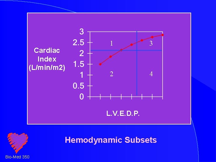
Cardiac Index (L/min/m 2) 1 3 2 4 L. V. E. D. P. Hemodynamic Subsets Bio-Med 350
ACUTE M. I. Pericarditis Pleuritic chest pain © Radiation to the trapezius ridge © Fever © Pericardial friction rub © Bio-Med 350
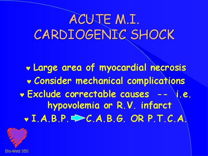
ACUTE M. I. CARDIOGENIC SHOCK Large area of myocardial necrosis © Consider mechanical complications © Exclude correctable causes -- i. e. hypovolemia or R. V. infarct © I. A. B. P. C. A. B. G. OR P. T. C. A. © Bio-Med 350
Bio-Med 350
Bio-Med 350
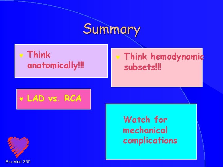
Summary © Think anatomically!!! © LAD vs. RCA © Think hemodynamic subsets!!! Watch for mechanical complications Bio-Med 350
THE END Bio-Med 350
- Slides: 45