Arrhythmia Arrhythmia is defined as loss of cardiac


- Slides: 28

Arrhythmia

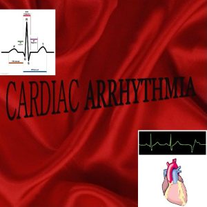
• Arrhythmia is defined as loss of cardiac rhythm, especially irregularity of heart beat. PATHOPHYSIOLOGY: SUPRAVENTRICULAR ARRHYTHMIAS: • Common supraventricular tachycardias requiring drug treatment are atrial fibrillation (AF) or atrial flutter, paroxysmal supraventricular tachycardia (PSVT), and automatic atrial tachycardias.

Atrial Fibrillation and Atrial Flutter: • AF is characterized as an extremely rapid (400 to 600 atrial beats/min) and disorganized atrial activation. • There is a loss of atrial contraction (atrial kick), and supraventricular impulses penetrate the atrioventricular (AV) conduction system in variable degrees, resulting in irregular ventricular activation and irregularly irregular pulse (120 to 180 beats/min).

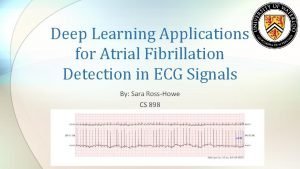
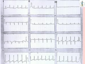
Atrial Fibrillation ECG Tracing

• Atrial flutter is characterized by rapid (270 to 330 atrial beats/min) but regular atrial activation. • The ventricular response usually has a regular pattern and a pulse of 300 beats/min. • This arrhythmia occurs less frequently than AF but has similar precipitating factors, consequences, and drug therapy.

Atrial Flutter Tracing

• The predominant mechanism of AF and atrial flutter is reentry, which is usually associated with organic heart disease that causes atrial distention (e. g. , ischemia or infarction, hypertensive heart disease, valvular disorders). • PSVT arising by reentrant mechanisms includes arrhythmias caused by AV nodal reentry, AV reentry incorporating an anomalous AV pathway, sinoatrial (SA) nodal reentry, and intra atrial reentry.

• Automatic atrial tachycardias such as multifocal atrial tachycardia appear to arise from supraventricular foci with enhanced automatic properties. • Severe pulmonary disease is the underlying precipitating disorder in 60% to 80% of patients.

VENTRICULAR ARRHYTHMIAS: Premature Ventricular Complexes: • Premature ventricular complexes (PVCs) are common ventricular rhythm disturbances that occur in patients with or without heart disease and may be elicited experimentally by abnormal automaticity, triggered activity, or reentrant mechanisms.

Two Ventricular Premature Complexes

Ventricular Tachycardia: • Ventricular tachycardia (VT) is defined by three or more repetitive PVCs occurring at a rate greater than 100 beats/min. • It occurs most commonly in acute myocardial infarction (MI); other causes are severe electrolyte abnormalities (e. g. , hypokalemia), hypoxemia, and digitalis toxicity. • The chronic recurrent form is almost always associated with underlying organic heart disease (e. g. , idiopathic dilated cardiomyopathy or remote MI with left ventricular [LV] aneurysm).

Monomorphic ventricular tachycardia

Torsades de pointes QRS complexes varying in amplitude, axis and duration.

• Incessant VT refers to VT occurring more frequently than sinus rhythm, so that VT becomes the dominant rhythm. • Monomorphic VT has a consistent QRS configuration, whereas polymorphic VT has varying QRS complexes. • Torsade de pointes (Td. P) is a polymorphic VT in which the QRS complexes appear to undulate around a central axis.

BRADYARRHYTHMIAS: • Asymptomatic sinus bradyarrhythmias (heart rate less than 60 beats/min) are common especially in young, athletically active individuals. • However, some patients have sinus node dysfunction (sick sinus syndrome) because of underlying organic heart disease and the normal aging process, which attenuates SA nodal function. • AV block or conduction delay may occur in any area of the AV conduction system. AV block may be found in patients without underlying heart disease (e. g. , trained athletes) or during sleep when vagal tone is high.

CLINICAL PRESENTATION • Supraventricular tachycardias may cause a variety of clinical manifestation ranging from no symptoms to minor palpitations and/or irregular pulse to severe and even life-threatening symptoms. • Patients may experience dizziness or acute syncopal episodes; symptoms of HF; anginal chest pain; or, more often, a choking or pressure sensation during the tachycardia episode. • Patients with bradyarrhythmias experience symptoms associated with hypotension such as dizziness, syncope, fatigue, and confusion. • If LV dysfunction exists, symptoms of congestive HF may be exacerbated.

DESIRED OUTCOME • The desired outcome depends on the underlying arrhythmia. • For example, the ultimate treatment goals of treating AF or atrial flutter are restoring sinus rhythm, preventing thromboembolic complications, and preventing further recurrences.

• Drugs may have antiarrhythmic activity by directly altering conduction in several ways. • Drugs may depress the automatic properties of abnormal pacemaker cells by decreasing the slope of phase 4 depolarization and/or by elevating threshold potential.


Acute Ventricular Tachycardia • If severe symptoms are present, synchronized DCC should be institute immediately to restore sinus rhythm. • Patients with mild or no symptoms can be treated initially with antiarrhythmic drugs. IV amiodarone is now recommended as first-line therapy in this situation. • Patients with chronic recurrent sustained VT are at extremely high risk for death; trial-and-error attempts to find effective therapy are unwarranted. • The automatic ICD (implantable cardioverterdefibrillator) is a highly effective method for preventing sudden death due to recurrent VT or VF.

• For an acute episode of Td. P, most patients require and respond to DCC. • However, Td. P tends to be paroxysmal and often recurs rapidly after DCC. • IV magnesium sulfate is considered the drug of choice for preventing recurrences of Td. P. • Agents that prolong the QT interval should be discontinued, and exacerbating factors (e. g. , hypokalemia, hypomagnesemia) corrected. Drugs that further prolong repolarization (e. g. , IV procainamide) are contraindicated. Lidocaine is usually ineffective.

• Treatment of sinus node dysfunction involves elimination of symptomatic bradycardia and possibly managing alternating tachycardias such as AF. • In general, long-term therapy of choice for patients with significant symptoms is a permanent ventricular pacemaker. • Patients who remain symptomatic may benefit from adding an α-adrenergic stimulant such as midodrine. • Other drugs that have been used successfully (with or without β-blockers) include fludrocortisone, anticholinergics (scopolamine patches, disopyramide), α-adrenergic agonists (midodrine), adenosine analogs (theophylline, dipyridamole), and selective serotonin reuptake inhibitors (sertraline, fluoxetine).

Adenosine: • Drug therapy involves blocking the AV node because most PSVT rhythms involve a re-entry circuit within this area. • An initial 6 -mg IV bolus is given; if this is unsuccessful within 2 minutes, it can be followed by one or two additional 12 -mg IV boluses, up to a maximum of 30 mg. • Because of its short half-life (9 seconds), adenosine should be administered as a rapid bolus (over 1– 3 seconds), followed immediately by a saline flush. • Nondihydropyridine calcium channel blockers, verapamil and diltiazem, can be used in patients with PSVT. • Verapamil (2. 5– 5 mg IV given over 2 minutes) achieves peak therapeutic effects in 3 to 5 minutes after dosing and can be repeated at 10 - to 15 -minute intervals to a maximum dose of 20 mg if needed.

• In high-risk patients with MI who are not candidates for β-blockade, alternative antiarrhythmic therapy with amiodarone can be considered. • Amiodarone is a class III antiarrhythmic agent but also has antiadrenergic, class I, and class IV activity. • Treatment with amiodarone (800 mg/day for 7 days followed by 400 mg/day for 6 days of the week for 1 year) was associated with a significant reduction in cardiac mortality and significant ventricular arrhythmias.
 Mit-bih arrhythmia database
 Arrhythmia
 Arrhythmia
 Which arrhythmia refers to rapid random inefficient
 Dysrhythmia vs arrhythmia
 The method of unit costing is adopted by
 Collection of well defined object
 Bed making introduction
 Anesthesia for cardiac tamponade
 Teixit muscular cardiac
 Comparison of skeletal cardiac and smooth muscle
 Pearson education
 Phase 2 cardiac rehab exercises
 Spread of cardiac impulse
 Orthorpnea
 Cardiac muscle cell vs skeletal
 Cardiac muscle striations
 Ucsf cardiac anesthesia
 What are the 5 heart sounds
 Brachioradialis posterior view
 Lesson 6: cardiac emergencies and using an aed
 Cardiac muscle tissue
 Neil moat
 Wandering atrial pacemaker criteria
 Negative inotropic drugs
 Cardiac output and stroke volume
 Gittata cardiaca
 Cardiac workload
 Cardiac distension
