Anomalies of the Head and Neck Dr Shathaly
. U")


 • incomplete fusion of the lateral lingual swellings results")


 palate and cleft uvula.")


- Slides: 30
Anomalies of the Head and Neck Dr. Shathaly Mustafa Majed. MSc. Human Morphology(Anatomy). U of K MSc molecular medicine U of K MD Surgery. SSMB MSc Medical Education Diploma in research methodology and biostatistics. UMST
Pharyngeal Region • Ectopic Thymic and Parathyroid Tissue. • Branchial Fistulas.
Ectopic Thymic and Parathyroid Tissue • glandular tissue derived from the pouches undergoes migration. • Thymic tissues may remain in the neck. • Parathyroid glands may descend to the thorax especially the inferior parathyroid gland.
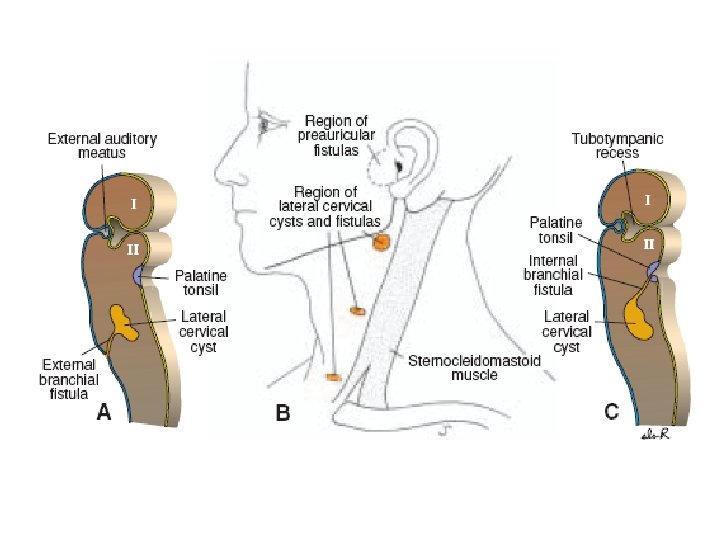
Branchial Fistulas • occur when the second pharyngeal arch fails to grow caudally over the third and fourth arches, leaving remnants of the second, third, and fourth clefts in contact with the surface by a narrow canal. • Such a fistula, found on the lateral aspect of the neck directly anterior to the sternocleidomastoid muscle.
Neural Crest Cells and Craniofacial Defects • Neural crest cells are important in : • Formation of much of the craniofacial region. • Conotruncal endocardial cushions, which separate the outflow tract of the heart into pulmonary and aortic channels.
Di. George anomaly • CATCH 22 : Ø Cardiac defects. Ø Abnormal facies. Ø Thymic hypoplasia. Ø Cleft palate. Ø Hypocalcemia. • deletion on the long arm of chromosome 22.
• Origin of the defects is caused by abnormal development of neural crest cells that contribute to formation of all of the affected structures.
Tongue • Ankyloglossia. • Macroglossia. • Microglossia.
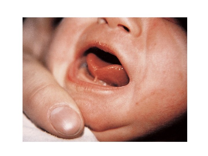
Ankyloglossia • the frenulum is short and extends to the tip of the tongue. • This interferes with its free protrusion and may make breast-feeding difficult.
Macroglossia • An excessively large tongue is not common. • It results from generalized hypertrophy of the developing tongue
Microglossia • An abnormally small tongue is extremely rare. • usually associated with micrognathia (underdeveloped mandible and recession of the chin).
Bifid or Cleft Tongue (Glossoschisis) • incomplete fusion of the lateral lingual swellings results in a deep midline groove in the tongue.
Thyroid Gland • Thyroglossal cyst. • Thyroglossal fistula. • Ectopic thyroid.
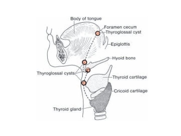
Thyroglossal cyst • it is a cystic remnant of the thyroglossal duct. • always near or in the midline of the neck.
Thyroglossal fistula • Sometimes a thyroglossal cyst is connected to the outside by a fistulous canal.
Ectopic thyroid tissue • may be found anywhere along the path of descent of the thyroid gland. • commonly found in the base of the tongue lingual thyroid.
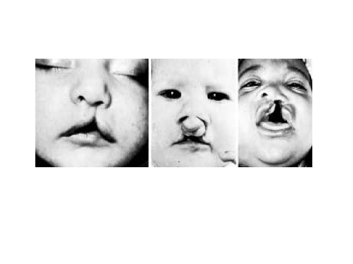
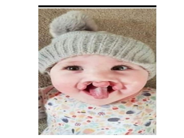
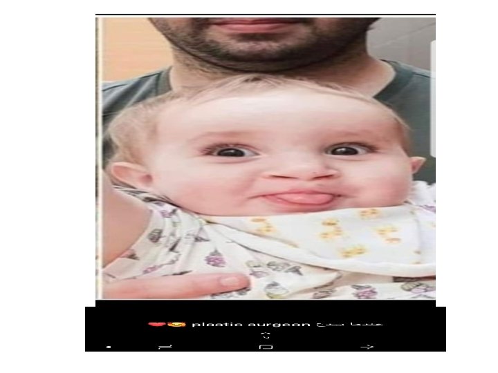
Facial Clefts • Cleft lip and cleft palate are common defects that result in abnormal facial appearance and defective speech. • The incisive foramen is considered the dividing landmark between the anterior and posterior cleft deformities. • anterior to the incisive foramen include lateral cleft lip, cleft upper jaw, and cleft between the primary and secondary palates
• due to a partial or complete lack of fusion of the maxillary prominence with the medial nasal prominence on one or both sides.
• posterior to the incisive foramen include cleft (secondary) palate and cleft uvula. • Cleft palate results from a lack of fusion of the palatine shelves
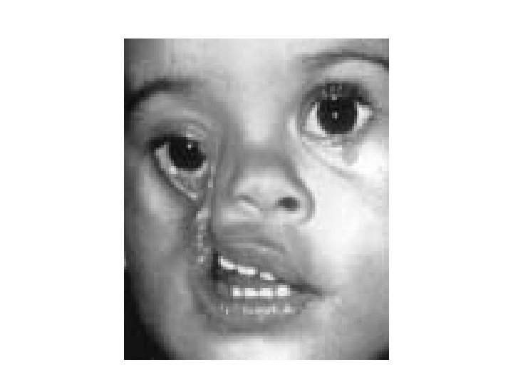
• Oblique facial clefts are produced by failure of the maxillary prominence to merge with its corresponding lateral nasal prominence
• Median cleft lip, a rare abnormality, is caused by incomplete merging of the two medial nasal prominences in the midline.
Tooth Abnormalities • Natal Teeth have erupted by the time of birth. Usually they involve the mandibular incisors, which may be abnormally formed. • Teeth may be abnormal in number, shape, and size. • They may be discolored by foreign substances, such as tetracyclines, or be deficient in enamel, a condition often caused by vitamin D deficiency (rickets).