Anatomy Physiology 2 Main Parts Lymphatic vessels Lymphoid

")


- Slides: 22
Anatomy & Physiology
� 2 Main Parts �Lymphatic vessels �Lymphoid tissues/organs � FUNCTIONS: �Drain excess fluid (edema) and returns it to blood �Plays essential roles in body defense and resistance to disease
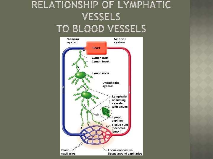
� Lymph—excess tissue fluid � Properties of lymphatic vessels �One way system toward the heart �No pump �Lymph moves toward the heart Transported through milking action of skeletal muscle Rhythmic contraction of smooth muscle in vessel walls helps pump the lymph through the system
� Lymph capillaries �Walls overlap to form flap-like minivalves to allow entrance for lymph Fluid leaks into lymph capillaries �Lymph Capillaries are anchored to connective tissue by filaments �Higher pressure on the inside of the capillary closes minivalves Fluid is forced along the vessel for examination � Examined by cells of immune system for any potential threats
� Lymphatic vessels collecting � Collect lymph from lymph capillaries � Carry lymph to and away from lymph nodes � Return fluid to circulatory veins near the heart Right lymphatic duct � Drains lymph to right arm/head/thorax Thoracic duct � Drains rest of body that R. Lymph duct doesn’t
� Harmful materials that enter lymph vessels �Bacteria �Viruses �Cancer cells �Cell debris
� Filter lymph before it is returned to the blood � Areas of high concentration? �Inguinal, � Defense axillary, and cervical regions cells within lymph nodes �Macrophages—engulf and destroy foreign substances �Lymphocytes—provide immune response to antigens � Nodes swell when fighting infection because nodes have engulfed the foreign substance to prevent it from spreading
� Most are kidneyshaped and less than 1 inch long � Cortex �Outer part �Contains follicles— collections of lymphocytes � Medulla �Inner part �Contains phagocytic macrophages
� Several other organs contribute to lymphatic function �Spleen �Thymus �Tonsils �Peyer’s patches
Blood filled organ which filters blood � Location: Left side of abdomen above diaphragm � Functions: � � Filters/cleanses blood of foreign substances � Destroy worn-out blood cells � Recycles components of worn out blood cells (i. e. hemoglobin) � Stores platelets � Protect body during hemorrhage
� Lymphoid mass found in throat � Function: Programs lymphocytes with the help of thymosin (hormone)
� Small, lymphoid tissue in back of throat � Function: Capture and remove any foreign pathogens(substan ces) �Reason they become swollen during colds/viruses
� Location: Wall of small intestine � Function: Capture and destroy bacteria �Prevent foreign pathogens from penetrating intestinal wall
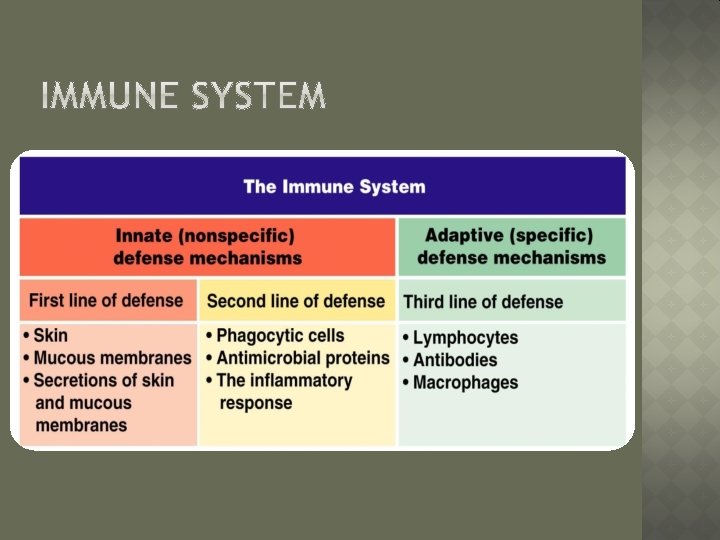
� The body is constantly in contact with bacteria, fungi, and viruses � The body has two defense systems foreign materials �Innate (nonspecific) defense system Attacks ANY foreign pathogen �Adaptive (specific) defense system Attacks SPECIFIC substances � Immunity—specific �-Immun=free resistance to disease
� AKA “non-specific defense system” � Innate body defenses are mechanical barriers to pathogens such as �Body surface coverings Intact skin Mucous membranes � Line all body cavities that are exposed (i. e. digestive, respiratory, urinary, and reproductive tracts) �Specialized human cells �Chemicals produced by the body
� Skin and mucous membranes �Physical barrier to foreign materials �Also provide protective secretions p. H of the skin is acidic to inhibit bacterial growth Sebum is toxic to bacteria Stomach releases hydrochloric acid to kill pathogens Saliva and lacrimal fluid contain lysozymes which kill bacteria Mucus traps microorganisms and prevent them from entering the digestive or respiratory tract
� Phagocytes �I. e. macrophage or neutrophil �Swallows foreign pathogen � Natural �Kill killer cells cancer and virus cells �Fight off these pathogens by detecting sugar molecules on pathogen’s surface
� Inflammatory response �Happens when tissue is injured � 4 signs: Redness, heat, inflammation, swelling pain �Histamine & Kinins causes: Vessels to dilate causing inflammation Activate pain receptors to alert body of problem Attract WBCs and phagocytes to clean cellular debris from tissue damage
� Antimicrobial � Fever proteins