Lymphatic System The Lymphatic System consist of Lymphatic


")


 consist")

. 3.")


- Slides: 35
Lymphatic System
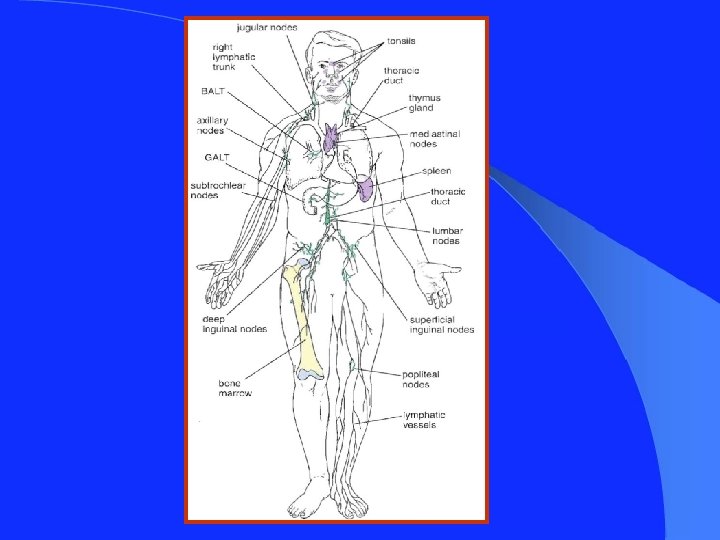
The Lymphatic System consist of: * Lymphatic Vessels. * Lymphoid Organs.
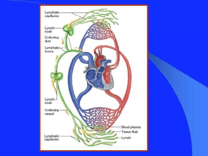
Lymphatic Vessels These vessels covey fluids from the tissues to the blood stream. L. V. found in most tissue and organs but absent from central nervous system, cartilage, bone and epidermis.
This L. V. including of: 1. Lymph capillaries vessels They are small thin walled and building ended tube. Its similarly blood capillaries vessels but the endothelial cells overlap each other but there is gap between them and no pericytes and continuous basal membrane is absent.
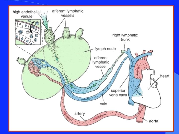
2. Lymphatic collecting vessels or larger lymphatic vessels The larger vessels have thicker walls in which the typical three layers can sometimes be distinguished the tunica intimae consists of endothelium and a thin layer of longitudinal elastic fibers, the tunica media composed of arranged circularly smooth muscle fibers and the tunica adventitia is the thickest and composed of longitudinal collagen elastic fiber and few smooth muscle fibers. These vessels posses valves which are more numerous than those in vein.
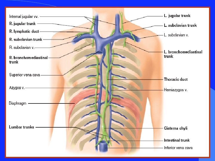
3. Lymphatic Trunks or Lymphatic Ducts There are only two lymph duct: a- Thoracic lymphatic duct. b- Right lymphatic duct. The walls of these duct resemble those of large veins, but are thinner and posses valves. The ducts drain into the venous system by joining the subclavian vein at the root of the neck. The right lymph ducts collected lymph from right parts of head and right parts of thorax, whereas the thoracic ducts collected lymph from the parts remnants of body.
Lymphoid Organs This organ including of: 1. Lymph Nodes. 2. Thymus. 3. Spleen. 4. Tonsils.
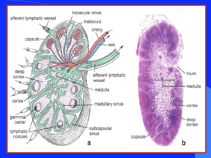
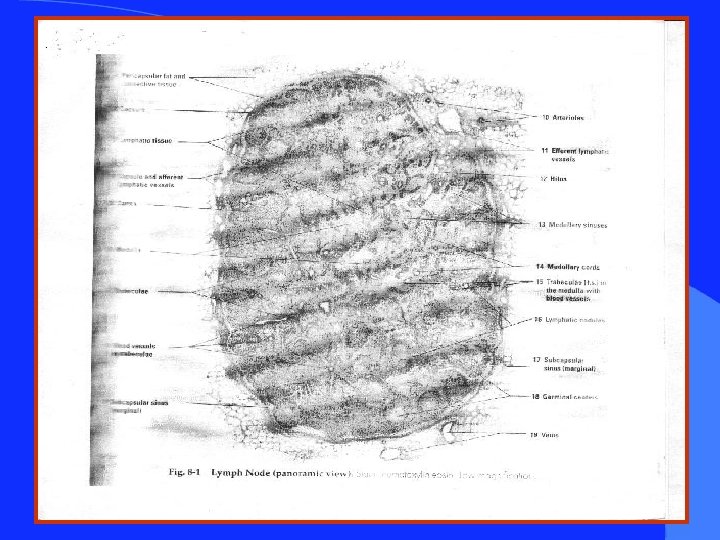
1 - Lymph Nodes These nodes found more in the axilla, groin, along great blood vessels of neck and in large number in thorax and abdomen.
The structure of lymph nodes consists of: 1. Cortex. It darker region composed of lymphocyte aggregate which is called "Lymph Nodules". The lymph nodules contain the germinal center, and covered by subcapsular sinus.
2. Medulla. It is lighter region and composed medullary cords (consists of lymphatic tissue) this region contain more lymph sinuses and blood capillaries and macrophages than the cortex. Lymph node is surrounded by thick connective tissue called "Capsule". Lymph nodes have a hilum where blood vessels and nerve enter and leave and the lymphatic vessels leave the node. The capsule sends septa into the node divided the paranchyme into incomplete compartment.
Function of Lymph Nodes 1. Product of lymphocytes and plasma cells. 2. Immunology function the lymphocytes in lymph nodes are able to recognize and reaction with foreign antigen.
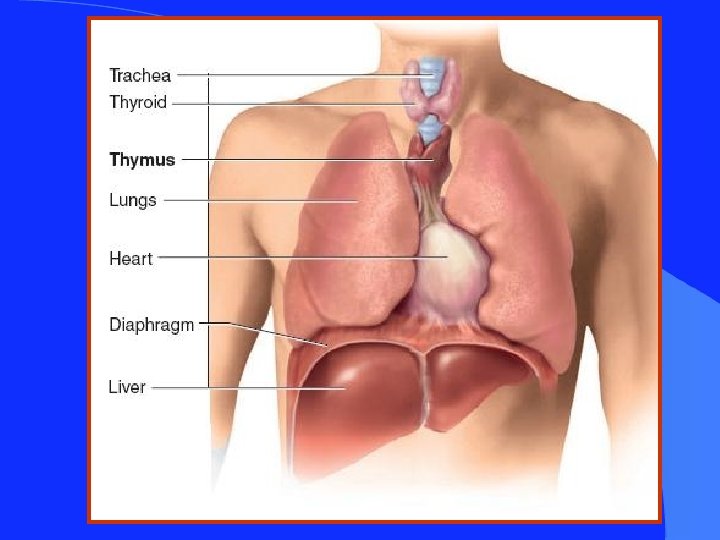
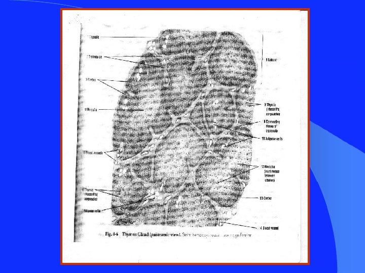
2 - Thymus Is a flattened, bilobed structure lying in upper part of the thorax between the strum and upper four costal cartilage. The weight of thymus a variant of the size between birth and old age. Thymus is surrounded by connective tissue called "Capsule" from which trabeculae (septa) extend into organ, practically subdivided into lobules composed into: 1. Cortex. The darker color. 2. Medulla. The lighter color.
Both cortex and medulla have the same cells type and which including: 1. Reticular Epithelial Cells. These cells more in medulla than cortex. 2. Lymphocytes or Thymocytes. These more in cortex than the medulla. 3. Macrophagus. Present in medulla more than the cortex. 4. Hassalls Corpuscle. They are rounded oval structure consists of concentrically arranged flattened epithelial.
Function of Thymus 1. Production of T-lymphocytes which are responsible for cellular immunity and help hormonal immune response. Lymphocyte production take place in cortex. 2. Filtration of blood. 3. Product the thymosin hormone. This hormone produced by the reticulo epithelial cells and influence the maturation of T-lymphocytes and stimulate their activity in the body.
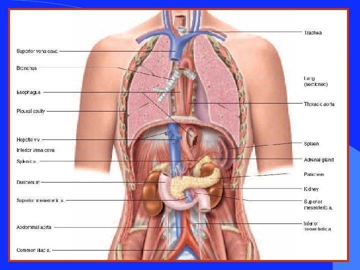
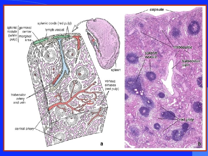
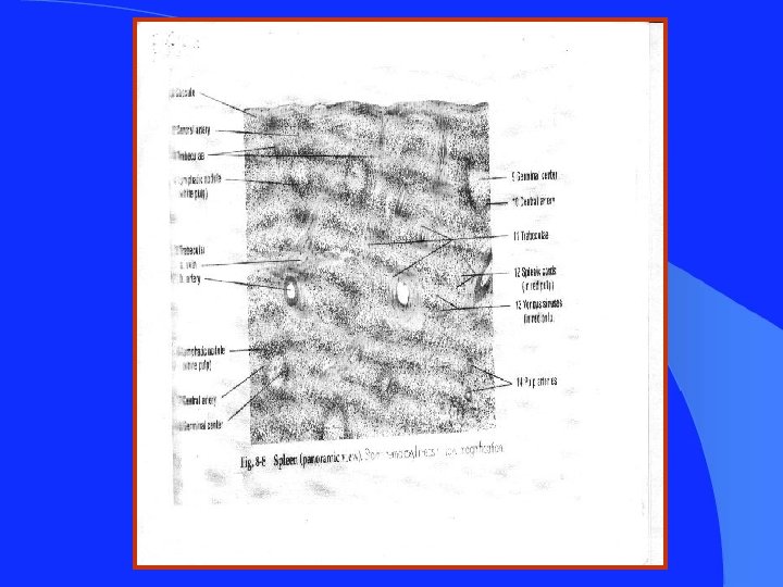
3 - Spleen Is the largest haemolymph organ in the body. It's situated in the abdomen under the left half of diaphragm. Its surrounded by connective tissue called "Capsule" contain few smooth muscle. The capsule sends trabeculae extend into the organ, particularly subdivided into lobules composed into: -
1. Red Pulp. This pulp occupies the centrical masses soft dark-red mass (histologically) consist of large irregular thin walled of blood vessels called "Splenic Sinusoides". These sinusoids separated by cord of reticular tissue called "Splenic Cords or Billroths". The red pulp contains monocytes, plasma cells, red blood cells and white blood cells and less number from lymphocytes.
2. White Pulp or Splenic Pulp. This pulp distributed in roughly spherical masses consist of lymphoid tissue. This pulp structure resemble the cortex of lymphoid nodules. Its filled with lymphocytes and some macrophage and plasma cells. The space between the traboculae is filled with lymphatic tissue. Spleen is covered by capsule except where it is attached by the peritoneum.
Function of Spleen 1. Filtration of blood. 2. Immune defense (forming the antibody). 3. Blood forming such lymphocyte + monocytes. 4. Blood storage. as
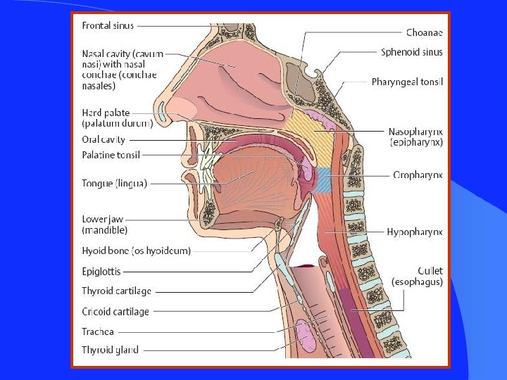
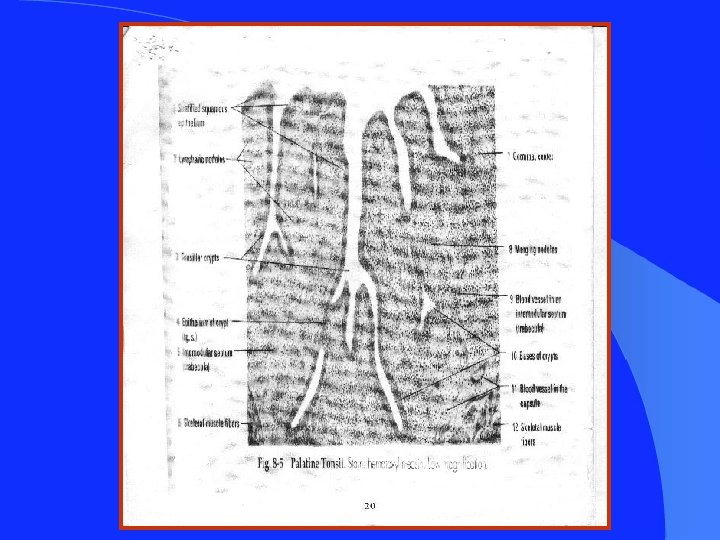
4 - Tonsils Aggregation of lymphoid tissue. They form a discontinuous ring. There are three types of tonsils: 1. Palatin tonsils. Located in the lateral walls of the pharynx. These tonsils are lined by a non karetinized stratified squamous epithelium tissue.
2. Lingual tonsils. Located in the posterior third of the tongue. This tonsils lined by a non karetinized stratified squamous epithelium tissue. 3. Pharyngeal tonsils. Located in the root of the nasopharynx. This lined by a with ciliated psedostratified epithelium tissue.
Structure of Tonsils All have the basically similar structure. These aggregation of lymphoid tissue resting in lamina propria. This lamina consist of lymphoid tissue containing lymph nodules. The surface is covered by epithelium tissue which extend down subdivided into (10 -20) deep pit called "Tonsillar Crypt". The crypts many branch.
Function of Tonsil 1. Formation of lymphocyte. 2. Defense of organism toward involving microorganism.