ACUTE AND CHRONIC DISEASES OF THE PHARYNX Department


- Slides: 33

ACUTE AND CHRONIC DISEASES OF THE PHARYNX Department of ENT diseases of Tashkent Medical Academy

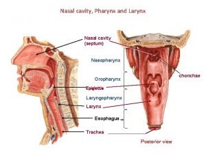
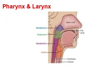
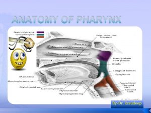
THE FLOORS OF THE PHARYNX The pharynx is a place of crossing over the respiratory and digestive tract. The lower boundary of the pharynx serves as a place of transition in her esophagus at 6 cervical vertebrae. There are three of the pharynx: Upper – nasopharynx Medium – oropharynx Lower – hypopharynx The pharynx connects the nose and mouth from above, from below the larynx and esophagus. The pharynx is formed by muscles fibrous membranes and inside is lined with mucous membrane. The length of the throat of her adult set to the lower end is 14 cm (12 -15), the transverse size of the average equal to 4. 5 cm

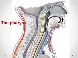
SAGITTAL SECTION OF THE PHARYNX

LIMFADENOIDNOE PHARYNGEAL RING OF PIROGOV-VALDEER I and II - palatine tonsil III - nasopharyngeal IV - lingual V and VI - tubal Besides, there is accumulation of tissue limfadenoidnoy on the back of the pharynx, in the lateral ridges and the lingual surface of the epiglottis.

THE FUNCTIONS OF THE PHARYNX Swallowing and sucking Voice-and speech production. Breath Defensive when eating and breathing

CLASSIFICATION OF ANGINA OF B. S. PREOBRAJENSKIY Catarrhal • Follicular • Lacunar • Fibrinous • Herpetic • Necrotizing (gangrenous) • Abscess (abscess intratonzillyarny) • Mixed forms

PHARYNGOSCOPE WITH CATARRHAL ANGINA On faryngoscopy swollen tonsils, a few, highly reddened, their surface is covered with a mucous discharge. The mucosa around the tonsils is more or less hyperemic, but diffuse hyperemia oropharynx is not available, which is typical for acute pharyngitis. In more severe cases are petechial hemorrhages in the mucosa.

PHARYNGOSCOPE WITH LACUNAR ANGINA On the swollen and reddened mucosa tonsils are formed from the depths of the lacunae tonsillis white or yellow caps, consisting of bacteria, epithelial cells and rejected by a large number of white blood cells. On the surface of the tonsils is often formed yellowish-white coating, which does not go beyond the tonsils. In the lacunar angina affected the entire fabric of the amygdala, which, because of the swell and increases in volume. Plaque formation in the gaps distinguish this form of diphtheria, in which, apart from the lacunae, prominent location and affected mucosa of the tonsils.

PHARYNGOSCOPE WITH FOLLICULAR ANGINA Reddened and swollen on the mucous membrane on both tonsils appears a significant amount of roundthe size of a pinhead, rising slightly yellowish or yellowish-white dots, which are festering follicles tonsils. Yellowish-white spots gradually increased and suppurate opened.

PHARYNGOSCOPE WITH FLEGMANOZ ANGINA Sharp protrusion tonsils, palatal arches and soft palate to the midline (globular formation on the one hand the throat), tongue is displaced in the opposite direction, intensity, and bulging bright redness in the area of greatest bulging with pressure-fluctuation, tongue coated with a thick coating and sticky saliva.

PLACE OF OPENING OF A PERITONSILLAR ABSCESS

PERITONSILLAR TISSUE INCISION

EXPANSION OF THE CAVITY

RETROPHARYNGEAL ABSCESS When viewed from the back of the throat or the feeling of a finger is determined by the fluctuating vapor protruding tumor. Abscess can spread to the region of large vessels of the neck or down on prespinal fascia into the chest cavity and cause purulent mediastinitis.

THE LOCATION AND SIZE OF THE OPENING OF RETROPHARYNGEAL ABSCESS

THE SCHEME OPENED RETROPHARYNGEAL ABSCESS An autopsy should be performed with care to avoid flowing of pus in the larynx and vertebral lesions and blood vessels. After opening the abscess immediately tilted his head down sick.

CHRONIC TONSILLITIS

CHRONIC TONSILLITIS Etiology Microbial flora (streptococcus and staphylococcus) Patogenesis Toxic component Allergic component Neuro-reflex component Violation of the immune processes Factors causing Recurrent angina Chronic source of infection (sinusitis, carious teeth and other) Reduction of general and local protective factors. Harmful environmental factors Classification Compensated form Decompensated form Simple Toxico-allergic form I degree Toxico-allergic form II degree Clinic Subjective symptoms Drying, scratch, foreign body sensation in the throat Recurrent sore throat Pain in the heart and large joints Allocation of purulent plugs Bad breath Objective symptoms Umbilicus adhesion tonsils with bows Purulent follicles in the tonsils Cchanges in septic arches (symptoms of Giza, Zach, Preobrajenskiy) Abnormal discharge in the gaps pus and caseous plug) Increase in regional lymph nodes Low-grade fever Changes in the forms of the tonsils (hypertrophy, atrophy)

Classification of chronic tonsillitis (Preobrajenskiy - Palchun) Chronic tonsillitis Toxico-allergic form Simple form I - degree II -degree Comorbidity Directly related to disease pathogenesis

CHRONIC TONSILLITIS Treatment Prophylaxis Rinsing and alkaline disinfectants Prevention and etiology causing f actors Lavage lacunae of tonsils Applique drugs (lubrication, Individual and social prevention forward into the gaps of tonsils) Physiotherapy (UV , ultrahigh, electrophoresis) Hyposensitization treatment General strengthening therapy Low-frequency ultrasound treatment Bilateral tonsillectomy Ultrasound and cryosurgery

CHRONIC PHARYNGITIS Etiology Microbial flora Pathogenesis Toxic component Allergic component Factors causing Smoking, drinking alkogol Increased dustiness and increase aerating Metabolic disorder, lack of vitamins Pocket of infection (chronic tonsillitis, sinusitis) Clinical forms Catarrhal Atrophic Hypertrophic Clinic Subjective symptoms Drying, scratch, foreign body sensation in the throat Persistent cough Pain when swallowing Objective symptoms Edema and hyperemia of the mucous membrane Muco-purulent discharge (in the back of the throat) Lymphoid appearance of granules in the rear and side walls of the pharynx Swelling and redness on the side walls of the pharynx Bad breath Low-grade fever

CHRONIC PHARYNGITIS Treatment Prophylaxis Rinsing the nose and Prevention and etiology ca using factors paranasal sinuses Alkaline inhalation and g Individual and social prevention argling lubrication of the pharynx drugs (lugol, Jõks, chlorhexidine) Drop in oil droplets nose Phonophoresis Vitamin therapy Galvanokaustika and cry osurgery

Adenoids (nasopharyngeal tonsil hypertrophy) Etiology Diagnosis frequent colds general inspection the metabolism of protein anterior and posterior rhinoscopy disturbance of endocrine function endoscopic examination of the harmful environmental factors nasopharynx genetic predisposition finger study nasopharyngeal Узига хос хусусиятлари in children 2 -12 years occurs more frequently with hypertrophy tonsils Differential diagnosis Degrees of hypertrophy nasopharyngeal tonsil I degree – димог 1/3 епади II degree – димог 2/3 епади III degree – димогни бутунлай епади with hypertrophic rhinitis with choanal polyp with juvenile angiofibroma Treatment adenotomia

Adenoids Clinic Subjective symptoms respiratory failure through the nose frequent colds hearing loss reduced memory and performance periodic headache sleep disturbances snoring Objective symptoms exterior signs of adenoidizm abnormal growth of the toothjaw system hypertrophy of tonsils signs of evaporation of the nasal mucosa mucus from the nose respiratory failure through the nose conductive hearing loss "Closed" snuffles

HYPERTROPHY OF TONSILS Etiology frequent colds the metabolism of protein disturbance of endocrine function harmful environmental factors genetic predisposition Special features in children 2 -12 years occurs more frequently with adenoids Degree of hypertrophy of tonsils I degree –closes the 1/3 between the arches and uvula II degree – closes the 2/3 between the arches and uvula III degree – covers more than 2 / 3 between arches and uvula Diagnosis pharyngoscope probing and palpation of the tonsillar Differential diagnosis with chronic tonsillitis Clinic Subjective symptoms speech disorder difficulty swallowing violation of mouth breathing sleep disturbances and snoring Objective symptoms hypertrophy of tonsils tonsil of soft consistency and a smooth surface in lakunes there is no abnormal discharge Treatment rinse binders and lubricating agents cauterizing (on hypertrophy I-II degree) tonzillotomiya (on hypertrophy II-III degree)

PHARYNGEAL FOREIGN BODY Nutrients bones (especially the fish-bone) solid food particles items needles, coins denture teeth, the particles of toys localization of tonsils in vallekula and pyriform sinuses In sides of the pharynx in tonsil tongue special features more common in older people and children Diagnosis anamnesis endoscopy ENT – organs X-ray Clinic Subjective symptoms stabbing pain when swallowing foreign body sensation Objective symptoms pain on palpation foreign body sensation hyperemia and edema around the foreign body hypersialosis shortness of breath low-grade fever Treatment removal of foreign body anti-inflammatory therapy gargling Prophylaxis pay attention to children health work

BURN THE THROAT Etiology unpleasant experience (attempt) trauma alcohol poisoning Types of burns chemical thermal electrical radial Process always occurs with burn the mouth and esophagus Clinic Subjective symptoms sharp pain when swallowing throat widening malaise, weakness headache Objective symptoms redness and swelling of the mucous membranes of the mouth and pharynx colorful attacks related chemical composition erosion, ulcers hypersialosis shortness of breath low-grade fever Diagnosis anamnesis endoscopy ENT – organs contrast radiography of the esophagus esophagoscopy Treatment neutralize poison antishock therapy and disintoxication anti-inflammatory therapy symptomatic treatment topical treatment Prophylaxis labeling of chemicals injury prevention in the industry health work

SYPHILIS OF THE PHARYNX Etiology Specific pathogen treponema pallidum Route of infection Oral Genital Особенности патогенеза Infection is usually an injury of the mucous membrane The process often one-sided Sick newborn infants may Diagnosis Endoscopy ENT – organs general examination Detection of the pathogen in the lesion focus serological diagnosis Differential diagnosis With angina With tuberculosis of the pharynx With leukoplakia With tumor Treatment Total specific treatment Local - rinse disinfectant (hydrogen peroxide, a decoction of camomile, etc. )

SYPHILIS OF THE PHARYNX Clinic Subjective symptoms Discomfort in the throat general malaise Objective symptoms primary period Chancre in the oral mucosa, the tongue, soft palate, at the bow, palatine tonsil; forms: erosive, ulcerative, unilateral regional lymphadenitis secondary period (6 -8 weeks) Pink-red flushing and swelling of the mucous membrane of the soft palate, arches, tonsils, at least - pustular - ulcerative syphiloderm, papules, including erosive, plaque on the tongue, hard palate, palatal bow; spotted (rozeoleznaya) or papular rash on the skin, lymph nodes poliadenit. Tertiary period Single gumma (ulcers, defects) in hard or soft palate

NASOPHARYNGEAL FIBROMA Objective symptoms Congestion and inflammation in the nasal cavity and the pharynx nodular, firm, bright red swelling in the nasopharynx Possible facial asymmetry Violation of nasal breathing "Closed" snuffles Complications Germination in the nasal cavity and paranasal sinuses Germination in the cranial cavity Germination in orbit Germination in pterygopalatine fossa hemorrhagic anemia Diagnosis Front and rear rhinoscopy A digital examination of nasopharynx Craniography and carotid angiography computed tomography biopsy Treatment Sclerotherapy Surgical removal of the tumor, with preliminary ligation of the external carotid artery Contributing factors Endocrine functions Tumor type basal Wing-maxillary Sphene-ethmoid Localization Pharyngeal fascia, the main body of the nasopharynx The periosteum of the cervical vertebrae or the sphenoid bone Особенности Develops, usually in boys aged 12 -20 years Clinic Subjective symptoms Progressive difficulty in nasal breathing Repeated nosebleeds Changes in voice Periodic headache Malaise, sleep disturbance

CANCER OF THE TONSILS Classification Т 1<2 см, Т 2>2 – 4 см, Т 3>4 см, Т 4 – spread to the bone and muscle; N 1 – lymph nodes are moving on the affected side N 2 – lymph nodes are moving in the opposite side or both sides N 3 – lymph nodes are still M 1 – presence of distant metastases ------------------------- Stage I – T 1 N 0 M 0 Stage II – T 2 N 0 M 0 Stage III – T 3 N 0 M 0 Stage IV – T 4 N 0 - 1 M 0 T 1 - 4 N 2 -3 M 0 T 1 - 4 N 0 -3 M 1 Contributing factors Smoking Carious teeth and other foci of infection (micro) trauma tonsils Diagnosis Endoscopy ENT – organs Palpation, probing cytological examination Biopsy Radiography of the thoracic cavity, cranium Differential diagnosis With benign tumors of the pharynx With paratonzillyarnym abscess With angina Simanovsky – Vincent With Hodgkin's With syphilis of the pharynx

CANCER OF THE TONSILS Clinic Subjective symptoms Awkwardness when swallowing Spontaneous pain in the throat, radiating to the jaw and the ear Violation of swallowing, choking Changes in voice Malaise Objective symptoms Seal with tonsil surface roughness Ulceration and infiltration of tonsils with the transition to a side wall of the pharynx and the tongue Restricting the mobility of tongue Lockjaw masticatory muscles The increase in regional lymph nodes Increased salivation with blood Fetid breath Treatment radiation therapy surgical treatment combined treatment Chemotherapy symptomatic treatment

 Iceberg phenomenon definition
 Global alliance against chronic respiratory diseases
 Differences between acute and chronic inflammation
 Chapter 18 common chronic and acute conditions
 Morphological types of inflammation
 Leukemia death rate
 Periradicular tissue
 Acute cholecystitis vs chronic cholecystitis
 Acute subacute chronic
 Gallbladder adenocarcinoma
 Acute vs chronic heart failure
 Development of respiratory system
 Epliglottis
 Pharynx to esophagus
 Parts of the nose
 Pharynx and larynx
 Modern lifestyle and hypokinetic diseases
 Venn diagram of communicable and noncommunicable diseases
 Section 19-3 diseases caused by bacteria and viruses
 Define a primary skin lesion and list three types
 Chapter 6 musculoskeletal system
 Chapter 24 sexually transmitted diseases and hiv/aids
 Chapter 22 genetics and genetically linked diseases
 Chapter 21 mental health diseases and disorders
 Chapter 17 reproductive system diseases and disorders
 Chapter 15 nervous system diseases and disorders
 Milady chapter 10
 Elsevier
 In what situation should a nail service not be performed?
 Icd 10 morbus hansen
 Cardiovascular system diseases and disorders chapter 8
 Periapical granuloma vs abscess
 Milady chapter 8 skin disorders and diseases
 Venn diagram of communicable and non-communicable diseases

