PHARYNX AND LARYNX The Pharynx Is fibromusculomembranous tube

PHARYNX AND LARYNX

The Pharynx Is fibromusculomembranous tube. Situated behind the nasal cavities, the mouth, and the larynx Funnel-shaped : slightly wider superiorly and narrower inferiorly. Continuous inferiorly with the esophagus opposite C 6 vertebra Common pathway for both air and food. Walls: lined by a mucosa contain skeletal muscles primarily used for swallowing. Flexible lateral walls �distensible �to force swallowed food into the esophagus.

Has regions: nasopharynx oropharynx laryngopharynx

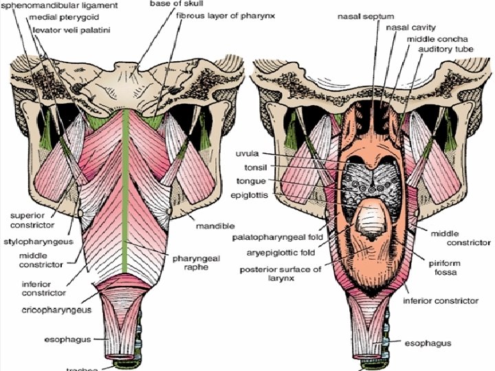
Nasopharynx Location: posterior to the nasal cavity. Extends from skull base superiorly to the soft palate inferiorly �Soft palate separates it from the posterior part of the oral cavity. Communicates inferiorly with the oropharynx through the velopharyngeal sphincter �The pharyngeal isthmus is the opening in the floor between the soft palate and the posterior pharyngeal wall. Related posteriorly to the base of the skull. Contains adenoid tissue and the orifices of the Auditory (Eustachian )tubes. Pharyngeal tonsil (Adenoids) posterior nasopharynx wall single

Auditory tubes paired In the lateral walls of the nasopharynx. The elevation ridge called the tubal elevation The pharyngeal recess is a depression in the pharyngeal wall behind the tubal elevation The salpingopharyngeal fold is a vertical fold of mucous membrane covering the salpingopharyngeus muscle. By means of the auditory tube, the mucous membrane is also continuous with that of the tympanic cavity. �connect the nasopharynx to the middle ear.
")
Oropharynx Location: behind the oral cavity (in front of 2 nd&3 rd Cervical vertebra) Extend: from the soft palate superiorly to tip of epiglottis (vallecula) inferiorly Communicates: Anteriorly with the oral cavity separated by the oropharyngeal isthmus Superiorly with the nasopharynx Inferiorly with the hypopharynx Boundaries: Posterior and lateral walls are formed by the superior and middle pharyngeal constrictors. On lateral wall is the palatine tonsils lie laterally between the anterior and posterior Pilars (the palatoglossal and the palatopharyngeal arches ) The floor is formed by the posterior one third of the tongue and the interval between the tongue and epiglottis. �In the midline is the median glossoepiglottic fold , and on each side the lateral glossoepiglottic fold. � The depression on each side of the median glossoepiglottic fold is called the vallecula.

Glossoepiglottic fold Lateral Glosso- Epiglottic Vallecula Lateral Glosso- Epiglottic

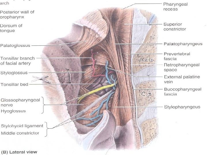
Palatine Tonsils Lymphoid tissues covered by mucous membrane, and projects into the pharynx. The surface is pitted by numerous small openings that lead into the tonsillar crypts. The tonsil is covered on its lateral surface by a fibrous capsule separated from the superior constrictor muscle by loose areolar tissue and the external palatine vein descends from the soft palate in this tissue to join the pharyngeal venous plexus. Lateral to the superior constrictor muscle lie the styloglossus muscle, the loop of the facial artery, and the internal carotid artery. Reaches maximum size during early childhood. And diminishes after puberty Blood Supply The tonsillar branch of the facial artery. The veins pierce the superior constrictor muscle and join the external palatine, the pharyngeal, or the facial veins. Lymph Drainage of the Tonsil The upper deep cervical lymph nodes, just below and behind the angle of the mandible Waldeyer's Ring of Lymphoid Tissue Surrounds the opening into the respiratory and digestive systems forms a ring. The lateral part of the ring is formed by the palatine tonsils and tubal tonsils. The pharyngeal tonsil in the roof of the nasopharynx forms the upper part, and the lingual tonsil on the posterior third of the tongue forms the lower part.
 Location: Behind the Larynx (in front of 3 rd to 6 th")
Laryngopharynx (Hypolarynx) Location: Behind the Larynx (in front of 3 rd to 6 th Cervical vertebra) Extend: From the tip of epiglottis superiorly to the lower border of cricoid cartilage Inferiorly It does not only lie behind the larynx BUT also projects laterally on each side of the larynx So it is formed of : Postcricoid region ( behind the larynx) Two pyriform fossa (on each side of the larynx): a depression in the mucous membrane on each side of the laryngeal inlet. The lateral wall is formed by the thyroid cartilage and the thyrohyoid membrane. Communicates: Anteriorly with the Larynx Superiorly with the oropharynx Inferiorly with the esophagus
")
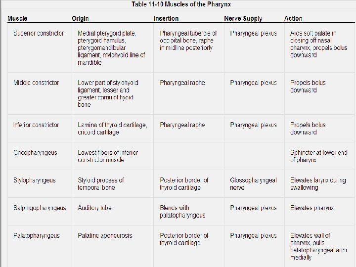
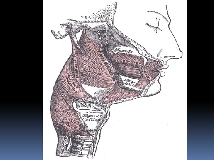
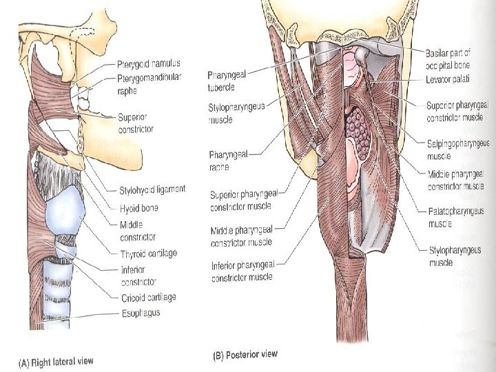
Muscles of the Pharynx The consist of : Superior, middle, and inferior constrictor muscles(circular) fibers Stylopharyngeus and salpingopharyngeus muscles (longitudinal) fibers Constrictor muscles extend around the pharyngeal wall to be inserted into a fibrous band or raphe that extends from the pharyngeal tubercle on the basilar part of the occipital bone of the skull down to the esophagus. The three constrictor muscles overlap each other like 3 cups The lower part of the inferior constrictor, which arises from the cricoid cartilage, is called the cricopharyngeus muscle. The fibers of the cricopharyngeus pass horizontally around the lowest and narrowest part of the pharynx and act as a sphincter. Killian's dehiscence is the area on the posterior pharyngeal wall between the upper propulsive part of the inferior constrictor and the lower sphincteric part, the cricopharyngeus.

Blood Supply From the External Carotid Artery & its branches 1. Tonsillar artery (from Facial Artery) 2. Ascending palatine artery (from Facial Artery) 3. Ascending pharyngeal Artery (from external carotid) 4. Descending palatine artery ( from Maxillary artery. 5. Dorsalis lingulae artery (from Lingual artery) Lymph Drainage of the Pharynx Directly into the deep cervical lymph nodes or indirectly via the retropharyngeal or paratracheal nodes into the deep cervical nodes

Nerve Supply Motor by X , Except : Stylopharyngeus : IX Tensor palati : V Sensory to mucous meebrane Nasopharynx: V via maxillary (V 2) Oropharynx: IX Laryngopharynx: X (internal alryngeal Autonomic: sympathetic: SCG Parasympathetic: through VII

The Larynx Provides a protective sphincter at the inlet of the air passages and is responsible for voice production. Situated below the tongue and hyoid bone and between the great blood vessels of the neck and lies at the level of the fourth, fifth, and sixth cervical vertebrae. Opens above into the laryngeal part of the pharynx, and below is continuous with the trachea. Is covered in front by the infrahyoid strap muscles and at the sides by the thyroid gland.

Larynx Structure: Is a frame of cartilage connected by ligaments, lined by mucous membrane, moved by muscles.

I. LARYNX: CARTILAGES A. THYROID CARTILAGE – Shield shaped has Sup. & Inf. Horns from upper & lower edges Superior horn is connected by a ligament to the posterior end of the greater horn of the hyoid bone. Medial surface of Inf. horns make synovial hinges joint with Cricoid Cartilage On the outer surface of each lamina is an oblique line for the attachment of muscles. Laryngeal Prominence = Adam’s Apple, more prominent in males

B. CRICOID; Complete ring of cartilage has narrow Arch ant. , broad Lamina post. = Signet ring Lamina articulates with inf. cornu laterally and with arytenoid cartilage posteriorly All these joints are synovial.

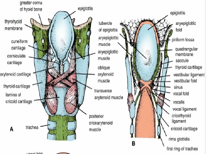
C. Arytenoids 2 pyramidal shaped cartilages above lamina have base froming synovial joints with upper border of Cricoid lamina permit Swivel = Rotate Sliding = Ab/Adduct have an apex articulates with the small corniculate cartilage Have a vocal process that projects forward and gives attachment to the vocal ligament. Have a muscular process that projects laterally gives attachment to the posterior and lateral cricoarytenoid muscles. Medial surface of each cartilage faces the other; Anterolateral surface has two depressions, separated by a ridge, for muscle (vocalis) and ligament (vestibular ligament) attachment.

D. Corniculate - nodules above arytenoids in aryepiglottic folds E. Cuneiform – rod shaped, above corniculate cartilages

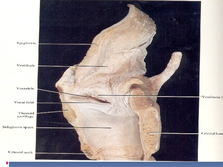
F. EPIGLOTTIS – leaf shaped cartilage posterior to root of tongue attached by its stem to the posterior aspect of the thyroid cartilage at the angle via thryo-epiglottic ligament and projects posterosuperiorly from its attachment to the thyroid cartilage connected to body of hyoid and post side of thyroid cartilage The inferior half of the posterior surface of the epiglottis is raised slightly to form an epiglottic tubercle.

II. LIGAMENTS OF LARYNX A. Structural ligaments - hold larynx, hyoid, trachea together 1. Thyrohyoid Membrane links larynx to hyoid pierced on each side by the superior laryngeal vessels and the internal laryngeal nerve, a branch of the superior laryngeal nerve Median thyrohyoid ligament – thickened midline part 2. Cricothyroid Membrane links thyroid to cricoid; Median cricothyroid ligament - thickened midline part 3. Cricotracheal ligament links cricoid to first tracheal cartilage

4. Quadiangular membrane: connect arytenoid to epiglottis Overlied by aryepiglottic Folds It is lower free edge is called vestibular ligament deep to Vestibular (False Vocal) Folds

5. Thyroepiglottic Ligament: links epiglottis to thyroid cartilage

B. FUNCTIONAL LIGAMENTS Conus Elasticus -Vibrating lips that arise entire upper edge of arch of cricoid 1. from Attach: ant. to Thyroid, post. to Arytenoid Functions: � Sound Production � Close Rima Glottidis 2. Vocal Ligaments - Upper free edges vocal folds 3. Rima Glottidis (glottis) - opening between ligaments deep to vocal Is bounded in front by the vocal folds and behind by the medial surface of the arytenoid cartilages. The glottis is the narrowest part of the larynx and measures about 2. 5 cm front to back in the male adult and less in the female. In children the lower part of the larynx within the cricoid cartilage is the narrowest part.

Inlet of the Larynx inlet looks backward and upward into the laryngeal part of the pharynx. The opening is wider in front than behind and is bounded in front by the epiglottis, laterally by the aryepiglottic fold of mucous membrane, and posteriorly by the arytenoid cartilages with the corniculate cartilages. The cuneiform cartilage lies within and strengthens the aryepiglottic fold and produces a small elevation on the upper border. The Piriform Fossa is a recess on either side of the fold and inlet. is bounded medially by the aryepiglottic fold and laterally by the thyroid cartilage and the thyrohyoid membrane

Cavity of the larynx Extends from the inlet to the lower border of the cricoid cartilage. It is divided into three regions: The vestibule, which is situated between the inlet and the vestibular folds The middle region, which is situated between the vestibular folds above and the vocal folds below The lower region, which is situated between the vocal folds above and the lower border of the cricoid cartilage below 1. Vestibule - inlet above false vocal folds 2. Vestibular (False Vocal) Folds - overlie vestibular ligaments is vascular and pink in color 3. Vocal (True Vocal) Folds - overlie vocal ligaments white in color 4. Ventricle- area between true and false vocal folds; lateral extension is Laryngeal Sinus The saccule of the larynx is a diverticulum of mucous membrane that ascends from the sinus. The mucous secretion lubricates the vocal cords.

View during laryngoscopy
 -")
III. MUSCLES OF LARYNX - well named A. Extrinsic muscles (ex. hyoid muscles) - Move whole larynx as in swallowing Many of these muscles acts indirectly as they are attached to the hyoid bone, which is attached to the thyroid cartilage by the thyrohyoid membrane. Elevation: digastric, stylohyoid, mylohyoid, geniohyoid, stylopharyngeus, salpingopharyngeus, and palatopharyngeus muscles Depression: sternothyroid, sternohyoid, and omohyoid muscles B. Intrinsic Muscles: 1)Two muscles modify the laryngeal inlet : Narrowing the inlet: The oblique arytenoid muscle Widening the inlet: The thyroepiglottic muscle 2) change tension in vocal lig changes pitch: increase tension raises pitch, decreased tension lowers pitch 2) open & close rima glottidis
 Cricothyroid M: Tense vocal ligament Increasing pitch 2) Thyroarytenoid M: Relaxes vocal ligaments")
1) Cricothyroid M: Tense vocal ligament Increasing pitch 2) Thyroarytenoid M: Relaxes vocal ligaments Decreases pitch 3) Posterior Cricoarytenoid Abducts vocal fold 4) Lateral Cricoarytenoid Adduct vocal folds 5) Arytenoid (Transverse and oblique arytenoid) Adduct vocal folds + approximate aryt. 6) Aryepiglotic M Pulls epiglottis down during swallowing Covers inlet to larynx - Not necessary in adult humans
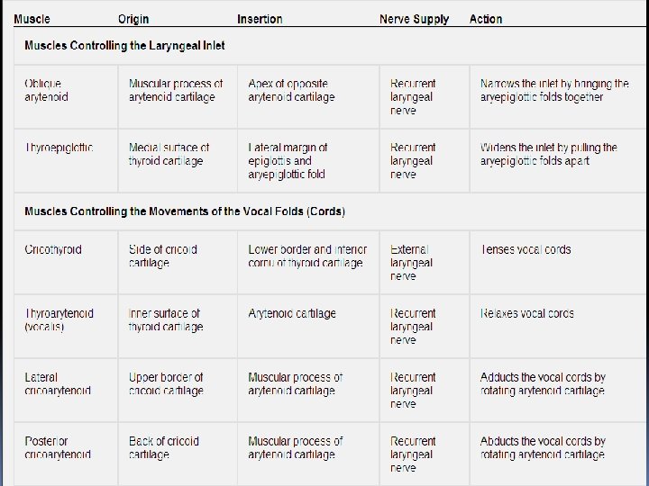

Nerve supply A. Superior Laryngeal N. divides to 1. Internal Laryngeal N. GVA Sensory to Larynx Above True Vocal Folds 2. External Laryngeal N. SVE Motor to Cricothyroid B. Recurrent Laryngeal N. GVA Sensory to Larynx Below True Vocal Folds SVE motor to all other Muscles of Larynx

Blood and Lymph supply Blood supply Upper half: Sup. Laryngeal A. from Sup. Thyroid artery Lower half: Inf. Laryngeal A. from Inf. Thyroid Artery Lymph drainage: Superior Deep Cervical Nodes Larynx above true vocal folds Inferior Deep Cervical Nodes Larynx below true vocal folds

Movements of the Vocal Folds With Respiration On quiet inspiration, the vocal folds are abducted and the rima glottidis is triangular in shape with the apex in front. On expiration the vocal folds are adducted, leaving a small gap between them. On deep inspiration, the vocal folds are maximally abducted and the triangular shape of the glottis becomes a diamond shape because of the maximal lateral rotation of the arytenoid cartilages

Sphincteric Function of the Larynx Has two sphincters in the larynx: one at the inlet and another at the rima glottidis. The sphincter at the inlet is used only during swallowing. As the bolus of food is passed backward between the tongue and the hard palate, the larynx is pulled up beneath the back of the tongue. The inlet of the larynx is narrowed by the action of the oblique arytenoid and aryepiglottic muscles. The epiglottis is pulled backward by the tongue and serves as a cap over the laryngeal inlet. The bolus of food, or fluids, then enters the esophagus by passing over the epiglottis or moving down the grooves on either side of the laryngeal inlet, the piriform fossae. In coughing or sneezing, the rima glottidis serves as a sphincter. After inspiration, the vocal folds are adducted, and the muscles of expiration are made to contract strongly. As a result, the intrathoracic pressure rises, and the vocal folds are suddenly abducted. The sudden release of the compressed air will often dislodge foreign particles or mucus from the respiratory tract and carry the material up into the pharynx, where the material is either swallowed or expectorated. In the Valsalva maneuver, forced expiration takes place against a closed glottis. In abdominal straining associated with micturition, defecation, and parturition, air is often held temporarily in the respiratory tract by closing the rima glottidis. After deep inspiration the rima glottidis is closed. The muscles of the anterior abdominal wall now contract, and the upward movement of the diaphragm is prevented by the presence of compressed air within the respiratory tract. After a prolonged effort the person often releases some of the air by momentarily opening the rima glottidis, producing a grunting sound.

Nerve injuries A. B. C. D. E. produces weakness of the voice because the vocal fold cannot be tensed. The cricothyroid muscle is paralyzed. results in the vocal fold on the affected side assuming the position midway between abduction and adduction. It lies just lateral to the midline. Speech is not greatly affected because the other vocal fold compensates to some extent and moves toward the affected vocal fold. results in both vocal folds assuming the position midway between abduction and adduction. Breathing is impaired because the rima glottidis is partially closed, and speech is lost. results in a greater degree of paralysis of the abductor muscles than of the adductor muscles. The affected vocal fold assumes the adducted midline position. results in bilateral paralysis of the abductor muscles and the drawing together of the vocal folds. Acute breathlessness (dyspnea) and stridor follow, and cricothyroidotomy or tracheostomy is necessary.

Voice Production in the Larynx The intermittent release of expired air between the adducted vocal folds results in their vibration and in the production of sound. The frequency, or pitch, of the sound is determined by changes in the length and tension of the vocal ligaments. The quality of the voice depends on the resonators above the larynx, namely, the pharynx, mouth, and paranasal sinuses. The quality of the voice is controlled by the muscles of the soft plate, tongue, floor of the mouth, cheeks, lips, and jaws. Normal speech depends on the modification of the sound into recognizable consonants and vowels by the use of the tongue, teeth, and lips. Vowel sounds are usually purely oral with the soft palate raised so that the air is channeled through the mouth rather than the nose. Speech involves the intermittent release of expired air between the adducted vocal folds. Singing a note requires a more prolonged release of the expired air between the adducted vocal folds. In whispering, the vocal folds are adducted, but the arytenoid cartilages are separated; the vibrations are given to a constant stream of expired air that passes through the posterior part of the rima glottidis.

Mucous Membrane of the Larynx The mucous membrane of the larynx lines the cavity and is covered with ciliated columnar epithelium. On the vocal cords, however, where the mucous membrane is subject to repeated trauma during phonation, the mucous membrane is covered with stratified squamous epithelium. Ø Note: Mucosa Tightly Attached to vocal folds Ø Anaphylactic Shock Swell Vestibular folds -Suffocation
- Slides: 43