Xiaoyu Che Atomic force microscopyAFM is one of

 is one of the foremost tools for imaging, measuring and")


 dynamic mode (non-contact mode and tapping mode) � Static")


- Slides: 17
Xiaoyu Che
� Atomic force microscopy(AFM) is one of the foremost tools for imaging, measuring and manipulating matter at the nanoscale � A type of scanning probe microscopy(SPM) � SPM: Forms images of surfaces using a physical probe that scans the specimen � Scanning Tunneling microscope(STM, the predecessor of AFM) is also a type of SPM.
� High-resolution mapping of surface topography, by far the biggest application of the AFM � Offers image resolution down to the atomic scale
� The information is gathered by “feeling” the surface with a mechanical probe. � A cantilever with a sharp tip, which is typically silicon or silicon nitride. � The tip radius of curvature is very small, on the order of nanometers to ensure accuracy. So how does it feels the sample?
Let’s Review Hooke’s law: The force F needed to extend or compress a spring by some distance x is proportional to that distance. Formula: F=-kx
� In atomic scale it is not spring-mass system anymore. (contact force, van der Waals force, capillary force, chemical bonding, electrostatic force, magnetic force, etc. ) That’s why it is called atomic force microscopy! � So when the tip approaches the surface it can “feel” these forces and the deflection is measured to be converted into image information.
� But how we measure the deflection? � Use a laser spot reflected from the top surface of the cantilever into an array of photodiodes. � Position sensitive detector(PSD) Angular displacement Differential amplifier Two closely spaced photodiodes
So that’s how it works.
� Static mode (contact mode) dynamic mode (non-contact mode and tapping mode) � Static mode: � Dynamic mode:
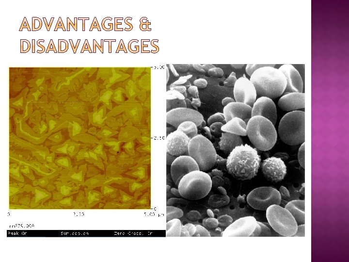
� Attractive forces can be quite strong, cause the tip to contact the surface. � Mainly used to image hard surfaces when the presence of lateral forces is not expected to modify the morphological features. � On crystalline surfaces such as mica, Au (111), salt crystals, etc. � Prone to noise and drift � low stiffness cantilevers are used to boost the deflection. � Si probes are more common Cd. F 2 films grown on a Ca. F 2 (111) substrate. Scan is taken in contact mode using a CSC 21 probe (now upgraded to HQ: XSC 11). Scan size 2 x 2 µm, height 2 nm. .
� The tip of the cantilever does not contact the sample surface. � Oscillated at either its resonant frequency (frequency modulation) or just above (amplitude modulation) � The van der Waals forces or any other long range force acts to decrease the resonance frequency of the cantilever. � Maintains a constant oscillation amplitude or frequency by adjusting the average tip-to-sample distance. � Tip-to-sample distance at each (x, y) data point , construct a topographic image of the sample surface.
� Does not suffer from tip or sample degradation effects � If a few monolayers of adsorbed fluid are lying on the surface of a rigid sample, the images may look quite different. � Frequency modulation: changes in the oscillation frequency provide information about tip-sample interactions. � Amplitude modulation: changes in the oscillation amplitude or phase provide the feedback signal for imaging
� In ambient conditions, most samples develop a liquid meniscus layer -> keep the probe tip close enough to the sample for short-range forces to become detectable while preventing the tip from sticking to the surface � The cantilever is driven to oscillate up and down near its resonance frequency. Images are produced by imaging the force of intermittent contacts. � Lessens the damage done to the surface and the tip.
� Three-dimensional surface profile. � Do not require any special treatments (such as metal/carbon coatings) that would irreversibly change or damage the sample, � Does not typically suffer from charging artifacts in the final image. � Can work perfectly well in ambient air or even a liquid environment. � Higher resolution than SEM, comparable in resolution to STM and TEM. � Can be combined with a variety of optical microscopy techniques. ]
� � � Single scan image size. The scanning speed of an AFM is also a limitation. Can be affected by nonlinearity, hysteresis, and creep of the piezoelectric material. The possibility of image artifacts, which could be induced by an unsuitable tip, a poor operating environment, or even by the sample itself. Cannot normally measure steep walls or overhangs.
� Lang, K. M. ; D. A. Hite, R. W. Simmonds, R. Mc. Dermott, D. P. Pappas, and John M. Martinis (2004). "Conducting atomic force microscopy for nanoscale tunnel barrier characterization". Review of Scientific Instruments 75: 2726– 2731. Bibcode: 2004 RSc. I. . . 75. 2726 L. doi: 10. 1063/1. 1777388. � G. Schitter, M. J. Rost (2008). "Scanning probe microscopy at video-rate" (PDF). Materials Today (UK: Elsevier) 11 (special issue): 40– 48. doi: 10. 1016/S 13697021(09)70006 -9. ISSN 1369 -7021. � R. V. Lapshin, O. V. Obyedkov (1993). "Fast-acting piezoactuator and digital feedback loop for scanning tunneling microscopes" (PDF). Review of Scientific Instruments (USA: AIP) 64 (10): 2883– 2887. Bibcode: 1993 RSc. I. . . 64. 2883 L. doi: 10. 1063/1. 1144377. ISSN 0034 -6748. � R. V. Lapshin (1995). "Analytical model for the approximation of hysteresis loop and its application to the scanning tunneling microscope" (PDF). Review of Scientific Instruments (USA: AIP) 66 (9): 4718– 4730. Bibcode: 1995 RSc. I. . . 66. 4718 L. doi: 10. 1063/1. 1145314. ISSN 0034 -6748. (Russian translation is available). � Cappella, B; Dietler, G (1999). Surface Science Reports 34 (1 -3): 1– 104. doi: 10. 1016/S 0167 -5729(99)00003 -5 http: //www. see. ed. ac. uk/~vkoutsos/Forcedistance%20 curves%20 by%20 atomic%20 force%20 microscop y. pdf. Bare URL needs a title. � Gross, L. ; Mohn, F. ; Moll, N. ; Liljeroth, P. ; Meyer, G. (27 August 2009). "The Chemical Structure of a Molecule Resolved by Atomic Force Microscopy". Science 325 (5944): 1110– 1114. doi: 10. 1126/science. 1176210. � Göddenhenrich, T. (Na. N undefined Na. N). "Force microscope with capacitive displacement detection". Journal of Vacuum Science & Technology A: Vacuum, Surfaces, and Films 8 (1): 383. doi: 10. 1116/1. 576401. � Giessibl, F. J. ; Trafas, B. M. (1 January 1994). "Piezoresistive cantilevers utilized for scanning tunneling and scanning force microscope in ultrahigh vacuum". Review of Scientific Instruments 65 (6): 1923. doi: 10. 1063/1. 1145232. Gavin M. King, Ashley R. Carter, Allison B. Churnside, Louisa S. Eberle, and Thomas T. Perkins (2009). "Ultrastable Atomic Force Microscopy: Atomic-Scale Stability and Registration in Ambient Conditions". Nano Letters 9: 1451– 1456. doi: 10. 1021/nl 803298 q. � M. Hoffmann, Ahmet Oral, Ralph A. G, Peter (2001). "Direct measurement of interatomic force gradients using an ultra-low-amplitude atomic force microscope". Proceedings of the Royal Society a Mathematical Physical and Engineering Sciences 457: 1161. Bibcode: 2001 RSPSA. 457. 1161 M. doi: 10. 1098/rspa. 2000. 0713. � � Hinterdorfer, P; Dufrêne, Yf (May 2006). "Detection and localization of single molecular recognition events using atomic force microscopy". Nature methods 3 (5): 347– 55. doi: 10. 1038/nmeth 871. ISSN 1548 -7091. PMID 16628204.