The Nervous System Part I Organization Spinal Cord


- Slides: 56
The Nervous System Part I: Organization, Spinal Cord, and Peripheral Nerves
Introduction • Most of the rapid changes in a vertebrate’s internal or external environments are mediated by the nervous system. • Activation of the correct combination of effectors so that the appropriate responses are made requires a processing of signals within the nervous system that is called coordination or integration. • The nervous system is constantly inundated with nervous signals – many are filtered as they pass to the higher centers so that some are suppressed and others enhanced.
• Sensory inputs from different types of receptors are combined to provide a more complete picture of the changes taking place. – And these inputs are combined with memories of past events before a response is determined • Often, information about the outcome of the response is fed back into the nervous system reinforcing, or altering the past memories. – Although much of vertebrate behavior is based on inherited neuronal connections, vertebrates learn from their experiences.
Cellular components of the Nervous System • There are two basic cellular components to the nervous system: 1. Neurons, which transmit signals to other neurons, glands, muscle cells, or receptor cells, and 2. Glial cells, which provide for structural and functional needs of the neuronal cells.
Neurons and Synapses • The primary functional units of the nervous system are the neurons, or nerve cells. • Neurons are arranged into two groups – Central Nervous System (CNS) • The brain and spinal cord, and – Peripheral Nervous System (PNS) • Containing the nerves between the CNS and the receptors and effectors.
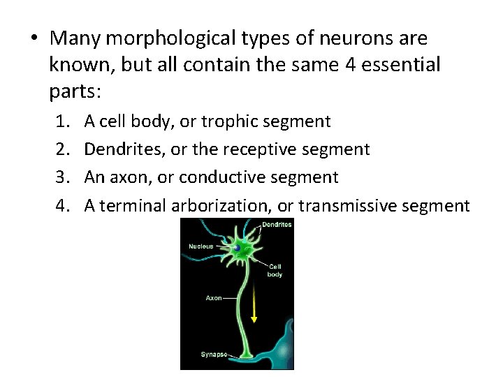
• Many morphological types of neurons are known, but all contain the same 4 essential parts: 1. 2. 3. 4. A cell body, or trophic segment Dendrites, or the receptive segment An axon, or conductive segment A terminal arborization, or transmissive segment
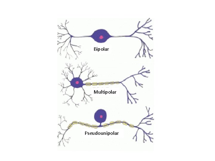
• The cell body contains the nucleus and metabolic machinery of the neuron, and is the site of protein and neurotransmitter synthesis. • The cell body varies in its position relative to the dendrite; – in multipolar motor neurons the cell body lies close to the dendrite and a long axon extends outward from the body; – in a bipolar cell the body lies in the middle of the axon; – or it may be set off to the side, as in the pseudounipolar sensory neuron (13 -1).
Bipolar Multipolar Pseudounipolar
• Dendrites receive stimuli and initiate the nerve impulse; • Axons are long, slender, cytoplasmic processes often called nerve fibers. • Conduct sensory – input from the dendrites of receptors to the CNS, – impulses within the CNS, – impulses with the effectors, and – motor impulses from the CNS to the effectors.
• The branching terminal aborizations, called telodendria make contact with the receptive segments of other neurons at special junctions, called synapses or at synapse • like myoneural junctions or axoglandular junctions. • A nerve terminal contains many minute synaptic vesicles, in which neurotransmitter is stored. – Many types of neurotransmitter are known • As a given rule, only one type of neurotransmitter is found in each neuron. • When a nerve impulse reached the synapse, – some of the neurotransmitter is released from the synaptic vesicle into the extracellular space, – it crosses the narrow synaptic cleft to bind with the receptor sites of the target cell; – And either excites or inhibits this cell.
• The polarity of communication of the nervous system is determined by the synapse locations. – Nerve impulses only travel in one direction, from presynaptic neuron to postsynaptic receptors. • Neurotransmitters can either be excitatory, partially depolarizing a cell membrane, or inhibitory, hyperpolarizing the membrane. – A threshold level of depolarization must be reached before an action potential is generated
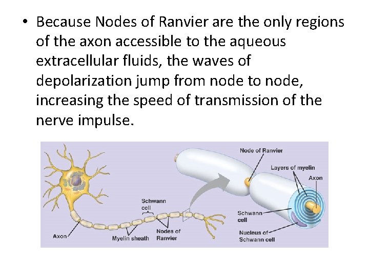
Schwann Cells and Neuroglia • Glia is the general term used to describe nonneuronal cells of the nervous system. • In gnathostomes, all axons are surrounded during their embryonic development by glial components called Schwann cells. – The inner tongue-like process of these cells grow around the axon many times so that multiple layers of the Schwann cell’s plasma membrane are laid down, forming the myelin sheath. – Gaps between successive Schwann cells interrupt the myelin sheath at areas called Nodes of Ranvier
• Because Nodes of Ranvier are the only regions of the axon accessible to the aqueous extracellular fluids, the waves of depolarization jump from node to node, increasing the speed of transmission of the nerve impulse.
• Schwann cells associated with many peripheral axons envelop the axon but do not grow into a sheath. – These cells are referred to an nonmyelinated. • Ex axons of autonomic nerves, • Many other types of glial cells, called neuroglia, occur in the CNS – They are 30 -50 times more numerous than neurons.
• Oligodendrocytes send out processes that wrap around and myelinate the axons within the CNS. – Regions of the CNS containing myelinated axons appear whitish when stained, and therefore, are called white matter, and contain bundles axons extending between different parts of the CNS. – Regions of non-myelinated axons, dendrites, and cell bodies are termed gray matter. – Dense aggregations of cell bodies within the gray matter are called nuclei, and act as land marks within the CNS because they indicate areas of processing of sensory inputs. • Star–shaped glail cells known as astrocytes are interposed among neuronal cell bodies. – They function to support adjacent cells and also to maintain the ionic composition of the extracellular fluid.
Organization of the Nervous System • The basic structure and plan of the nervous system is similar in all vertebrates. • Because of the way they develop; the spinal cord and brain of a vertebrate are hollow and lined by a non-nervous and partially ciliated ependymal epithelium. – The adult spinal cord has a small central canal, which enlarges in parts of the brain to form a series of interconnected ventricles. • Cerebrospinal fluid slowly circulates within these spaces.
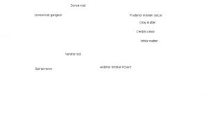
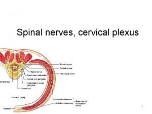
• Bilateral features of the nervous system are often referred to by the contrasting terms ipsilateral and contralateral. – These terms are especially important in describing the targets of the particular tracts of nerve cells, • Typical spinal nerves attach to the cord at segmental intervals by dorsal and ventral roots. – In most vertebrates, the dorsal and ventral roots of a body segment unite slightly peripheral to the spinal cord to form a spinal nerve • Dorsal, ventral, and communicating rami extend to different parts of the body from these nerves. • Most of the cranial nerves appear to be serially homologous to either a ventral or dorsal root of a typical spinal nerve.
The Three Connectional Categories of Neurons and their Distribution • Regardless of the differences in their morphologies, all neurons belong to one of three categories: 1. Primary sensory, or afferent neurons that carry impulses from free nerve endings or specialized receptor cells into the CNS; 2. Motor, or efferent neurons that carry signals from the CNS to the effectors, such as muscles or glands. 3. Interneurons which are confined to the CNS • They receive input from afferent neurons, integrate this information, and send it to the periphery through efferent neurons.
Basic Neuronal Circuitry • Activities of the nervous system are controlled by many types of circuit cells among sensory neurons, interneurons, and motor neurons. • There are three basic neuronal pathways common to all vertebrates; 1. reflexes, 2. ascending pathways, 3. descending pathways. • Much activity is controlled by reflexes, mediated by cells located in the spinal cord.
• Touching a hot stove initiates a reflex termed a 3 neuron reflex arc because it includes three types of neurons: A sensory neuron, an interneuron, and a motor neuron. – In contrast, the knee-jerk reflex requires only two types of neurons because the sensory neurons synapse directly with motor neurons. • Reflexes like the 2 - and 3 -neuron reflex arc involve neurons that are only on one side of the body and one body segment. • Other reflexes include neurons that cross the midline of the body and affect many body segments. • Axons that cross the midline are called commisures, or decussations.
• Reflexes also may involve neurons that ascend or descend along the body axis and affect many body segments; these are described as intersegmental reflexes. – For example, the coordinated movements of swimming and walking are integrated reflexively by pools of intersegmental and decussating neurons. • Many reflexes in the brain and spinal cord are products of long evolutionary history, they are innate reflexes. • Others called, conditioned reflexes, develop during thee lifetime of the animal’s repetitive experiences. – Conditioned reflexes are not inherited directly, although the capacity for them to develop is.
• Many sensory impulses ascend from the spinal cord or brainstem to higher centers in the brain. • Groups of axons with similar functions terminate in the same part of the brain and ascend together in common tracts that are described by their points of origin and termination. • Most sensory impulses decussate on their way to higher centers so that impulses originating on the left side of the body terminate on the right side of the brain, – The decussation is not always complete.
Spinal Cord • The spinal cord lies between the brain and spinal nerves, but the cord is far more than a simple passage way for nerve impulses between receptors and effectors and the brain. • Some interneurons of the spinal cord span from one segment of the trunk or tail to one or more other segments and thus are known as intersegmental neurons. – Their presence makes possible considerable sensorymotor integration within the spinal cord. • The rhythmic and coordinated movements of swimming and walking are controlled and integrated reflexively by pools of intersegmental and decussating spinal nerves, these are central pattern generators.
• The complexity of the spinal cord, and the degree to which the brain exerts control over spinal activity, increases during the course of vertebrate evolution. • Hagfishes and lamprey have relative simple spinal cords; – none of their axons are myelinated and there is no distinction between gray and white matter. • In gnathostomes, the spinal cord is larger, well vascularized, and more rounded. – The evolution of a highly vascular nervous systems was an important part of vertebrate evolution allowing for more complex nervous systems to evolve.
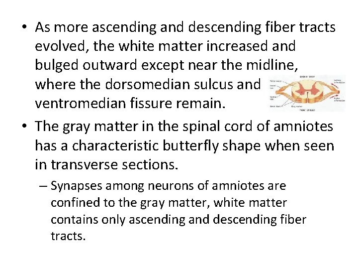
• As more ascending and descending fiber tracts evolved, the white matter increased and bulged outward except near the midline, where the dorsomedian sulcus and ventromedian fissure remain. • The gray matter in the spinal cord of amniotes has a characteristic butterfly shape when seen in transverse sections. – Synapses among neurons of amniotes are confined to the gray matter, white matter contains only ascending and descending fiber tracts.
• The brains of birds and mammals exert more control over body activities than do the brains of other vertebrates – tracts to and from the brain are correspondingly more numerous. • The degree of vascularization of the spinal cord also increases, and the number of neuroglial cells that service the neurons also is greater in birds and mammals.
• The spinal cord in most vertebrates is nearly as long as the vertebral column, although in mammals, frogs, and a few teleosts it is shorter because it grows more slowly than the rest of the body (13 -13 a). – As a consequence, the more caudal spinal nerves form a bundle, known as the cauda equina,
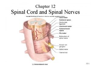
• Vertebrates with well-developed limbs have a large number of neurons supplying them, and the diameter of the spinal cord is correspondingly greater in the cervical and lumbar regions – Where most nerves to the limbs originate. • The spinal cord of mammals is protected by: – Thin pia mater, which overlies the neural tissue; – The highly vascularized arachnoid layer outside of it; and – The tough dura matter, which ensheaths the entire cord and roots of the spinal nerves.
Spinal Nerves • The spinal nerves of vertebrates usually attach by roots to the spinal cord, • However, in lampreys, the homologues of the dorsal and ventral roots form distinct dorsal and ventral spinal nerves. • The ventral nerves of lampreys are segmentally spaced and lead directly into the myomeres, they only carry somatic motor neurons. • The dorsal nerves of lamprey are intersegmental and pass between the myomeres to the body surface and gut regions, and contain both somatic and visceral sensory neurons. • Most visceral motor neurons reach the viscera of lampreys through a cranial nerve.
• The presence of a distinct dorsal and ventral spinal nerve, each with its own type of neurons, is believed to be a plesiomorphic character of vertebrates. • In all other craniates, including hagfishes, the distinct dorsal and ventral spinal nerves of a trunk segment unite to form a distinct spinal nerve. • The originally distinct dorsal and ventral nerves are now referred to as the dorsal and ventral roots of the spinal nerve. – All sensory nerves continue to enter through the dorsal root, and somatic motor fibers continue to leave through the ventral root.
Cranial Nerves of Gnathostomes as Represented by Chondrichthyans
Sensory Nerves Unique to the Head • These include four sensory nerves and two general categories of nerves found only in the head. • Terminal Nerve (0): The special somatic sensory terminal nerve may be a part of the chemosensory system regulating certain aspects if reproduction in response to olfactory pheromones.
• Olfactory Nerve (I): A special somatosensory nerve that returns chemosensory fibers from the nasal sac. • Optic Nerve (II): They are interneurons and carry impulses from the retina to the brain. – Because the retina develops as a part of the brain the optic nerve is technically a brain tract and not a nerve. • Somatoacoustic, Vestibulochochlear, or Octaval Nerve (VIII): The different names are recognized in different groups. – Nerve VIII can be characterized as a special somatic sensory nerve that returns fibers from all parts of the inner ear.
• Lateral Line Nerves (Not Numbered): The vast majority of amniotes have lateral line and electroreceptive organs innervated by a series of 6 lateral line nerves that develop from distinct lateral line placodes on the surface of the head. • Gustatory Nerves (Not Numbered): These are specialized visceral sensory nerves that carry taste sensations. The gustatory fibers become associated with cranial nerves VII, IX, and X.
Ventral Cranial Nerves • Those cranial nerves designated as ventral cranial nerves resemble the spinal nerves of the lamprey because they contain primarily somatic motor neurons going to many somatic muscles of the head. • This group consists of those cranial nerves that supply the extrensic ocular muscles and the epibranchial and hypobranchial muscles. • Some also carry somatic sensory proprioreceptive neurons, which return information about the degree of contraction of the extrensic ocular muscles.
• Oculomotor Nerve (III): The oculomotor nerve supplies the 4 extrensic ocular muscles: dorsal rectus, medial rectus, ventral rectus, and inferior oblique. • Trochlear Nerve (IV): The trochlear nerve supplies the superior oblique muscle. • Abducens Nerve (VI): The abducens nerve supplies the lateral rectus muscle. • Occipital Nerve: Epibranchial and hypobranchial muscles of fishes, which are located in the caudal region of the head and beneath the pharynx, are innervated by 3 -4 occipital nerves and by a variable number of anterior spinal nerves.
Dorsal Cranial Nerves • The next 4 cranial nerves (V, VII, IX, X) appear to be comparable with the dorsal spinal nerves of lampreys. • They are mixed nerves that contain some combination of sensory and motor neurons. • All supply somatic motor neurons to the branchiomeric muscles of the visceral arches, and they are sometimes referred to as branchiomeric nerves.
• Glossopharyngeal Nerve (IX): These nerves carry somatic motor neurons to the muscles of the third visceral arch, and returns visceral sensory neurons from the adjacent part of the gill pouch. In mammals, the free nerve endings and specialized receptors that are the source of the sensory information lie on the back of the tongue and pharynx. • Vagal Nerve (V): The vagal nerve has 4 branches that supply the branchiomeric muscles of the last 4 visceral arches and return visceral sensory neurons from the last 4 gill pouches.
• Facial Nerve (VII) and Trigeminal Nerve (V): These nerves carry somatic motor neurons to the branchiomeric muscles of the hyoid and mandibular arches, respectively. The hyomandibular nerve also carries visceral sensory neurons, and the mandibular ramus carries general somatic sensory neurons from the skin over the lower jaw.
Changes in the Cranial Nerves of Amniotes • The pattern of the cranial nerves we have seen in chondrichthyans persists with predictable modifications in amniotes. • A few evolutionary changes have occurred in the nerves returning from the special sensory organs.
• These changes include: – Most terrestrial verts have a vomeronasal organ, and they acquire with it a vomeronasal component to the olfactory nerve. – In most non-mammalian vertebrates, most of the fibers of the optic nerve decussate in the optic chiasm, located at the point of attachment of the optic nerves to the brain. • In many mammals, only half of the fibers decussate. This change may correlate with overlapping visual fields. – A cochlea is well developed in some sauropsids, especially birds, as well as in mammals, so nerve VIII in amniotes is often termed the vestibulocochlear nerve. – The lateral line system is entirely lost in amniotes, so the six lateral line nerves are absent.
The Brain • Craniates differ from early choanates in having a markedly enlarged brain; – The brain is not a completely new craniate feature. – We find a well-developed brain in many protostomes, including the octopus and arthropods.
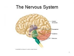
Embryonic Development and Regions of the Brain • The brain develops by an enlargement of the cranial end of the neural tube. • First, three brain expansions can be recognized: – a forebrain, or prosencephalon, – a midbrain, or mesencephalon, – and the hindbrain, or rhombencephalon.
• The prosencephalon first gives rise to the optic vessicles, which will form the retina of the eye. – The telencephalon, develops soon after as a pair of rostral extensions from the prosencephalon. – These differentiate into the cerebral hemispheres (which together constitute the cerebrum) and into paired olfactory bulbs. • The rest of the prosencephalon remains in the midline and differentiates as the diencephalon.
• The lateral walls of the diencephalon thicken to form the thalamus. – As they enter the brain the optic nerves partially decussate forming the optic chiasm, just rostral to the hypothalamus. • The mesencephalon remains undivided. – A narrow cerebral aqueduct of Sylvius passes through it and connects the third ventricle with the fourth ventricle in the rhombencephalon.
• The dorsal part of the mesencephalon, known as the tectum enlarges; – As a pair of optic lobes, in nonmammalian vertebrates – And, in mammals, is similarly differentiated into the paired superior and inferior colliculi. • This part of the mesencaphalon is known as the tegmentum.
• The rostral 1/3 of the roof of the rhombencephalon differentiates into the metencephalon, – The most particular part is the cerebellum. • During the embryonic development of birds and mammals, neuroblasts migrate from the cerebellum into the ventral part of the rhombencephalon and differentiate into the pontine and other nuclei, • These transmit messages between the cerebrum and cerebellum. – This region is know as the pons – A pons does not differentiate in reptiles and anamniotes, so all of the rhombencephalon, except the cerebellum, forms the 5 th brain region, the myencephalon, which leads to the spinal cord
Major Trends in Tetrapod Brain Evolution • The brain of fishes and tetrapods vary enormously in size and complexity. – But the brain regions of tetrapods, the structures they contain, and their basic organizational features are the same as in fish. • The cerebrum, thalamus, mesencephalic tectum, and cerebellum were the regions most affected as vertebrates adapted to a terrestrial life and became more active with a wider range of behavioral responses.
Amphibian Brain • The amphibian brain is little different from the brain of fishes. – Locomotor movements are not as complex as in fishes, and the cerebellum is therefore smaller. • Most adult amphibians live on land, and have lost the lateral line system and associated regions of the brain.
The Brian of Amniotes • The evolution of the cerebrum, dorsal pallium, and tectum are closely related. • The expanded cerebral hemispheres have grown partially caudal in reptiles and birds, and completely caudal in mammals covering the dorsal and lateral surfaces of the diencephalon.
• The expansion is closely linked to an expansion of the dorsal thalamus, which relays more somatosensory, optic, and auditory impulses. – This area becomes increasingly important as a sensory integration center. • The cerebrum is characterized by containing three neuronal layers: such a pattern is called an allocortex, the more advanced mammals have a 6 layered isocortex, or neocortex.
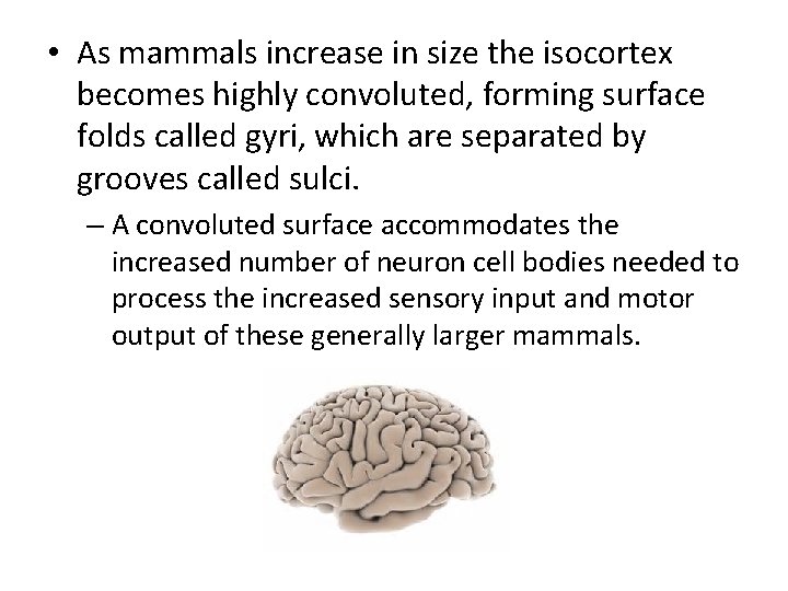
• As mammals increase in size the isocortex becomes highly convoluted, forming surface folds called gyri, which are separated by grooves called sulci. – A convoluted surface accommodates the increased number of neuron cell bodies needed to process the increased sensory input and motor output of these generally larger mammals.
The Cerebellum • All amniotes have lost the lateral line and electroreceptive systems, so the cerebellum no longer receives input from these systems, but it still has enlarged greatly relative to the cerebellum of amphibians and fishes. • Amniotes have developed an extraordinary wide range of motion of the head, trunk, tail, limbs, and digits as they have adapted to the more difficult problems of support, locomotion, and feeding in the terrestrial environments.
• The cerebellum is particularly large in birds and mammals, and its surface are is increased by the formation of many tightly packed, leaf-like folds called folia. – The auricles of the cerebellum remain vestibular centers in mammals. – The body of the cerebellum continues to receive an extensive input from most sensory modalities. • A prominent feature of the avian and mammalian cerebellum is a pair of lateral expansions of the cerebellar body, which form the cerebellar hemispheres that connect with the isocortex.
• An important overall effect of the cerebellum is to influence the extent, duration, and timing of muscle contractions. – These factors are essential for the smoothness of muscle contractions, for maintaining balance, and for muscle coordination. • Beyond this there is evidence that the cerebellum plays a role in motor learning and the ability to execute precise, carefully timed, and complex motor movements.
FIN ally