The cells of the nervous system and neurotransmitters


.")


 Impulse arrives at synapse with inhibitory")


 to trigger")
 blocks/inhibits/acts as an antagonist to the re -uptake proteins. or (Cocaine) prevents the")
")

- Slides: 54
The cells of the nervous system and neurotransmitters at synapses Key Area 4
Learning Intentions q Describe the structures and functions neurons. q Name the different types of neurons. q Describe the structure and function of the myelin sheath. q Explain how nerve impulses are transmitted between neurons.
Cells of the Nervous system • The nervous system consists of a complex network of nerve cells called neurons which receive and transmit electrical impulses from one part of the body to another. – they are a means of communication and control. • In addition, glial cells support and maintain the neurons • There are 3 types of neurones: Øsensory Øinter Ømotor
Structure of Neurons • The 3 types of neurone share the same basic structures: 1. a cell body These are thread like cytoplasmic 2. one axon extensions (called fibres) of the cell body. 3. several dendrites
Direction of nerve impulse The direction in which a nerve impulse travels is always: dendrites Sensory neuron cell body Inter neuron axon Motor neuron
Dendrites nerve fibres which receives impulses and passes them toward. S the cell body. Cell body contains the nucleus and most of the cytoplasm. Synthesises neurotransmitters. Axon a single nerve fibre which carries impulses Away from a cell body onto the next neurone in the sequence or to the effector. The axon ends at a junction called the synapse.
Sensory Neuron These neurones pass information from sense receptors to neurones in the central nervous system (CNS). A single dendrite receives information from sense receptors and transmits a nerve impulse towards the cell body. From the cell body, a single axon carries the nerve impulse into the spinal cord where the impulse is transmitted to the dendrites of an inter neurone. e. g. in the skin
Inter Neuron Carry impulses from sensory neurone to the motor neurone. Found completely within the CNS. These vary in shape but, in general, have several dendrites and an axon extending from the cell body, allowing impulses to be passed to a large number of other neurons.
Motor Neuron Transmit impulses from the CNS to an effector organ (muscle or gland). These the cell body and several short dendrites lie embedded in the CNS. One axon extends from the cell body, passing out of the CNS to reach the effector organ. Some axons of motor neurones are extremely long (over 1 metre), extending from the spinal cord to the muscles and glands at the tips of your fingers and toes.
Sense receptor CNS effector
Glial Cells • There are several types of glial cells which have numerous functions. • They do not transmit nerve impulses but are essential to provide neurons with…. 1. Some provide a physical support for neurons 2. Some carry out the process of myelination More about this in a minute
Glial cells Glial cell neuron is supported physically and chemically by glial cells Glial cell produces myelin sheath and physically supports the neuron
FYI: Nerves with no myelin sheath are described as being unmyelinated Myelination • The axons of nerve fibres are insulated by a layer of fatty material called the myelin sheath. • The myelin sheath is formed from special glial cells which wind their membrane around the axon many times. • The myelin sheath greatly increases the speed at which impulses are transmitted along a neuron (from node to node). Glial cell Nucleus of glial cell
Nucleus of glial cell axon Myelin sheath
Quick Research homework… • Find out about glial cells and the blood brain barrier (see page 254 of textbook).
Myelination & Development • Myelination is not complete at birth but continues from birth into adolescence. • As a result responses to stimuli in the first two years of life are not as rapid or co-ordinated as those of an older child or adult.
Myelination & Disease • Certain diseases damage or destroy the myelin sheath causing a loss of co-ordination. • These include: Multiple Sclerosis (MS), Poliomyelitis (polio) or Tay-Sachs disease (TSD) Find out more about these conditions on page 255
Synaptic Cleft • Neurons are separated from each other by tiny microscopic gaps. • The junction where two neurons meet (axon on one cell and dendrite of the next) is called the synapse. • The synapse allows the transmission of nerve impulses between neurons. • The narrow space which separates the plasma membranes of the two neurones is called the synaptic cleft. • Neurons connect with other neurons at the synaptic cleft.
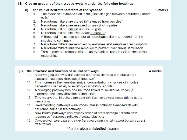
Neurotransmitters • The nerve cell before the synapse is called the presynaptic neuron. • The one after the synapse is called the postsynaptic neuron. • Neurons also connect with muscle fibres and endocrine glands cells via spaces similar to synaptic clefts. • For a nerve impulse to cross the synaptic cleft, chemicals called neurotransmitters have to enter the synapse. Can you name any from a pervious unit? acetylcholine noradrenaline (norepinephrine) • Neurotransmitters relay messages within and out with the brain from neuron to neuron. • Neurotransmitters are stored in vesicles.
When a nerve impulse arrives at the presynaptic membrane and reaches the axon endings it stimulates several vesicles which move to and fuse with the membrane. The vesicles release their neurotransmitters into the synaptic cleft (good example of exocytosis!). The neurotransmitters then diffuse across the cleft and bind with receptor sites on the membrane of the postsynaptic membrane (bit like a lock and key) which transmits the impulse through the postsynaptic neuron. Motor neuron The plasma membranes of the two neurons at a synapse are very close to one another and the axon endings (synaptic knob) contain a rich supply of vesicles full of one type of neurotransmitter. inter neuron Chemical transmission at a synapse
Key Points: Chemical transmission at a synapse postsynaptic neuron is activated and impulse is transmitted along the neuron Neurotransmitter combines with receptor on. . neurotransmitter is released into cleft Impulse reaches nerve (axon) endings of presynaptic neuron Use the jumbled statement to help you complete your summary flow diagram
Key Points: Chemical transmission at a synapse Direction of a nerve impulse Impulse reaches nerve (axon) endings of presynaptic neuron Arrival of impulse causes vesicles to fuse with presynaptic membrane Learn Me! neurotransmitter is released into cleft Neurotransmitter diffuses across synaptic cleft Neurotransmitter combines with receptor on. . Nerve endings of postsynaptic neuron is activated and impulse is transmitted along the neuron
Synapse animations. . http: //www. bbc. co. uk/schools/gcsebitesize/science/aqa_pre _2011/human/thenervoussystemrev 2. shtml https: //www. youtube. com/embed/Xfea. Mb. TKd. V 8? modestbr anding=1 Enzyme that degrades neurotransmitter
Learning Intentions q Describe the two different ways neurotransmitters can be removed from the synaptic cleft. q Explain excitatory and inhibitory signals. q Explain weak stimuli and summation of stimuli. q Explain how neurons are arranged into different types of neural pathways
Removal of neurotransmitters After the synapse… • The postsynaptic membrane remains excited for a short period of time. • Neurotransmitters are rapidly removed from the synaptic cleft once the impulse has been transmitted • This prevents continuous stimulation of postsynaptic neurons. (It also allows the neuron to respond to new signals, making precise control possible). • Removal of neurotransmitters can be achieved in two different ways…
Removal of Neurotransmitter Enzyme degradation Some neurotransmitters can be broken down into non-active products by enzymes in the postsynaptic membrane e. g. acetylcholinesterase non-active products. The non-active products are reabsorbed by the presynaptic neurone and can be resynthesised or stored in vesicles.
Removal of Neurotransmitter Re-uptake of neurotransmitters Other neurotransmitter are reabsorbed directly by the presynaptic membrane and stored in vesicles for future use e. g. noradrenaline (norepinephrine). Energy for both of these processes is released from mitochondria which are abundant in the pre-synaptic cytoplasm.
Excitatory and Inhibitory signals • We know that neurotransmitters bind with receptors. • It is the type of receptor which determines whether a signal is excitatory or inhibitory. • A neuron can have many synapses with other neurons. • It is the sum of the excitatory and inhibitory signals that determines the overall effect and whether the impulse is passed on (‘fired’).
Impulse arrives at synapse with excitatory receptors (E) Impulse arrives at synapse with inhibitory receptors (I) Impulse passes to next neuron if the sum of the excitatory signals exceeds the inhibitory ones
Example of inhibitory/excitatory effects: Acetylcholine The diagram shows how the release of the same neurotransmitter at parasympathetic nerve endings results in two different effects. Parasympathetic nerves l b a t o s l See a Release of acetylcholine at a parasympathetic nerve’s neuroeffector junction with the heart, combines with receptor sites of the type which pass on an inhibitory signal. As a result the rate andofforce of contraction Release acetylcholine at a of cardiac muscle decreases. parasympathetic nerve’s neuroeffector junction with the digestive tract’s muscle, combines with receptor sites of the type which pass on an excitatory signal. As a result the rate of peristalsis increases. 7 5 2 e g a p e 19. 2 on Therefore, the type of signal which is transmitted (either inhibitory or excitatory), depends on the type of RECEPTOR SITE present in the postsynaptic membrane with which the neurotransmitter combines.
Threshold In order for an nerve impulse to be fired there is a minimum number of neurotransmitter molecules needed to transmit the nerve impulse across the synaptic cleft. This is called the threshold. Weak stimuli fail to release enough neurotransmitter molecules and so the impulse fails to be transmitted across the synaptic cleft to the postsynaptic neuron. Summation If many weak stimuli reach a target neuron together, then their effect can be summated (added together) so that threshold is reached an impulse can be triggered. Learn!
Frequency of impulses Impulses are ‘all or none’. If enough neurotransmitter is released to reach threshold the impulse is fired. Each nerve impulse transmitted is EQUAL in size. The number of impulses transmitted per second i. e. the frequency, determines the intensity of the stimulus. For example, in relation to a sound stimulus: Loud stimulus High impulse frequency The converse applies to a quiet stimulus. Many impulses Brain perceives the sound as ‘loud’
Weak Stimuli Key point • Summation of weak stimuli can release enough neurotransmitter so that threshold is reached an impulse is triggered.
Related Topic Find out about…. Working with your partner, each of you should research one of the following…. . • Curare • Strychnine …. . then be ready to share key points. See page 259
Testing your knowledge v Collect a white board. v Test yourself by trying the questions on page 259 of the textbook, without your notes.
Extended Answer Question • Describe the function and mechanism of neurotransmitter action at the synapse. (7)
Marking Scheme
Complex Neural Pathways Enable the nervous system to carryout its many complex functions. What does diverge mean? Diverging neural pathways This is where one neuron is linked to several Toneurons. branch out from a common point This allows impulses from the CNS to be sent to several destinations (effectors/ muscles) at the same time. Diagrams must show direction of impulse
Examples of a diverging pathways • Fine motor control e. g. of the hands when playing the piano and temperature control. Motor area in the cerebrum Nerve impulses sent to several muscles at the same time Hypothalamus detects e. g. drop in temperature Nerve impulses sent to several effectors at the same time • • EFFECTORS Upper arm muscles Forearm muscles Hand muscles Finger muscles EFFECTORS • sweat glands • skin arterioles • skeletal muscles Permits finger muscles to work in a co-ordinated way (fine motor control) Permits effectors to work together in a co-ordinated way in order to increase body temperature
Exam question Explain how diverging pathways allow humans to perform a task such as threading a needle (2 marks). • Nerve impulse is transmitted to several points/destinations/effectors at the same time/simultaneously (1 mk) • This permits finger muscles to work in a coordinated way/permits fine motor control (1 mk)
“Summation” Converging Neural pathways This has several neurons come together, and link to one neuron. What does converge mean? This allows neurotransmitters from a number of nerve cells to be brought together making it more likely that the threshold is reached and the impulse is triggered. To come together and meet at a common point Converging neural pathways increase the sensitivity to excitatory or inhibitory signals. Diagrams must show direction of impulse.
Example of a converging neural pathway Rods in Retina of eye The nerve impulse transmitted by one rod in dim light is weak and would fail to reach threshold so the transmission of the impulse would not occur. However the convergent arrangement of several rods allow many impulses to be transmitted at the same time releasing enough neurotransmitter to reach threshold. The impulse crosses the synapse and is then transmitted via the optic nerve to the brain.
Reverberating neural pathways Neurons later in a neural pathway possess axon branches that form synapses with neurones earlier in the pathway. • This can be used to send the impulse back through the circuit. • Impulses can therefore be recycled back through the pathway continuously which brings about repeated activities e. g. breathing movements which repeats for a lifetime. Impulse is recycled allowing continuous stimulation Breathing muscles continuous in their action
Extended Answer Question
Testing your knowledge v Collect a white board. v Test yourself by trying the questions on page 267 of the textbook, without your notes.
Learning Intentions q To investigate neurotransmitters, mood and behaviour. q To investigate the action of recreational drugs.
Neurotransmitters, mood and behaviour & The mode of action of recreational drugs Collect: • Pupil handout • A 3 info sheet (extract from bright red book) • Textbook pages 267 -278 • Take your time to read the available resources and study the diagrams – make brief notes for each section on your handout.
Find out about…. Working with your group, each of you should research one of the following…. . • • • Cocaine Cannabis Ecstasy (NDMA) Alcohol Nicotine …. . then be ready to share key points. See page 272 -274 onwards
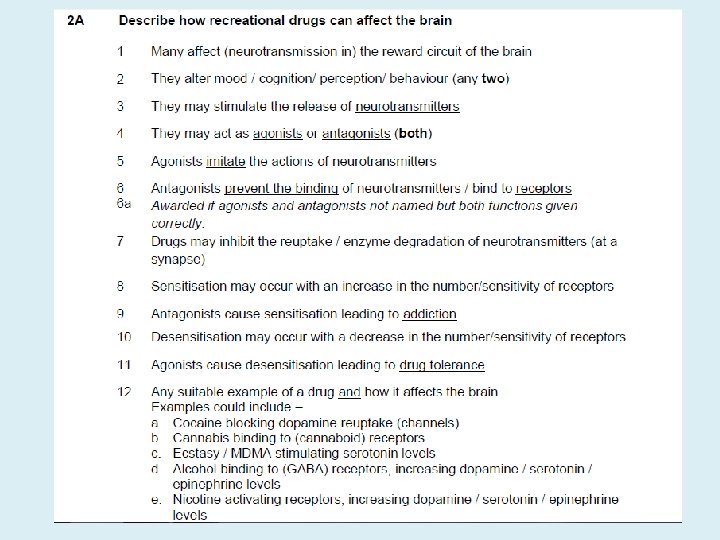
Practice Questions Enough / increased/more/a higher concentration of dopamine /neurotransmitter (is released) to trigger an impulse/reach threshold. or Many/a series of weak stimuli trigger an impulse/reach threshold. provides energy/ATP to make/ release neurotransmitter or dopamine/form vesicles/allow vesicles to move/allow vesicles to fuse with the membrane/to reuptake neurotransmitter.
(Cocaine) blocks/inhibits/acts as an antagonist to the re -uptake proteins. or (Cocaine) prevents the proteins from reabsorbing/taking up/ removing dopamine. 1 mark Dopamine/neurotransmitter remains in the synapse/at the receptors. or Dopamine/neurotransmitter continues to stimulate receptors/fire impulses. 1 mark
Extended Answer Question Describe how recreational drugs can affect the brain. (8)
Testing your knowledge v Collect a white board. v Test yourself by trying the questions on page 279 of the textbook, without your notes!