Biochemistry of neurotransmitters Dr Mamoun Ahram Neuroscience 2014

Biochemistry of neurotransmitters Dr. Mamoun Ahram Neuroscience 2014

References • This lecture • Mark’s Basic Medical Biochemistry, 4 th ed, pp. 908918 • http: //what-whenhow. com/neuroscience/neurotransmitters-theneuron-part-1/

What is a neurotransmitter? • A chemical substance that: – Is synthesized and stored in a presynaptic neuron (the enzymes needed for its synthesis must be present in the neuron), – Is released at a synapse following depolarization of the nerve terminal (usually dependent on influx of calcium ions), – binds to receptors on the postsynaptic cell and/or presynaptic terminal, – elicits rapid-onset and rapidly reversible responses in the target cell, – Is removed or inactivated from the synaptic cleft.
 –")
Types of neurotransmitters • Small-molecule – Amines (acetylcholine, epinepherine, dopamine, histmaine, etc. ) – Amino acids (glutamate, aspartate) • Neuropeptides • Gases (nitric oxide)

Note the differences • • • Onset and duration of action Concentration for action and receptor binding Concentration of [Ca+] for release Site of synthesis, modification Fate

NEUROPEPTIDES

Introduction • More than 50 neuropeptides have been described – Behavior – Pain perception – Memory – Appetite – Thirst – Temperature – Homeostasis – Sleep

Neuropeptides: neurohormones or neurotransmitters? • Neurohormones: when neurons secrete their peptides into the vascular system to be transported to a relatively distant target • Neurotransmitter: Many axon terminals of neurosecretory cells secrete their products at the synapse to directly affect a post synaptic cell • Neuropeptides can do both – depends on nerve terminal
 • Packaging into largedense core")
Stages of action • Synthesis (ER and Golgi apparatus) • Packaging into largedense core vesicles (with modifying enzymes) • Transport (fast-axonal transport) • Release • Action (prolonged) • Termination by diffusion and degradation

Synthesis is sequential

Synthesis is tissue-specific

Synthesis is controlled by alternative splicing

Role of calcium

Neuropeptides • • • The endogenous opiates Neuropeptide Y Galanin Pituitary adenylate cyclase–activating peptide (PACAP) Melanocyte-stimulating hormone (MSH) Neurokinin A (NKA) Substance P (SP) Neurotensin Calcitonin-gene–related protein (CGRP) Vasoactive intestinal polypeptide (VIP)

Substance P Member of tachykinins family 11 -amino acid peptide Act via neurokinin receptors 1, 2, or 3 (GPCR) and Gs Functions: vasodilation, inflammatory response, pain perception • Pathological connection: eczema, fibromyalgia, GI infection • •
 • • Member of the neurotrophin family 118 -acid polypeptide")
Nerve growth factor (NGF) • • Member of the neurotrophin family 118 -acid polypeptide hormone Act via tyrosine kinase receptors Affect tissue immune reactivity

Neuropeptide Y • A 36 amino acid peptide • Acts via G-protein coupled receptors (decrease in c. AMP, decrease in Ca+2 , and increase in K+ conductance) • Function: augments the vasoconstrictor effects of norepinephrine

SMALL-MOLECULE NEUROTRANSMITTERS

Types of small-molecule neurotransmitter • Nitrogen-containing molecules – amino acids and their derivatives – intermediates of glycolysis and the Krebs cycle (TCA cycle)

Stages of action • Synthesis of enzymes – Cytosol – ER-Golgi apparatus (packaging into large-dense core vesicles) • Transport of enzymes (slow and fast-axonal transport) • Synthesis in pre-synaptic terminal • Packaging in synaptic vesicles • Release • Action (short) • Termination by diffusion, reuptake, or inactivation
![[Ca+] = 2 m. M [Ca+] = 50 -100 u. M [Ca+] = 0.](http://slidetodoc.com/presentation_image/2feb0a2ea9e57d804c94f741a00a1c98/image-21.jpg "[Ca+] = 2 m. M [Ca+] = 50 -100 u. M [Ca+] = 0.")
[Ca+] = 2 m. M [Ca+] = 50 -100 u. M [Ca+] = 0. 1 u. M

Synaptic vs. large dense core vesicles
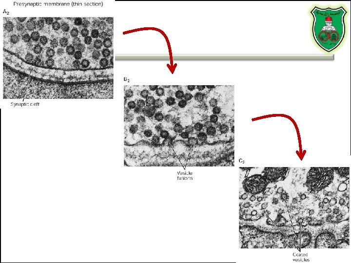

Role of clathrin and dynamin

SPECIFIC EXAMPLES OF NEUROTRANSMITTER SYNTHESIS
 – Pyrodoxal phosphate")
But first, note • Role of cofactors – S-adenosylmethionine (methyl transfer) – Pyrodoxal phosphate (vitamin B 6): transamination, decarboxylation – Tetrahydrobiopterin (BH 4)

TYROSINE-DERIVED NEUROTRANSMITTERS Dopamine, norepinephrine, and epinephrine

Diet/ liver phenylalanine hydroxylase Rate-limiting step Vitamin B 12 or folate Pyridoxal phosphate vesicular

50%
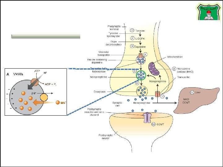
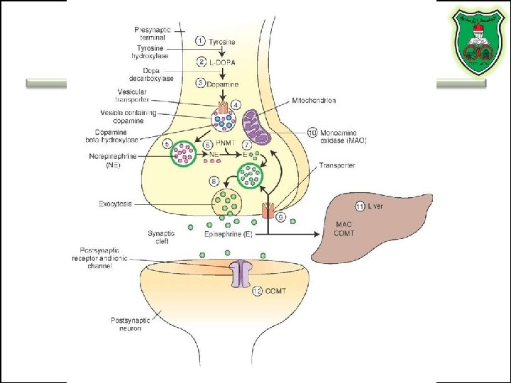

COMT and MAO Parkinson’s disease

Regulation • Tyrosine hydroxylase – Short term • Inhibition by free cytosolic catecholamines • Catecholamines compete with BH 4 binding to enzyme • Activation by depolarization – Tight binding to BH 4 following phosphorylation by PKA, CAM kinases, PKC – Long-term (plus dopamine -hyroxylase)

TRYPTOPHAN-DERIVED NEUROTRANSMITTERS Serotonin and melatonin
, like Prozac® inhibit")
BH 4 Serotonin Antidepressants , called selective serotonin re-uptake inhibitors (SSRIs), like Prozac® inhibit the reuptake process resulting in prolonged serotonin presence in the synaptic cleft. M AO 5 -hydroxyindoleacetic acid urine

Melatonin • Serotonin synthesized in the pineal gland serves as a precursor for the synthesis of melatonin, which is a neurohormone involved in regulating – sleep patterns – Seasonal and circadian (daily) rythyms – Dark-light cycle

GLUTAMATE AND ASPARTATE

Glutamate and aspartate • Nonessential amino acids • Do not cross BBB – must be synthesized in neurons • Main synthetic compartments – neurons – glial cells • Both are excitatory neurotransmitters.

Synthesis of glutamate • Two pathways: • Glycolysis Krebs cycle Glu Transamination or Dehydro dehydrogenation transaminase 1 • Glutamine (deamination) • Another source: aspartate 2 glutaminase • Removal – excitatory amino acid carrier 1 (EAAC 1) – glutamate transporter-1 (GLT 1) and glutamate—aspartate transporter (GLAST) 3 GABA -KG Glutamine synthetase
")
Sources of glutamate (supplementary)

Physiological and clinical implications • Excitatory amino acids may be involved in learning and memory processes, as well as motor functions. • Chronic neuropathological conditions such as amyotrophic lateral sclerosis [ALS] (also known as Lou Gehrig’s disease). – degeneration of the motor neurons in the anterior horn of the spinal cord, brainstem, and cerebral cortex. • Prolonged stimulation of neurons by excitatory amino acids results in neuronal death or injury. • Overexcitatory effects result in Alzheimer’s disease.

Aspartate • A vesicular uptake mechanism for aspartate has not yet been demonstrated, somewhat weakening the case for considering aspartate to be a neurotransmitter • Precursor: oxaloacetate (transmamination)

Glycine • The major inhibitory neurotransmitter in the spical cord • Synthesized from serine by serine hydroxymethyltransferase through 3 -phosphoglycerate • Removal: high-affinity transporter

OTHERS
 in many brain regions. –")
GABA • GABA is present in high concentrations (millimolar) in many brain regions. – These concentrations are about 1, 000 times higher than concentrations of the classical monoamine neurotransmitters in the same regions. • The GABA shunt is a closed-loop process with the dual purpose of producing and conserving the supply of GABA.

GABA shunt

Synthesis of acetylcholine • Choline + acetylcoenzyme-A by choline acetyltransferase in cytoplasm • Transported into and stored in vesicles. • Removal: hydrolysis by acetylcholinesterase Diet Membrane PL

Histamine • it does not penetrate the blood—brain barrier and, hence, must be synthesized. Pyridoxal phosphate X Astrocytes Neuron

Inactivation of histamine
 • • Glutamate is released (1) and acts on NMDA receptors")
Nitric oxide (NO) • • Glutamate is released (1) and acts on NMDA receptors located on the postsynaptic neuron (2) Ca 2+ enters the postsynaptic neuron and binds with calmodulin activating NOS (3) resulting in formation of NO and citrulline from L-arginine (4). No stimulates guanylate cyclase forming c. GMP (5), which results in a physiological response (6) No can diffuse out: a) to the presynaptic terminal (7) prolonging effect and b) into adjacent neurons (8) and glial cells (9) stimulating guanylate cyclase

Is NO a neurotransmitter? • Yes, but: – It is not stored in vesicles – It is not released by calcium-dependent exocytosis (it diffuses) – Its inactivation is passive (there is no active process that terminates its action) • It decays spontaneously – It does not interact with receptors on target cells • Its sphere of action depends on the extent to which it diffuses, and its action is not confined to the conventional presynaptic-postsynaptic direction. – NO acts as a retrograde messenger and regulates the function of axon terminals presynaptic to the neuron in which it is synthesized.
 • Half-life: 2 -4 seconds • NO is inhibited by hemoglobin")
Nitric oxide (NO) • Half-life: 2 -4 seconds • NO is inhibited by hemoglobin and other heme proteins which bind it tightly
 – Neurons and epithelial")
NO synthase • Isoform I (n. NOS or c. NOS) – Neurons and epithelial cells – activated by the influx of extracellular calcium • isoform II (i. NOS) – Macrophages and smooth muscle cells – induced by cytokines • and isoform III (e. NOS) – Endothelial cells lining blood vessels – activated by the influx of extracellular calcium • All three isoforms require BH 2 as a cofactor and nicotinamide adenine dinucleotide phosphate (NADPH) as a coenzyme
- Slides: 53