SPOTS Aarti Sardana 2 nd Yr Resident Deptt


")


 and epidural")


 greater than 3 mm (a")


 o Monoarticular presentation in the middle & elderly aged.")

- Slides: 64
SPOTS Aarti Sardana 2 nd Yr Resident, Deptt of Radiodiagnosis SSG Hospital Date: 14 -03 -08
1.
CROSSED FUSED ECTOPIA o KUB scout demonstrates no left renal shadow and an abnormal contour to the right renal shadow. CT and IVP show crossed fused ectopia of the left kidney. The bilateral collecting systems are intact and patent. The proximal ureter of the right kidney courses lateral to the ectopic left kidney. There is no evidence of pelvicaliectasis. The bladder appears normal in size and contour. No abnormal masses or renal calculi are present
Discussion o More common in males than in females. o This anomaly occurs as a result of poor ascent of the kidneys o o o from the pelvis to the upper abdomen. In most cases, fusion occurs between the lower pole of the orthotopic kidney and the upper pole of the ectopic kidney. More often the left kidney is ectopic than the right. The ectopic kidney will not rotate appropriately, and as a result, the renal pelvis is anterior in orientation rather than medial. This predisposes to ureteral obstruction, and hydronephrosis. Usually asymptomatic, however, it does result in increased incidence of renal stones within the ectopic kidney due to ureteral stasis. The ectopic kidney is more prone to trauma, particularly if it resides anterior to the spine. The most commonly associated serious congenital abnormality is imperforate anus (%4).
2.
o. Corkscrew Esophagus
Diffuse Esophageal Spasm o It is characterized by intermittently abnormal esophageal motility and chest pain, which may be accompanied by dysphagia and even food impaction. o Cause of DES is not known, though diffuse fragmentation of vagal filaments, increased endoneural collagen, and mitochondrial fragmentation are described. o This results in a functional imbalance between excitatory and inhibitory postganglionic pathways.
o Radiographic features: Ø Primary peristalsis present in upper esophagus but intermittently absent in smooth muscle segment. Ø Non peristaltic contractions which are repetitive, simultaneous, and may obliterate esophageal lumen causing “cork-screw or rosary bead” appearance. o However these findings may be minimal or non-specific & correlation with clinical symptoms & manometry is needed.
3.
o. Osteoid Osteoma of the Spine
Discussion o Osteoid Osteoma is a benign osteoblastic tumor of bone. Very frequently seen between the ages of 10 and 30 with male to female ratio of three to one. o The hallmark of Osteoid Osteoma is dull and localized intermittent bony aching pain which increases at night. o The nidus may be radiolucent or may contain calcifications with variable surrounding sclerotic reaction. o Surgery is the method of choice for treatment. o CT Scan is very helpful in cases of Osteoid Osteoma of spine, pelvis and femoral neck. o 70% in long bones, 20% in hands and feet. o The vertebral column, innominate bone, skull, ribs, clavicle and scapula are uncommon sites of the disease.
4.
Diagnosis Proximal Femoral Focal Deficiency (PFFD)
o Spectrum of conditions characterized by partial absence and shortening of the proximal portion of the femur
Levinson’s Classification of PFFD
HEAD PRESENT BONY CONNECTION BETWEEN COMPONENTS OF FEMUR PRESENT NORMAL ACETABULUM & SUBTROCHANTERIC VARUS TYPE A ABSENT ACTEABULUM NORMAL OR DYSPLASTIC WITH BONY TUFT TYPE B
HEAD ABSENT OSSICLE PRESENT COMPLETELY ABSENT FEMORAL SEGMENT SHORT WITH PROXIMALTAPERING TYPE C FEMORAL SEGMENT SHORT & DEFORMED TYPE D
5.
The ring-around-the-artery sign EXPLANATION o Pneumomediastinum refers to an abnormal presence of air in the mediastinum. o It manifests as air outlining normal anatomic structures. o When air collects around the mediastinal (extrapericardial) portion of the right pulmonary artery, air is seen as a lucency along or surrounding the artery on a lateral chest radiograph. o This is the reported appearance of the ringaround-the-artery sign
Other related signs of pneumomediastinum o Thymic sail sign : air can outline and elevate thymus o Tubular artery sign : outline of the ascending aorta, the aortic arch, o o o the major branches of the aorta Double bronchial wall sign : the trachea and proximal bronchi Air anterior to the pericardium may be the only radiographic manifestation, and it requires a lateral view for detection Continuous diaphragm sign : Mediastinal air outlining the superior surface of the diaphragm and separating it from the heart. On a lateral view, the same mediastinal air creates the continuous left hemidiaphragm sign Naclerio's V sign is produced by air outlining the descending thoracic aorta and extending laterally between parietal pleura and medial left hemidiaphragm. The extrapleural sign is the result of air trapped between parietal pleura and the hemidiaphragm (9).
Continuous diaphragm sign & continuous left hemidiaphragm sign
6.
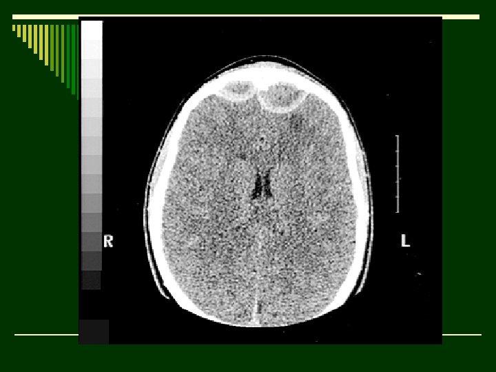
Diagnosis o Pott's puffy tumor (frontal sinusitis, subgaleal abscess, frontal bone osteomyelitis) and epidural abscesses Discussion o First described by Sir Percivall Pott in 1760. o This initial description included the subgaleal abscess and associated osteomyelitis. o Pott observed that the condition could be seen secondary to underlying infection in the sinuses (most often frontal or mastoid) or after injury. o Streptococcus, Haemophilus influenzae, Staphylococcus and Klebsiella
o Significantly decreased in the post-antibiotic era. o M/c: 10 and 20 years. o The resultant epidural abscess is thought to be related to the emissary veins that connect the frontal and dural sinuses. Epidural abscess is five times less common than subdural abscess, characteristically shows rim enhancement, and is a surgical emergency. Classically, a subdural abscess can be distinguished from an epidural abscess by its ability to cross sutures, a more cresentic than lentiform appearance, and an inabiltiy to cross the falx.
7. o. Incidental finding in someone who had not lived in a city all his life.
DIAGNOSIS o. CALCIFIED HYDATID CYST IN BOWEL
8.
Gas in the biliary tree o Causes for this appearance: o Previous instrumentation or surgery: ERCP and sphincterotomy for bile duct stones is a common cause, and biliary bypass surgery (eg. choledochojejunostomy) produces a similar appearance. o Passage of a stone through the common bile duct may allow reflux of air. o Fistula: Most commonly due to inflammation secondary to stones. The stone may pass into the duodenum, or into the transverse colon. In the former, the stone often impacts in the distal small bowel causing small bowel obstruction (gallstone ileus), and the combination of obstruction and biliary gas should suggest this (whether the stone is visible or not). Tumour may also cause a fistula. o Infection: emphysematous cholecystitis produces air in the gallbladder, and this may also outline the biliary tree. It is commoner in diabetics.
9.
o. Hair-on-end appearance : Thalassemia
10.
Hydrocephalus, air-encephalography. o The ventricles are outlined by air that has been exchanged for cerebro- spinal fluid, via lumbar puncture. The patient is anaesthetised erect. After sub-arachnoid air injection, change of position of the patient allows views to show different structures. In this view, air has outlined the posterior sections of the dilated lateral ventricles and the 'low' density above the pituitary fossa is air in the posterior part of the third ventricle. o Additional densities are iodinated oil run-up from the lumbar region in a previous assessment of the posterior fossa and now remains in recesses in the basal cisterns. o Very expensive and complex machines designed to allow invasive procedures to be performed, while moving the patient in different planes and allowing X-rays to be taken, at the same time. Compared with Computed tomography in a usually ambulant patient the information from these complex examinations now seems a limited return for the investment.
11.
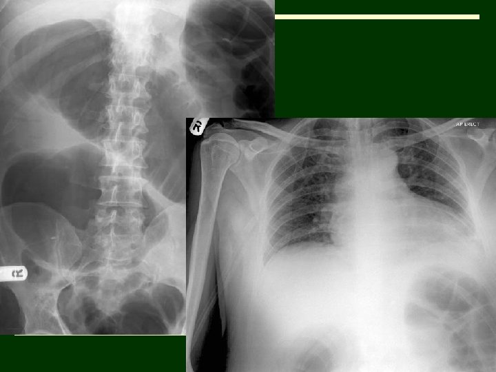
o. Lipohaemarthrosis, fractured neck of humerus
12. o 50 year old man underwent an above-knee amputation to remove his gangrenous leg. He developed nausea and dyspneoa on the 4 th postoperative day. The abdomen, though distended, was not tender and the bowel sounds were audible. Digital rectal examination was unremarkable.
o(A subsequent CT scan of the abdomen did not reveal a mechanical cause of the condition and colonoscopic decompression was therapeutic)
Diagnosis o. Acute colonic pseudo-obstruction or Ogilvie syndrome
Discussion o Acute colonic pseudo-obstruction or Ogilvie syndrome is a condition with clinical and radiological features of colonic obstruction without any evidence of a mechanical cause. o Abdominal distension in this patient accounted for the respiratory distress and the dilated large bowel loops seen on the chest radiographs guided towards the correct diagnosis. o Pathophysiology is not clearly understood, though it is thought to be due to an imbalance in the autonomic innervation leading to a functional bowel obstruction. o Ogilvie syndrome typically occurs in patients hospitalized with a significant illness e. g. severe cardio-respiratory disorders, sepsis, electrolyte imbalance and postoperatively. o Left untreated it can progress to colonic perforation and peritonitis. Plain abdominal film is the initial imaging investigation of choice. Colonoscopy can be both diagnostic and therapeutic
Differential o Mechanical colonic obstruction o Toxic megacolon o Mesenteric ischaemia
13.
Scheuerman's disease o There is irregularity of the vertebral end- plates at the vertebral disc margins, particularly anteriorly. It corresponds to the region of the limbus, a rim apophysis of bone with underlying cartilage, and the mechanical consequences of the attachment of the capsular ligament of the joint that is the intervertebral disc.
14. Post Gd fat sat. axial T 1 W
optic nerve tram-track sign in c/o optic nerve sheath Meningiomas
o The optic nerve tram-track sign is most commonly described in relation o o to optic nerve sheath Meningiomas tend to cause segmental or diffuse circumferential thickening of the optic nerve sheath. Once intravenous contrast material has been administered, the optic nerve can be seen on CT or MR images as an unenhanced central linear structure (negative defect) surrounded by the enhanced Meningioma. On transverse or Sagittal images this produces a tram-track sign, which is composed of two enhanced areas of tumor separated from each other by the negative defect of the optic nerve. The corresponding finding on coronal images is a doughnut configuration. Though it is less common, the tram-track sign may be evident at unenhanced CT when there is linear calcification of the optic nerve sheath Meningioma.
15.
Diagnosis o Achondrogenesis
o ACHONDROGENESIS – o Types of achondrogenesis include o Achondrogenesis Type I - a recessive lethal chondrodysplasia. o Achondrogenesis Type II – also k/a hypochondrogenesis. o A diagnosis of achondrogenesis is more likely when the patient also exhibits areas of absent ossification in the axial skeleton, micromelia, and hydrops.
o Other findings o Severe lack of ossification of the vertebral bodies o o (especially caudally). Small deformed iliac bones. Absent or poor ossification of pubic and ischial bones, calcaneus, and talus. Tubular bones that are strikingly short & malformed with wide, cupped ends. Short ribs with cupped and flared ends.
16.
Diagnosis o. Bone infarct
17.
Melorheostosis “Flowing wax" appearance
18. o 2 yr old male child with vomitting.
USG images
o Hypertrophic pyloric stenosis. o MT (serosa to mucosa) greater than 3 mm (a correlation between MT and the patient's age exists; ) - the most reliable sonographic sign. o Target sign on transverse images of the pylorus. o Pyloric channel length greater than 16 mm o Pyloric thickness (serosa to serosa) of greater than 11 mm
19.
COMPLETE CLOSED FUNNEL RETINAL DETACHMENT
Differential diagnosis of complete closed funnel retinal detachment CLOSED FUNNEL RD o Triangular membrane with apex at optic disc and base over the ora serrata. o Never attached to the posterior capsule of the lens. o Thin attachment at the optic disc PHPV (persistent hyperplastic primary vitreous) o Triangular membrane with apex at the posterior capsule and base over the optic disc. o Thin attachment to the posterior capsule of the lens. o Broad based attachment at the optic disc.
20.
Calcific supraspinatus tendonitis in a c/o HADD
Calcium hydroxypatite deposition disease (HADD) o Monoarticular presentation in the middle & elderly aged. o It is characterized by homogenous cloud like periarticular calcification most commonly affecting the shoulder in & around the supraspinatus tendon. o E/O: - Repetitive minor trauma with a cycle of necrosis & inflammation leading to dystrophic calcification. o Pain is related to the release of crystals into the surrounding tissues most notably joints & bursae.
Thank you