
What is Breast Cancer n n Abnormal cells


What is Breast Cancer ? n n Abnormal cells develop from normal cells in the breast to form tumors Tumor cells have genetic defects that allow the cells to grow too rapidly and to invade normal tissues Malignant cells Normal cells
Breast Cancer: Statistics Breast cancer is the most common cancer in American women n 20% of all cancers in women n Commonest cause of death - 35 -55 y n 185, 000 new cases every year n Lifetime risk for the average woman; approximately one in nine will develop breast cancer n
Risks n Finding of pre-malignant changes in breast tissue n Never having children n Having first child after age 30 n Obesity - Alcohol - Estrogen therapy
What is the risk for developing breast cancer? n n n Age 35 - 55 years History of breast cancer in a close relative (mother/sister/daughter) 90% of women with breast cancer have no relatives with the disease
Stages of breast cancer n n The rate at which a cancer will grow is not predictable - some tumors grow slowly, others rapidly The earlier a tumor is identified, the better the chance for cure 95% of women with cancers less than 1/2 inch in size are cured Advanced cases are from cure our aim is palliation
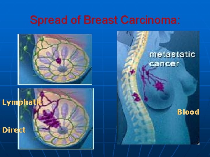
Spread of Breast Carcinoma: Lymphatic Blood Direct

TNM Staging Tumor n Tx primary tumor can not be assessed n n n Tis In situ carcinoma & paget’s disease T 0 no palpable mass T 1 tumor < or = 2 cm n T 1 a < or = 0. 5 cm no deep fixation n T 2 b 0. 5 – 1 cm + deep fixation n T 3 c 1 – 2 cm + deep fixation T 2 tumor 2 – 5 cm n T 2 a no deep fixation n T 2 b deep fixation T 3 tumor 5 – 10 cm n T 3 a no deep fixation n T 3 b deep fixation T 4 tumor of any size n T 4 a direct chest extension n T 4 b skin ( Peau d’orange, skin nodule & ulceration) n T 4 c T 4 a + T 4 b n T 4 d inflammatory breast cnacer
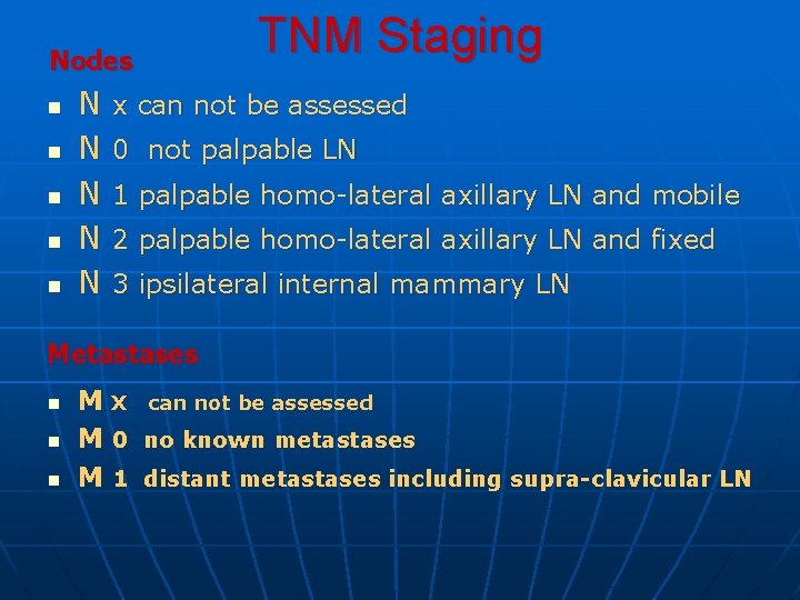
Nodes n n n N N N TNM Staging x can not be assessed 0 not palpable LN 1 palpable homo-lateral axillary LN and mobile 2 palpable homo-lateral axillary LN and fixed 3 ipsilateral internal mammary LN Metastases n n n M X can not be assessed M 0 no known metastases M 1 distant metastases including supra-clavicular LN
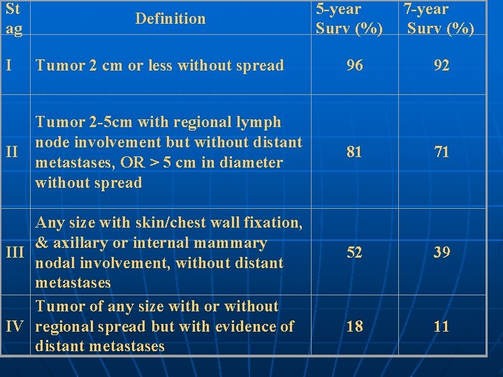
St ag Definition 5 -year Surv (%) 7 -year Surv (%) I Tumor 2 cm or less without spread 96 92 II Tumor 2 -5 cm with regional lymph node involvement but without distant metastases, OR > 5 cm in diameter without spread 81 71 52 39 18 11 Any size with skin/chest wall fixation, & axillary or internal mammary III nodal involvement, without distant metastases Tumor of any size with or without IV regional spread but with evidence of distant metastases
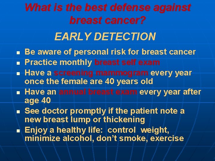
What is the best defense against breast cancer? EARLY DETECTION n n n Be aware of personal risk for breast cancer Practice monthly breast self exam Have a screening mammogram every year once the female are 40 years old Have an annual breast exam every year after age 40 See doctor promptly if the patient note a new breast lump or thickening Enjoy a healthy life: control weight, minimize alcohol, don’t smoke, exercise
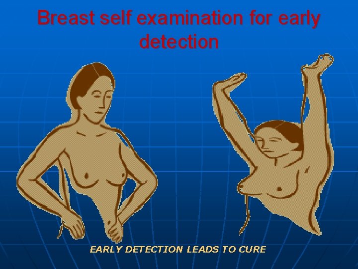
Breast self examination for early detection EARLY DETECTION LEADS TO CURE
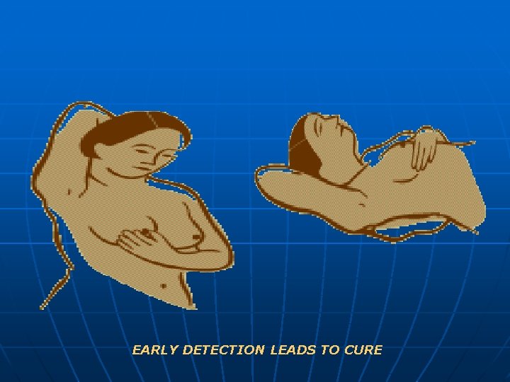
EARLY DETECTION LEADS TO CURE
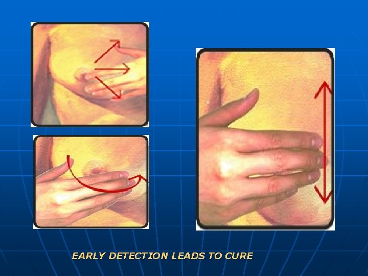
EARLY DETECTION LEADS TO CURE
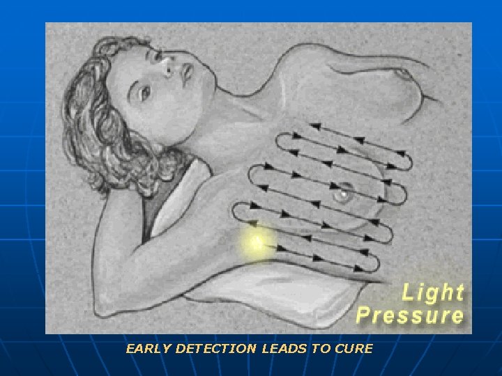
EARLY DETECTION LEADS TO CURE
What if a female feel a lump in her breast? n n The patient should go to her doctor immediately for a physical examination; and don’t wait for annual examination The doctor may refer the patient for breast imaging - a mammogram A biopsy may be recommended, depending on the features of the lump Many lumps in the breast are NOT cancer; benign fibrocystic disease and benign tumors are common EARLY DETECTION LEADS TO CURE
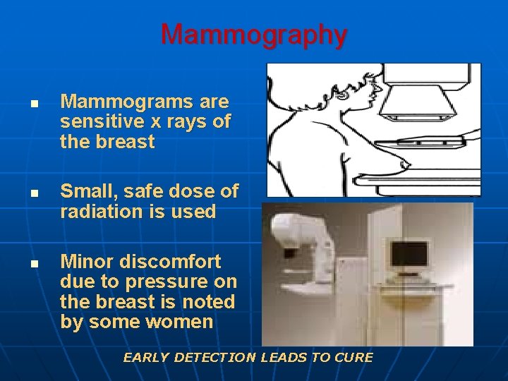
Mammography n n n Mammograms are sensitive x rays of the breast Small, safe dose of radiation is used Minor discomfort due to pressure on the breast is noted by some women EARLY DETECTION LEADS TO CURE
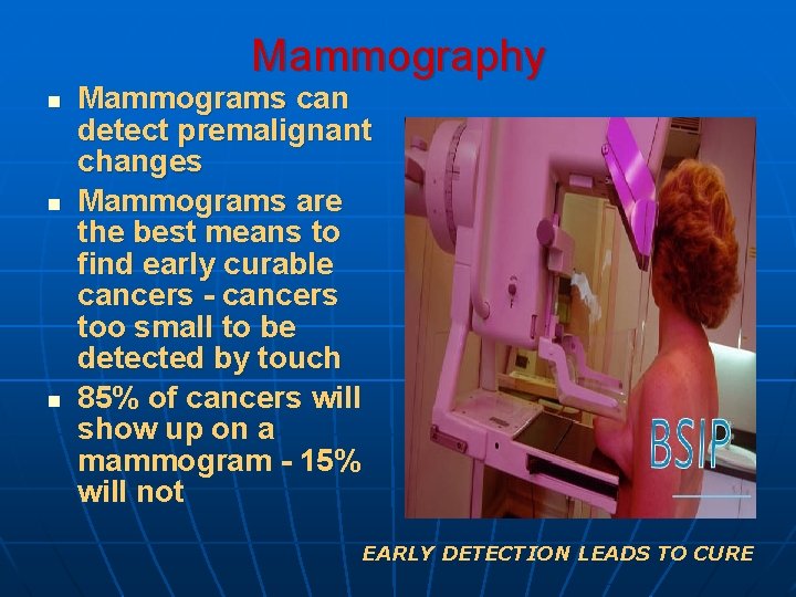
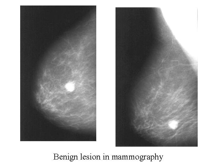
Mammography n n n Mammograms can detect premalignant changes Mammograms are the best means to find early curable cancers - cancers too small to be detected by touch 85% of cancers will show up on a mammogram - 15% will not EARLY DETECTION LEADS TO CURE
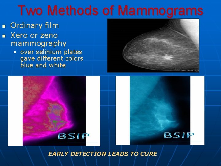
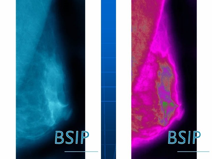
Two Methods of Mammograms n n Ordinary film Xero or zeno mammography • over selinium plates gave different colors blue and white EARLY DETECTION LEADS TO CURE
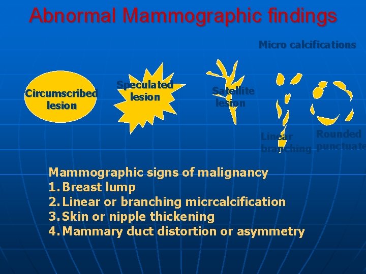
Abnormal Mammographic findings Micro calcifications Circumscribed lesion Speculated lesion Satellite lesion Rounded Linear branching punctuate Mammographic signs of malignancy 1. Breast lump 2. Linear or branching micrcalcification 3. Skin or nipple thickening 4. Mammary duct distortion or asymmetry
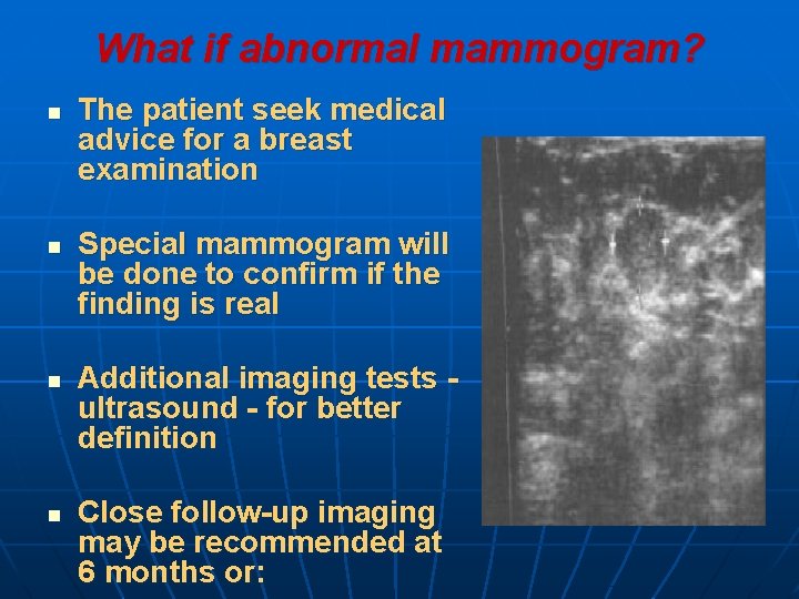
What if abnormal mammogram? n n The patient seek medical advice for a breast examination Special mammogram will be done to confirm if the finding is real Additional imaging tests ultrasound - for better definition Close follow-up imaging may be recommended at 6 months or:
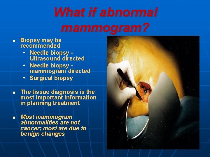
What if abnormal mammogram? n n n Biopsy may be recommended • Needle biopsy Ultrasound directed • Needle biopsy mammogram directed • Surgical biopsy The tissue diagnosis is the most important information in planning treatment Most mammogram abnormalities are not cancer; most are due to benign changes
How is the diagnosis of breast cancer made? n n n A suspicious sign on a mammogram or a lump in the breast will indicate the need for a tissue biopsy A biopsy of the abnormal tissue is required to make the diagnosis of breast cancer A careful physical examination will give signs about the stage of the cancer • size of tumor in breast • lymph node enlargement under arm • status of the opposite breast
How is the diagnosis of breast cancer made? n Features of the tumor: • Type of breast cancer - ductal, lobular, etc • Grade of the tumor • Hormone receptor status • Indicators of biologic growth rate potential • c-erb
Can breast cancer be prevented? n n n Recent studies showed that women with an increased risk for breast cancer can have that risk decreased by 40 - 50% by taking the anti-estrogen medication tamoxifen Tamoxifen benefited women with: • the breast cancer gene • age greater than 55 years • premalignant changes in previous biopsies Side effects • very small increase in risk of cancer of the uterus • blood clots
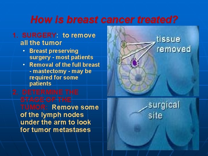
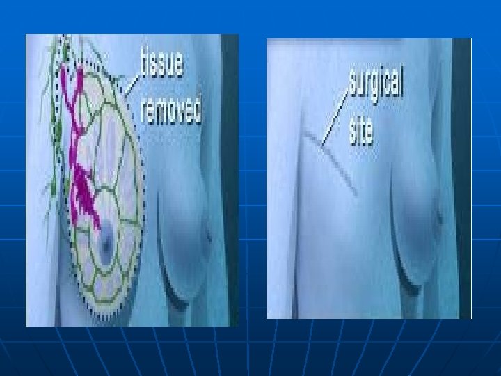
How is breast cancer treated? 1. SURGERY: to remove all the tumor • Breast preserving surgery - most patients • Removal of the full breast - mastectomy - may be required for some patients 2. DETERMINE THE STAGE OF THE TUMOR: Remove some of the lymph nodes under the arm to look for tumor metastases
How is breast cancer treated? 3. • • ADJUVANT THERAPY: Medical therapy to decrease the chance of tumor recurrence to improve the chances for cure Chemotherapy - many different therapies Hormonal therapy - tamoxifen, aromatase inhibitors
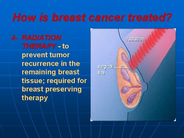
How is breast cancer treated? 4 - RADIATION THERAPY - to prevent tumor recurrence in the remaining breast tissue; required for breast preserving therapy

- Slides: 32