Pathological Myopia Pathological Myopia Clinical refractive error 6


- Slides: 25
Pathological Myopia
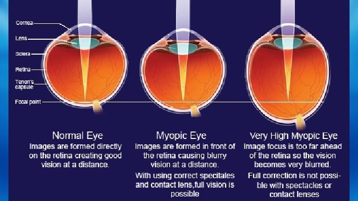
Pathological Myopia Clinical : refractive error >6 D. • Myopia with degenerative changes especially In the posterior segment. • Myopia caused by pathological axial elongated Of the eye ball. Specific : a rapidly progressive error which starts in childhood at 5 -10 years of age & results in high Myopia
Synonyms l l l Degenerative myopia Progressive myopia Malignant Myopia High degree myopia Magna myopia
Etiology l The rapid growth of axial growth of eye ball is witnessed as the main cause for the pathological Myopia which is unequivocal & outside the normal biological variants of development. l No satisfactory theory has emerged to explain this. l It is definitely linked with : (1) Role of heredity. (2) Role of general growth 1. Role of heredity : it is confirmed that genetic factors play a major role in the etiology.
l It is presumed that heredity linked growth of retina is the determination in the development of myopia. l The retina due to its distensibility stretches with retina. l But the choroid cannot & undergoes degeneration which in turn cause generation of retina.
Symptoms l l l l Image infection Anisometric amblyopia Subnormal visual acuity Visual field defects Impaired dark adaption Abnormal color discrimination Suboptimal binocularity
Signs l l l Prominent eye ball Cornea is large Anterior chamber is deep Large and sluggish pupils Visual field contraction On fundus examination : ü Large and pale optic disc ü Tilted optic nerve papillary atrophy ü Temporal myopic crescent/peripapillary
Signs ü ü ü ü ü Crescent Blond fundus Chorioretinal atrophy Peripheral vitreous detachment Lacquer cracks Lattice degeneration Peripheral retinal holes Macular holes Choroid neovascularization
üLarge and pale optic disc :
ü Tilted optic nerve with peripapillary atrophy :
Temporal myopic crescent/ peripapillary
ü Tigroid/Blond fundus :
ü Choroid retinal atrophy :
ü Posterior vitreous detachment
ü Lacquer cracks :
ü Lattice degeneration :
ü Cobblestone degeneration :
ü Foster-fuchs spots :
ü Peripheral retinal holes :
ü Macular holes :
ü Choroid neovascularisation :
Complications : l l l l Retinal detachment Complicated cataract Vitreous & choroidal hemorrhage Strabismus fixus convergence Normotensive glaucoma Myopic foveoschisis Posterior staphyloma
Treatment :
Thank you