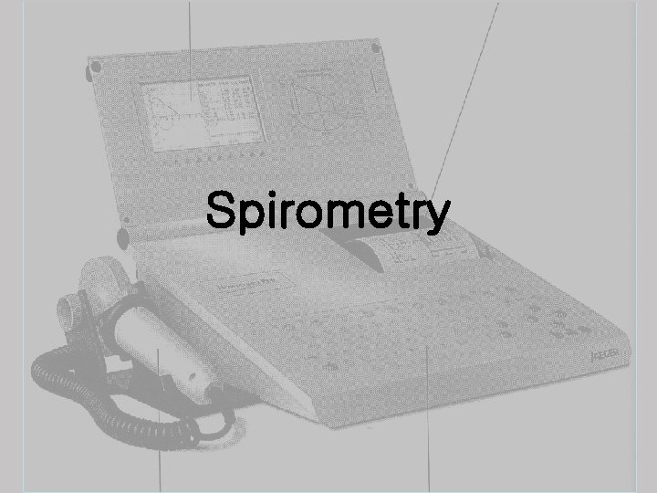
Spirometry ED 1 2 3 Asthma COPD Trauma


- Slides: 29
Spirometry
ED에서의 유용성 1. 2. 3. Asthma COPD Trauma
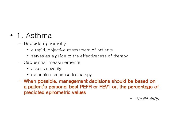
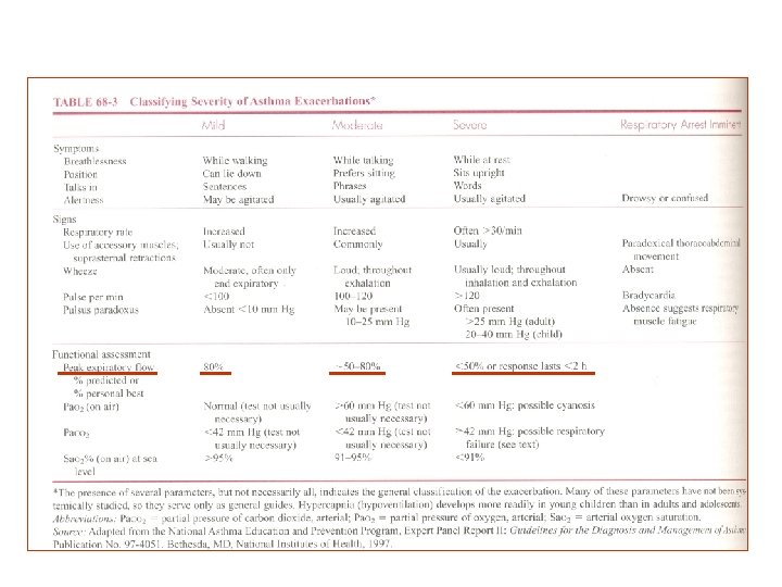
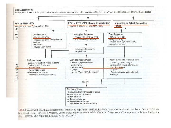
• 1. Asthma – Bedside spirometry • a rapid, objective assessment of patients • serves as a guide to the effectiveness of therapy – Sequential measurements • assess severity • determine response to therapy – When possible, management decisions should be based on a patient’s personal best PEFR or FEV 1 or, the percentage of predicted spirometric values – Tin 6 th 469 p
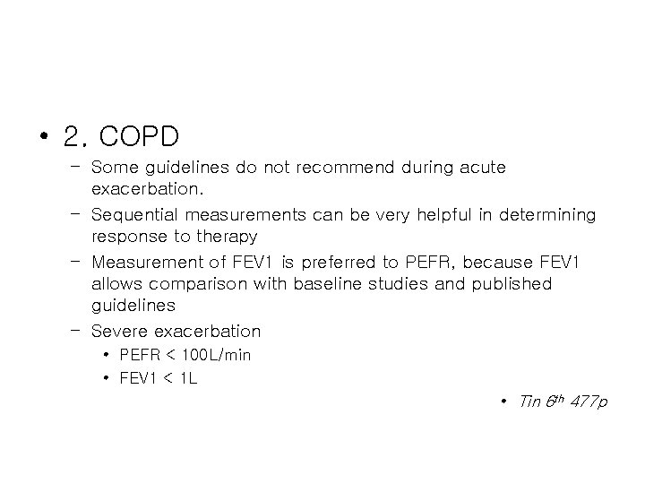
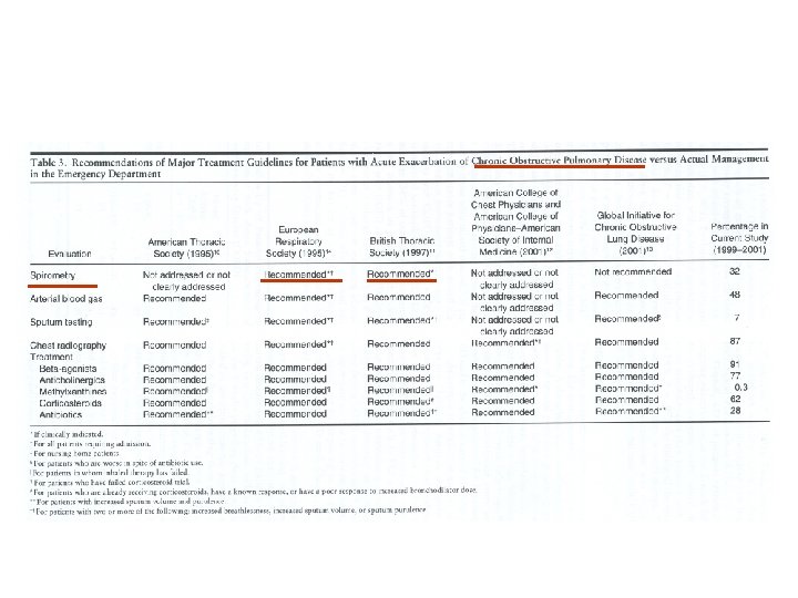
• 2. COPD – Some guidelines do not recommend during acute exacerbation. – Sequential measurements can be very helpful in determining response to therapy – Measurement of FEV 1 is preferred to PEFR, because FEV 1 allows comparison with baseline studies and published guidelines – Severe exacerbation • PEFR < 100 L/min • FEV 1 < 1 L • Tin 6 th 477 p
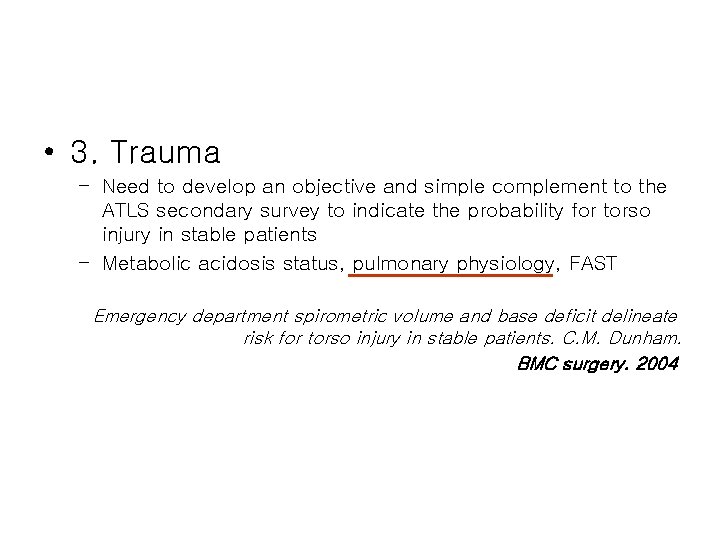
• 3. Trauma – Need to develop an objective and simple complement to the ATLS secondary survey to indicate the probability for torso injury in stable patients – Metabolic acidosis status, pulmonary physiology, FAST Emergency department spirometric volume and base deficit delineate risk for torso injury in stable patients. C. M. Dunham. BMC surgery. 2004
How to do? ? 1. 2. 3. Spirometry Flow/volume measurement after bronchospasmolysis
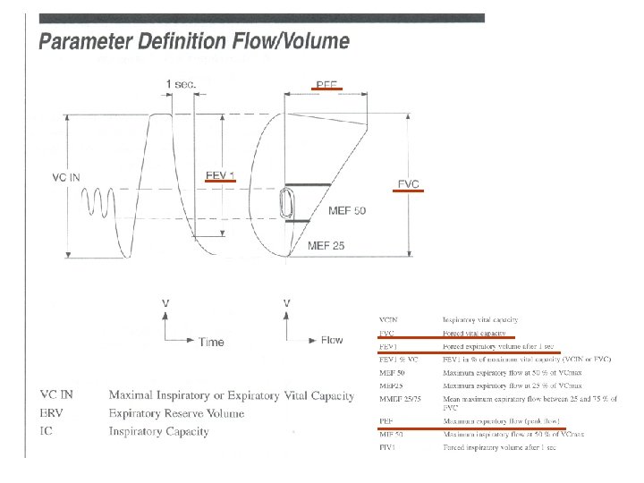
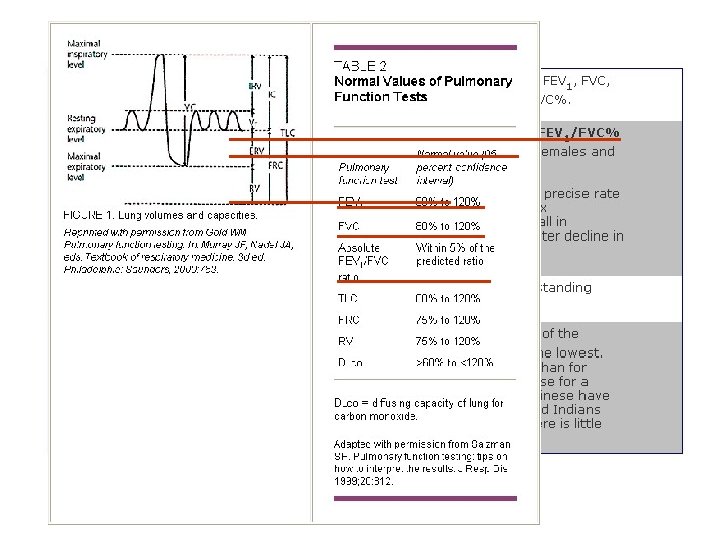
Parameter definition
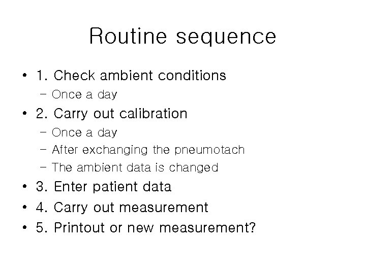
Routine sequence • 1. Check ambient conditions – Once a day • 2. Carry out calibration – Once a day – After exchanging the pneumotach – The ambient data is changed • 3. Enter patient data • 4. Carry out measurement • 5. Printout or new measurement?
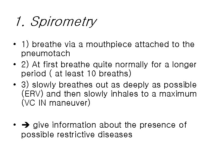
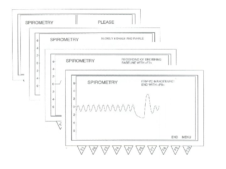
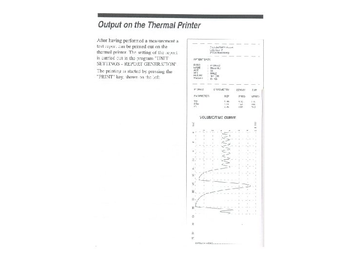
1. Spirometry • 1) breathe via a mouthpiece attached to the pneumotach • 2) At first breathe quite normally for a longer period ( at least 10 breaths) • 3) slowly breathes out as deeply as possible (ERV) and then slowly inhales to a maximum (VC IN maneuver) • give information about the presence of possible restrictive diseases
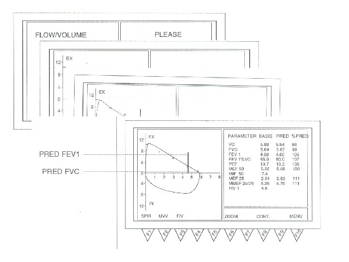
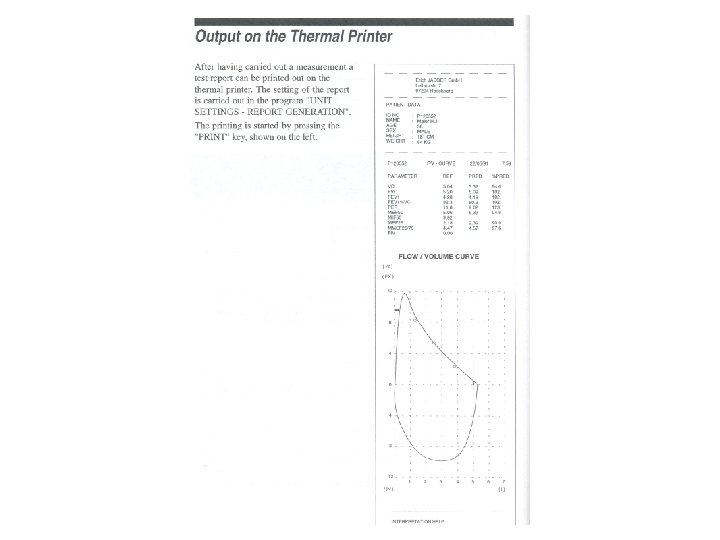
2. flow/volume • 1) Breathe quite normally • 2) Exhale slowly as deeply as possible, inhale slowly to a maximum and exhales much and as deeply as possible(FEV 1, MEF 50) and inhale to a maximum(MIF 50) • give information about the presence of possible restrictive or obstructive diseases of the respiratory organs and extrathoracic stenoses
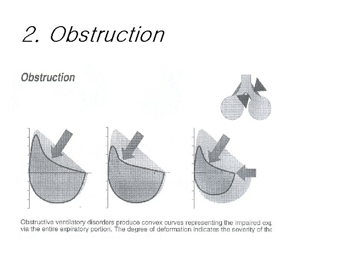
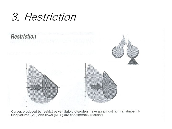
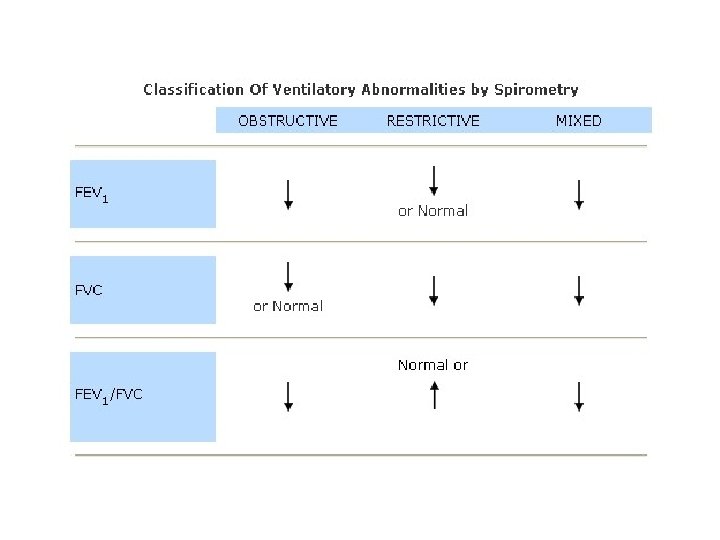
Typical flow/volume curves -interpretation 1. 2. 3. 4. 5. Normal Obstruction Restriction Emphysema Upper airway obstruction
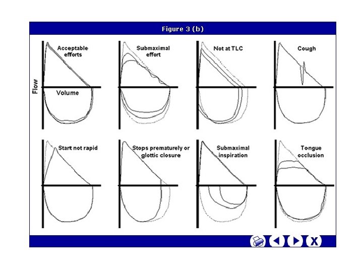
Problematic examples
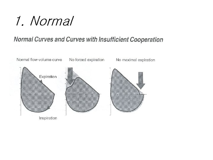
1. Normal
2. Obstruction
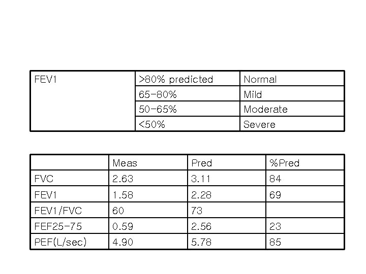
FEV 1 >80% predicted 65 -80% 50 -65% <50% Normal Mild Moderate Severe Meas Pred %Pred FVC 2. 63 3. 11 84 FEV 1 1. 58 2. 28 69 FEV 1/FVC 60 73 FEF 25 -75 0. 59 2. 56 23 PEF(L/sec) 4. 90 5. 78 85
3. Restriction
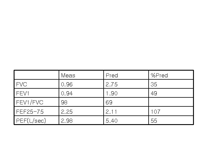
Meas Pred %Pred FVC 0. 96 2. 75 35 FEV 1 0. 94 1. 90 49 FEV 1/FVC 98 69 FEF 25 -75 2. 25 2. 11 107 PEF(L/sec) 2. 98 5. 40 55
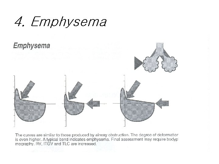
4. Emphysema
5. Upper airway obstruction
Interpretation algorithm