Special senses Audition 1 Audition Audition the sense


- Slides: 34

Special senses – Audition 1

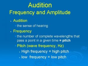
Audition �Audition, the sense of hearing, involves the transduction of sound waves into electrical energy, which then can be transmitted in the nervous system. �Sound is produced by waves of compression and decompression, which are transmitted in elastic media such as air or water. �These waves are associated with increases (compression) and decreases (decompression) in pressure. 2

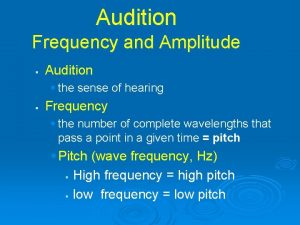
Sound waves �Frequency is measured in cycles per second (hertz, Hz). �Intensity is measured in decibels (d. B), a relative measure on a log scale. 3

Structure of the ear 4

Structure of the ear 1. Outer ear • consists of the pinna and the external auditory meatus (auditory canal). • directs the sound waves into the auditory canal. • It is air filled. 5

2. Middle ear • Is air filled • Contains the tympanic membrane and the auditory ossicles (malleus, incus and stapes). The stapes inserts into the oval window, a membrane between the middle ear and the inner ear. • Sound waves cause the tympanic membrane to vibrate. In turn, the ossicles vibrate, pushing the stapes into the oval window and displacing fluid in the inner ear. 6

3. Inner ear • Consists of: 1. a bony labyrinth, which consists of three semicircular canals (lateral, posterior and superior). 2. a membranous labyrinth, which consists of a series of ducts called the scala vestibuli, scala tympani, and scala media. • Is fluid filled • The fluid outside the ducts is perilymph; the fluid inside the ducts is endolymph. 7

�The cochlea and the vestibule are formed from the bony and membranous labyrinths. �The cochlea, which is a spiral-shaped structure composed of three tubular canals or ducts, contains the organ of Corti. • The organ of Corti contains the receptor cells and is the site of auditory transduction. 8

9

10

Inner ear �Structure of the cochlea: three tubular canals �(a) The scala vestibuli and scala tympani contain perilymph, which has a high [Na+] (similar to ECF). �(b) The scala media contains endolymph, which has a high [K+] and low [Na+]. • The scala media is bordered by the basilar membrane, which is the site of the organ of Corti. 11

Location and structure of the organ of Corti • The organ of Corti lies on the basilar membrane. • It contains the receptor cells (inner and outer hair cells) for auditory stimuli. Cilia protrude from the hair cells and are embedded in the tectorial membrane. • Inner hair cells are arranged in single rows and are few in number. • Outer hair cells are arranged in parallel rows and are greater in number than the inner hair cells. • The spiral ganglion contains the cell bodies of the auditory nerve [vestibulocochlear nerve, cranial 12

Steps in auditory transduction by the organ of Corti 13

�Sound waves are directed toward the tympanic membrane vibrates the ossicles vibrate and the stapes are pushed into the oval window this movement displaces fluid in the cochlea the sound energy is amplified by 1) the lever action of the ossicles and 2) the concentration of sound waves from the large tympanic membrane onto the small oval window. 14

1. Sound waves cause vibration of the organ of Corti. 2. The cell bodies of hair cells contact the basilar membrane, while their cilia are embedded in the tectorial membrane. Because the basilar membrane is more elastic than the tectorial membrane, vibration of the basilar membrane causes the hair cells to bend by a shearing force as they push against the tectorial membrane. 15

3. Bending of the cilia causes changes in K+ conductance of the hair cell membrane. • Bending in one direction causes an increase in K+ conductance and depolarization; bending in the other direction produces a decrease in K+ conductance and hyperpolarization. 4. The oscillating potential that results (receptor potentials of the auditory hair cells) is called the cochlear microphonic potential. 16

5. Depolarization opens voltage-gated Ca 2+ channels in the presynaptic terminals of the hair cells Ca 2+ enters the presynaptic terminals release of glutamate action potentials form in the afferent cochlear nerves transmit this information to the CNS. • When the hair cells are hyperpolarized, the opposite events occur, and there is decreased release of glutamate. 6. The oscillating potential of the hair cells causes intermittent firing of the cochlear nerves. 17

18

Central auditory pathways 19

� Fibers ascend through the lateral lemniscus to the inferior colliculus to the medial geniculate nucleus of the thalamus to the auditory cortex which is transverse temporal gyri in the temporal lobe � Fibers may be crossed or uncrossed. As a result, a mixture of ascending auditory fibers represents both ears at all higher levels. � The input from each ear is bilaterally represented in the ascending auditory system pathway at the level of the lateral lemniscus and above. Thus, the representation of auditory space is complex, even at the brainstem level. Consequently, unilateral deafness may occur with isolated lesions of the cochlear nuclei or more peripheral structures. Central lesions do not cause unilateral deafness, although they may interfere with overall sensitivity to speech or with sound localization. 20

�There is tonotopic representation of frequencies at all levels of the central auditory pathway. �Discrimination of complex features (e. g. , recognizing a patterned sequence) is a property of the cerebral cortex. 21

Vestibular system 22

Vestibular system �Is used to maintain equilibrium or balance by detecting angular and linear acceleration of the head. �Sensory information from the vestibular system is then used to provide a stable visual image for the retina (while the head moves) and to make the adjustments in posture that are necessary to maintain balance. 23

Structure of the vestibular organ 24

Structure of the vestibular organ �It is a membranous labyrinth within the bony labyrinth, consisting of three perpendicular semicircular canals, a utricle, and a saccule. • The semicircular canals are arranged perpendicular to each other and they detect angular or rotational acceleration. 25

Structure of the vestibular organ • Each semicircular canal, filled with endolymph, contains an enlargement at one end called an ampulla. • Each ampulla contains vestibular hair cells, which are covered with a gelatinous mass called a cupula. �The utricle and saccule detect changes in the position of the head with respect to gravity (linear acceleration). 26

Structure of the vestibular organ �The canals are filled with endolymph and are bathed in perilymph. �The receptors are hair cells located at the end of each semicircular canal. Cilia on the hair cells are embedded in a gelatinous structure called the cupula. A single long cilium is called the kinocilium; smaller cilia are called stereocilia. 27

Steps in vestibular transduction - angular acceleration 28

1. During counterclockwise (left) rotation of the head, the horizontal semicircular canal and its attached cupula also rotate to the left. Initially, the cupula moves more quickly than the endolymph fluid. Thus, the cupula is dragged through the endolymph; as a result, the cilia on the hair cells bend. 29

2. If the stereocilia are bent toward the kinocilium, the hair cell depolarizes (excitation). If the stereocilia are bent away from the kinocilium, the hair cell hyperpolarizes (inhibition). Ø Therefore, during the initial counterclockwise (left) rotation, the left horizontal canal is 30

3. After several seconds, the endolymph “catches up” with the movement of the head and the cupula. The cilia return to their upright position and are no longer depolarized or hyperpolarized. 31

4. When the head suddenly stops moving, the endolymph continues to move counterclockwise (left), dragging the cilia in the opposite direction. ØTherefore, if the hair cell was depolarized with the initial rotation, it now will hyperpolarize. If it was hyperpolarized initially, it now will depolarize. ØTherefore, when the head stops moving, the left horizontal canal will be inhibited and the right horizontal canal will be excited. 32

Structure of the vestibular organ 33

Central Vestibular Pathways � The vestibular afferent fibers project to the brain -stem through the vestibular nerve. The cell bodies of these afferent fibers are located in Scarpa's ganglion. The afferent fibers terminate in the vestibular nuclei, which are located in the rostral medulla and caudal pons. Afferent fibers from different parts of the vestibular apparatus end in different vestibular nuclei and also give off collaterals to the cerebellum. 34
 What is the difference between somatic and special senses
 Messiners
 Building vocabulary activity: the special senses
 Building vocabulary activity: the special senses
 Chapter 17 special senses answer key
 Somatic and special senses
 Special senses quiz
 Chapter 15 special senses
 The cones of the retina are coursera quiz answers
 Special senses anatomy
 Learning exercises chapter 11 medical terminology
 Special senses the eyes and ears
 Extraocular muscles
 Thermoreceptors
 Emmentropia
 Anatomy and physiology chapter 8 special senses
 The general and special senses chapter 9
 What are the special senses
 Signal conclusion
 Cn v test
 General and special senses
 Cranucle
 Modiolus
 5 basic tastes
 Dominant genetic variance
 Narrow sense heritability vs broad sense heritability
 12 guideposts michael shurtleff
 Blind orchestra auditions study
 Audition by michael shurtleff summary
 Audition script
 Adobe audition loops
 Audition cs
 Mock trial audition tips
 Adobe audition 4
 Adobe audition pitch shifter
