RUSH PROTOCOL Rapid Ultrasound for Shock and Hypotension



- Slides: 33
RUSH PROTOCOL Rapid Ultrasound for Shock and Hypotension
Ultrasound (US)-- “resuscitative. ” �Patients with hypotension or shock �Ultrasound is ideal for the evaluation of critically ill patients in shock, and ACEP guidelines �Direct visualization of pathology and differentiation of shock states. �The RUSH Protocol first introduced in 2006 by Weingart SD et al, and later published in 2009. It was designed to be a rapid and easy to perform US protocol (<2 min) by most emergency physicians.
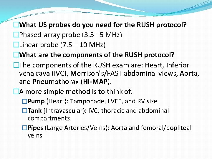
�What US probes do you need for the RUSH protocol? �Phased-array probe (3. 5 - 5 MHz) �Linear probe (7. 5 – 10 MHz) �What are the components of the RUSH protocol? �The components of the RUSH exam are: Heart, Inferior vena cava (IVC), Morrison’s/FAST abdominal views, Aorta, and Pneumothorax (HI-MAP). �A more simple method is to think of: �Pump (Heart): Tamponade, LVEF, and RV size �Tank (Intravascular): IVC, thoracic and abdominal compartments �Pipes (Large Arteries/Veins): Aorta and femoral/popliteal veins
Summary Table Resuscitation 2013 conference
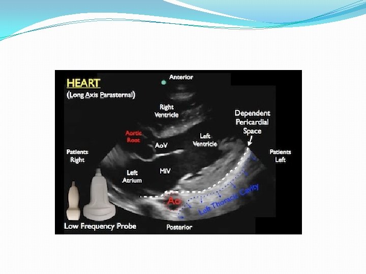
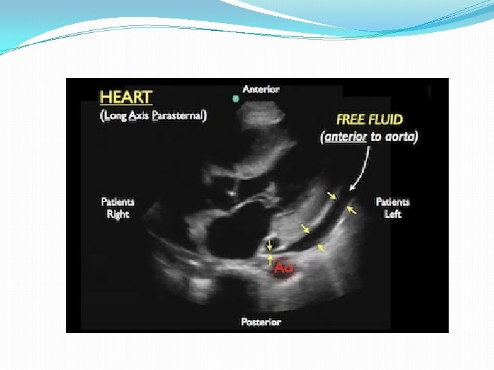
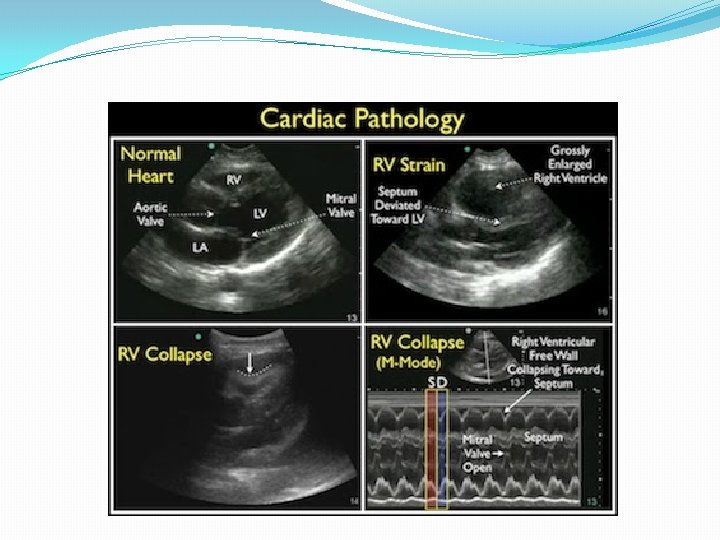
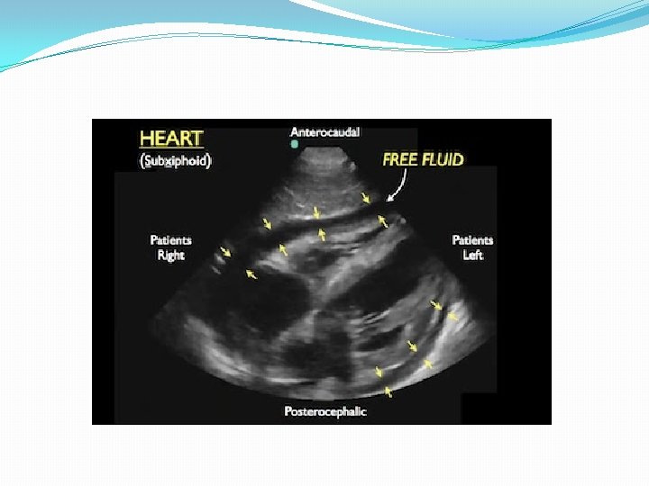
How do you evaluate the PUMP? �Component: Heart (parasternal long axis view) �Probe: Phased array probe (3. 5 - 5 MHz) �Location: Just left of the sternum, 3 rd and 4 th intercostal space �Finding: Pericardial effusion (tamponade) �Small effusions are best identified posterior to left ventricle (dependent portion of pericardium) �Can find compression of the right ventricle (Singh S et al Sens 92%, Spec 100%, PPV 100%)
�Finding: Left ventricular ejection fraction estimation �Look at anterior leaflet of mitral valve, which should normally touch septum �<30% difference of LV size between systole and diastole indicates severely decreased LV function �Finding: Right ventricular strain �Normally RV should be 60% of LV size (If RV = LV size, this is abnormal) �Lodato JC et al: If Mc. Connell Sign (reduction in RV free wall motility with sparing of the apex) is present, specificity for PE is 96%, but sensitivity is 16%.
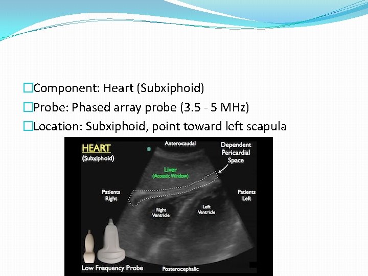
�Component: Heart (Subxiphoid) �Probe: Phased array probe (3. 5 - 5 MHz) �Location: Subxiphoid, point toward left scapula
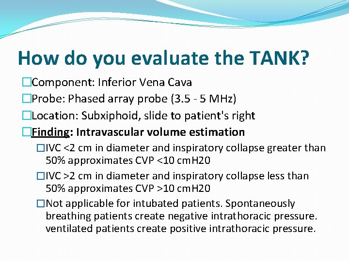
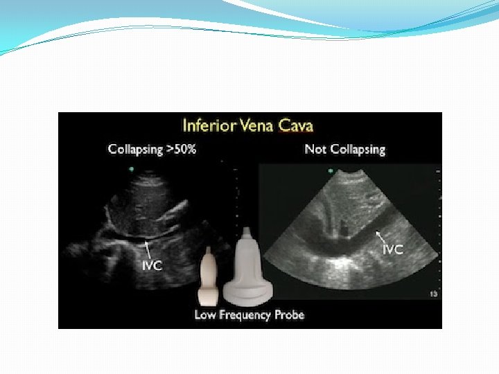
How do you evaluate the TANK? �Component: Inferior Vena Cava �Probe: Phased array probe (3. 5 - 5 MHz) �Location: Subxiphoid, slide to patient's right �Finding: Intravascular volume estimation �IVC <2 cm in diameter and inspiratory collapse greater than 50% approximates CVP <10 cm. H 20 �IVC >2 cm in diameter and inspiratory collapse less than 50% approximates CVP >10 cm. H 20 �Not applicable for intubated patients. Spontaneously breathing patients create negative intrathoracic pressure. ventilated patients create positive intrathoracic pressure.
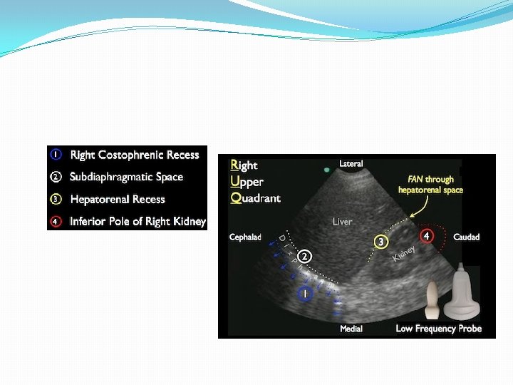
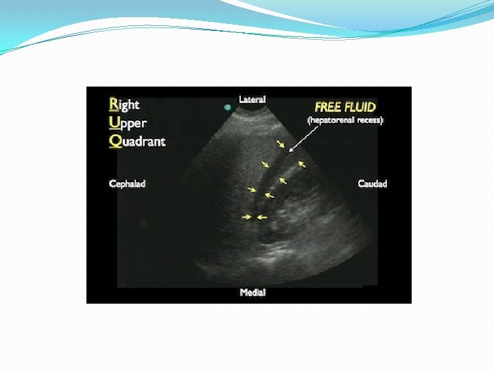
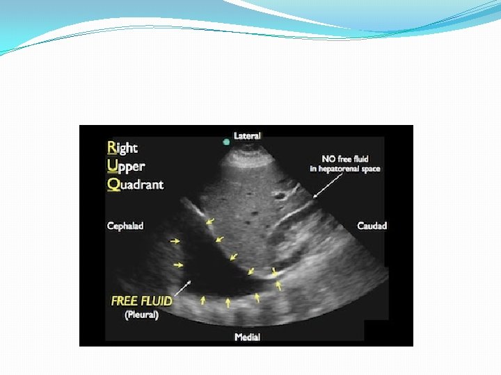
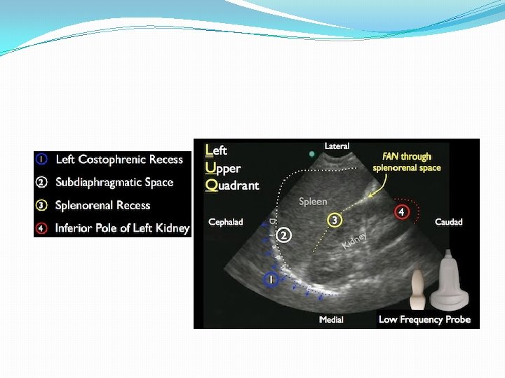
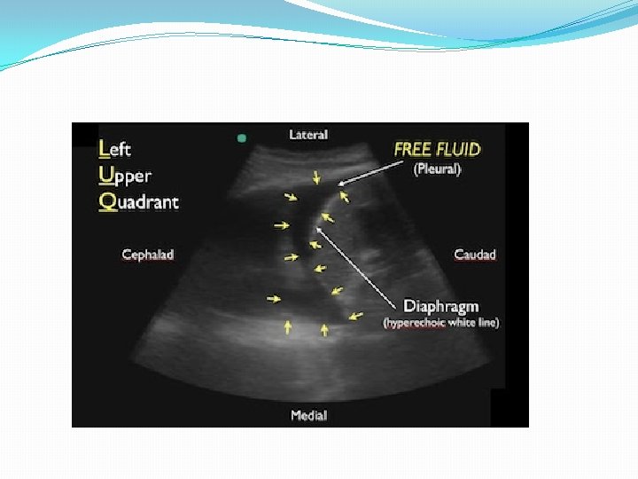
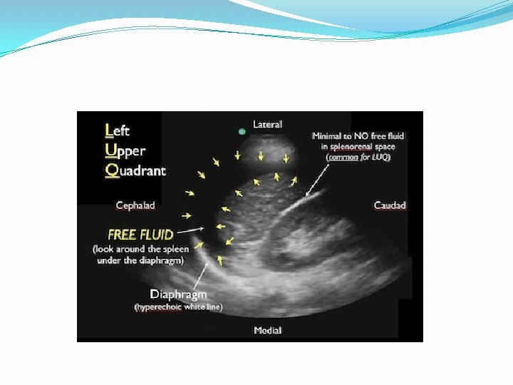
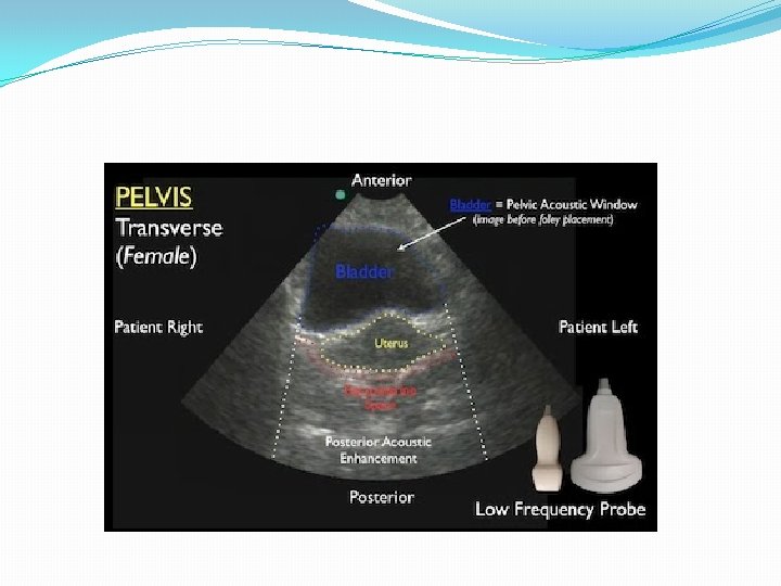
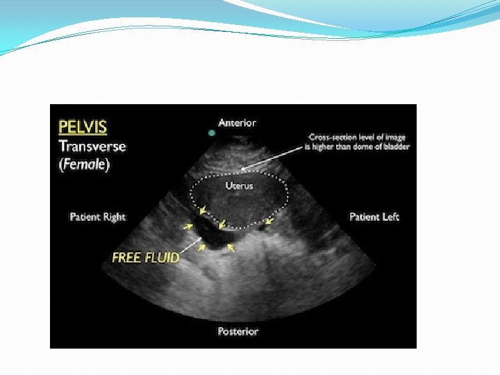
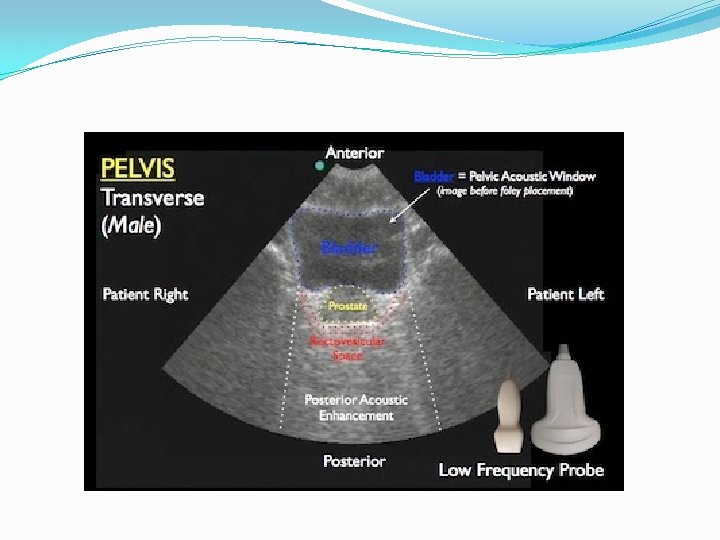
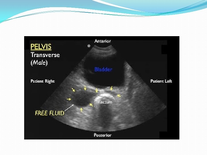
�Component: FAST abdominal views �Probe: Phased array probe (3. 5 - 5 MHz) �Location: Hepatorenal recess, Splenorenal recess, and bladder �Finding: Internal blood loss
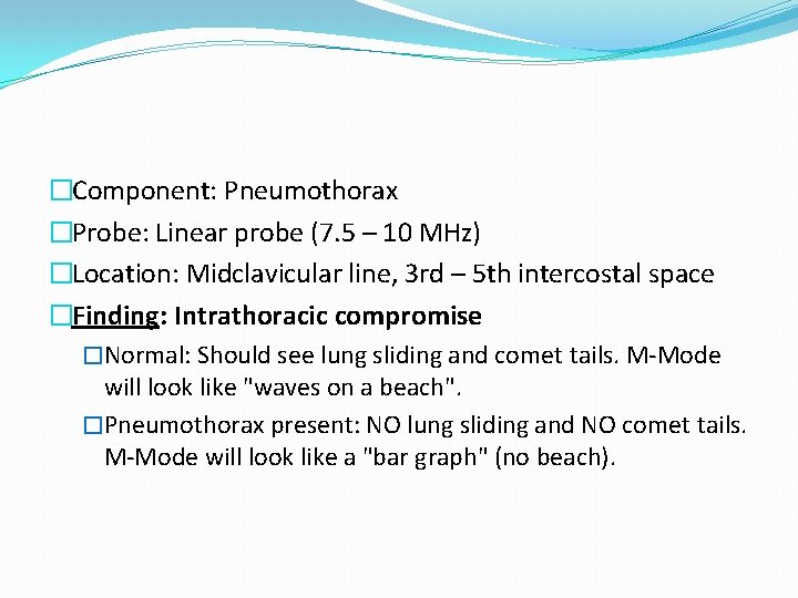
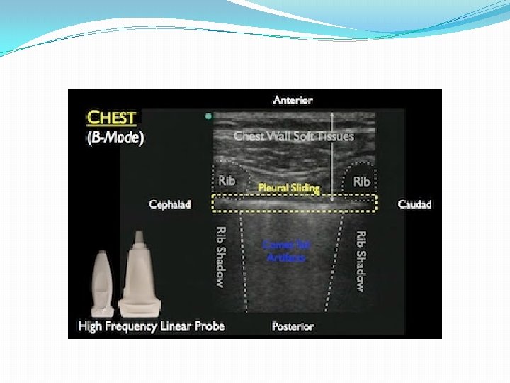
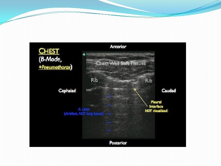
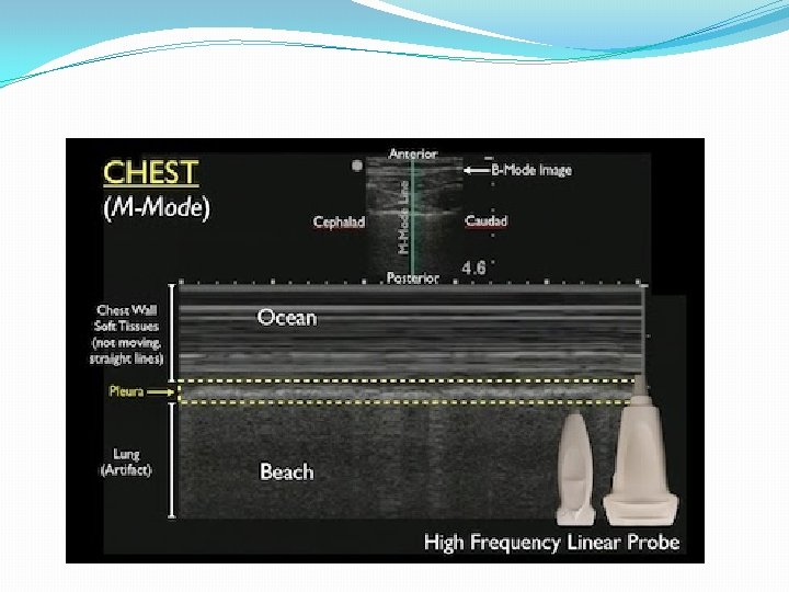
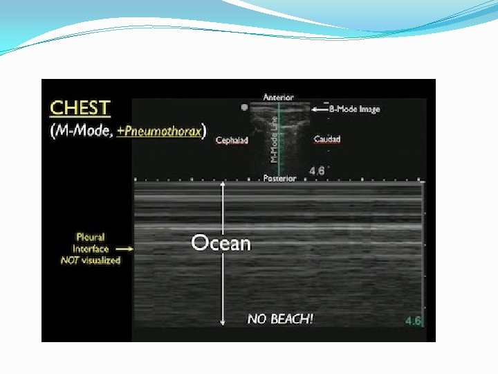
�Component: Pneumothorax �Probe: Linear probe (7. 5 – 10 MHz) �Location: Midclavicular line, 3 rd – 5 th intercostal space �Finding: Intrathoracic compromise �Normal: Should see lung sliding and comet tails. M-Mode will look like "waves on a beach". �Pneumothorax present: NO lung sliding and NO comet tails. M-Mode will look like a "bar graph" (no beach).
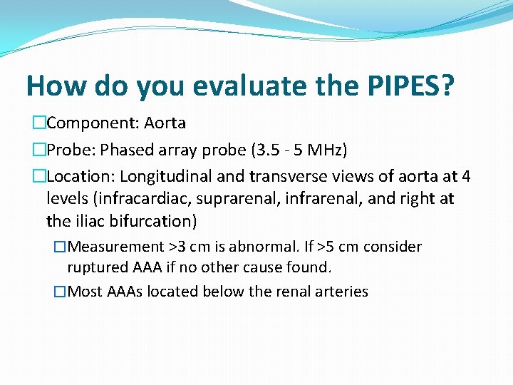
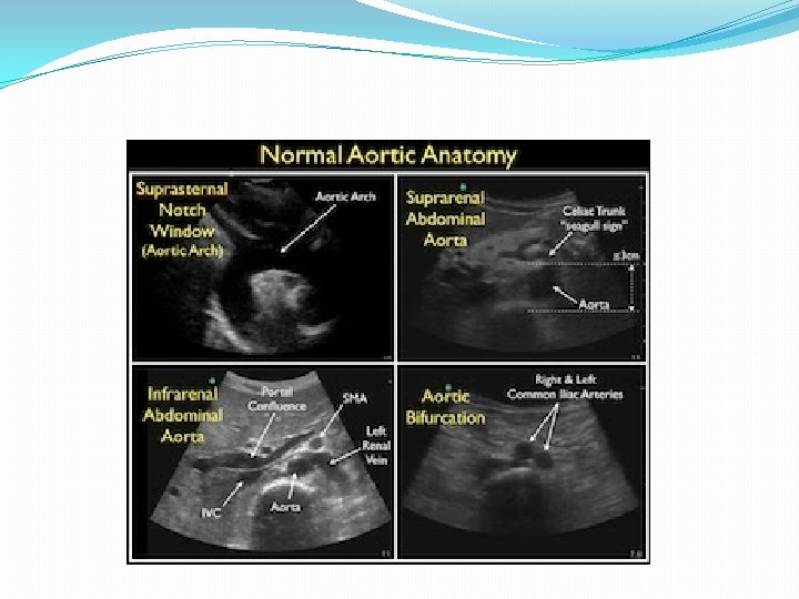
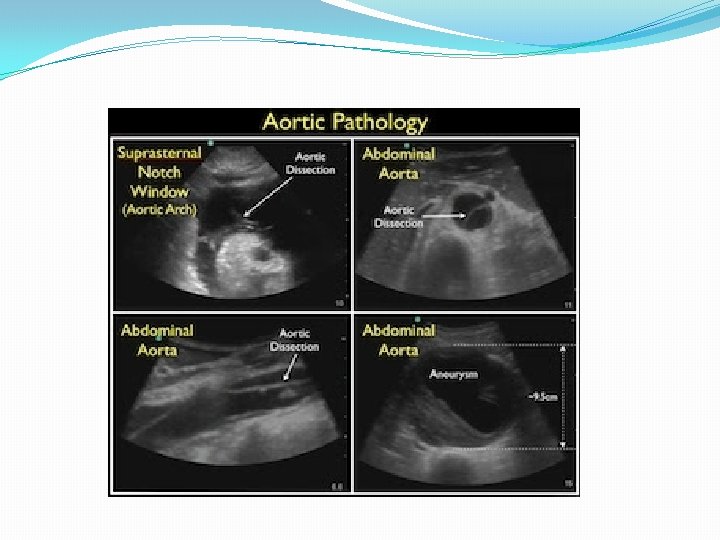
How do you evaluate the PIPES? �Component: Aorta �Probe: Phased array probe (3. 5 - 5 MHz) �Location: Longitudinal and transverse views of aorta at 4 levels (infracardiac, suprarenal, infrarenal, and right at the iliac bifurcation) �Measurement >3 cm is abnormal. If >5 cm consider ruptured AAA if no other cause found. �Most AAAs located below the renal arteries
RUSH protocol to medical patients EFAST exam to trauma patients.