
Mathematics in Molecular and Cellular Biology Many thanks
Mathematics in Molecular and Cellular Biology Many thanks to Martin Burger for inviting me to his ‘Swan Song’ in Linz! I am here for Wolfgang Nonner Many thanks to Mike Fill for help with slides 1
Multiscale Models of Nerve and Muscle also called Physiology of Nerve and Muscle 2
From Structure to Function By Fundamental Physical Laws 3
From Structure ANATOMY to Function PHYSIOLOGY using fundamental physical laws PHYSIOLOGY 4
What is Multiscale Analysis is also called Physiology 5

Multiscale MATHEMATICAL Analysis has rarely been possible until now 6
Multiscale MATHEMATICAL Analysis has rarely been possible until now Except for Nerve Cells Hodgkin, Huxley and Katz 7
Multi. Scale Analysis of Nerve Function is more complete than of ANY other cell/tissue in Biology Multiscale Mathematical Analysis is not available for any other tissue or cell, although many are working to change this! 8
From Anatomy to Physiology using Biophysics & Biochemistry From Structure to Function using Fundamental Physical Laws 9
PHYSIOLOGY of Nerve and Skeletal Muscle 10
Multiscale MATHEMATICAL Analysis of Muscle has not been attempted but structures are nearly all known now, so it may be possible 11
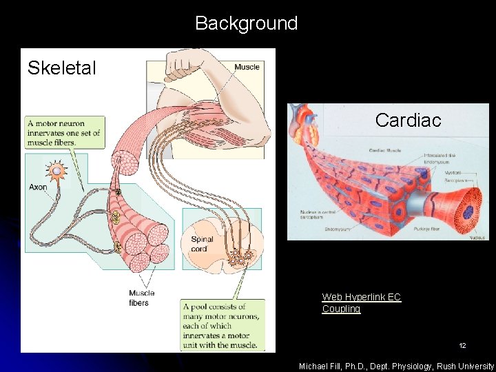
Background Skeletal Cardiac Web Hyperlink EC Coupling 12 Michael Fill, Ph. D. , Dept. Physiology, Rush University
Skeletal Muscle • Functional unit is the Motor Unit controlled by one neuron • Motor Unit has 1 to ~100 muscle cells called muscle fibers • Fundamental Contraction of fiber is Twitch • Twitch is fast and brief • Twitch is controlled by the action potential of the muscle fiber 13
Cardiac Muscle • Dreadful Design UNintelligent Design • Nervous control is indirect • Fundamental Contraction of fiber is long and slow • Fundamental Contraction is controlled by the action potential 14
Skeletal Muscle • • Excellent Design Intelligent Design Nervous control is direct Fundamental Contraction of fiber is short Fundamental Contraction is controlled by the action potential • Role of myelin • Why a synapse? • Penalty for synapse 15
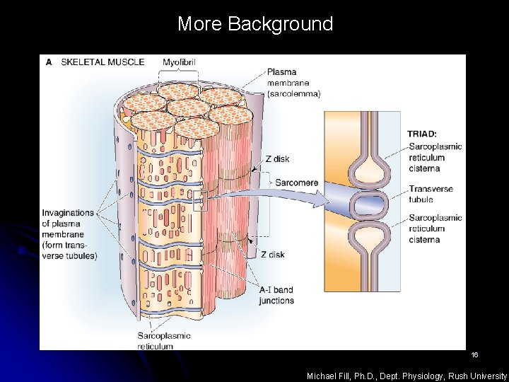
More Background 16 Michael Fill, Ph. D. , Dept. Physiology, Rush University
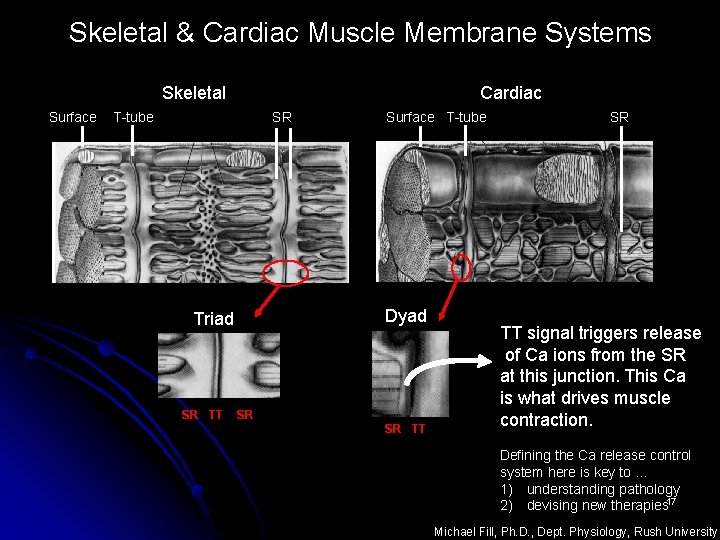
Skeletal & Cardiac Muscle Membrane Systems Skeletal Surface Cardiac T-tube SR Dyad Triad SR TT Surface T-tube SR SR TT signal triggers release of Ca ions from the SR at this junction. This Ca is what drives muscle contraction. Defining the Ca release control system here is key to … 1) understanding pathology 2) devising new therapies 17 Michael Fill, Ph. D. , Dept. Physiology, Rush University
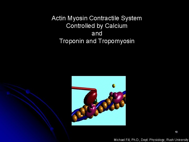
Actin Myosin Contractile System Controlled by Calcium and Troponin and Tropomyosin 18 Michael Fill, Ph. D. , Dept. Physiology, Rush University
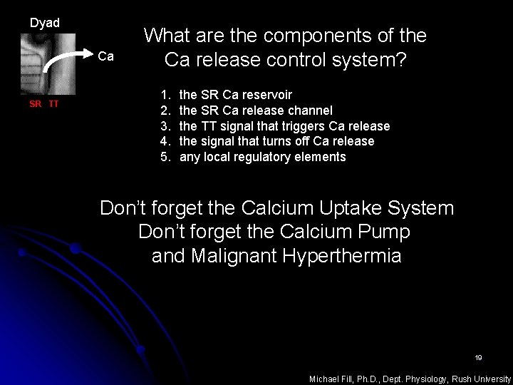
Dyad Ca SR TT What are the components of the Ca release control system? 1. 2. 3. 4. 5. the SR Ca reservoir the SR Ca release channel the TT signal that triggers Ca release the signal that turns off Ca release any local regulatory elements Don’t forget the Calcium Uptake System Don’t forget the Calcium Pump and Malignant Hyperthermia 19 Michael Fill, Ph. D. , Dept. Physiology, Rush University
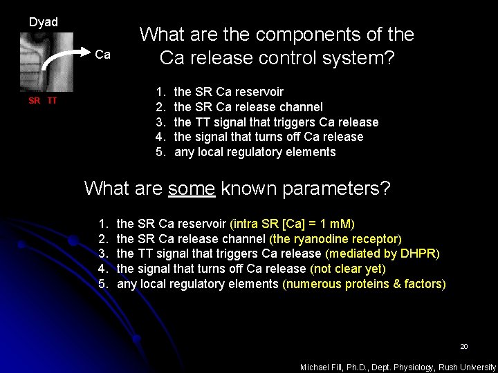
Dyad Ca What are the components of the Ca release control system? 1. 2. 3. 4. 5. SR TT the SR Ca reservoir the SR Ca release channel the TT signal that triggers Ca release the signal that turns off Ca release any local regulatory elements What are some known parameters? 1. 2. 3. 4. 5. the SR Ca reservoir (intra SR [Ca] = 1 m. M) the SR Ca release channel (the ryanodine receptor) the TT signal that triggers Ca release (mediated by DHPR) the signal that turns off Ca release (not clear yet) any local regulatory elements (numerous proteins & factors) 20 Michael Fill, Ph. D. , Dept. Physiology, Rush University
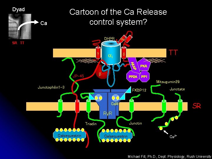
Dyad Cartoon of the Ca Release control system? Ca DHPR SR TT g a 1 a 2 TT d PP 2 A PP 1 P AKA PKA b JP-45 Mitsugumin 29 Junctophilin 1 -3 FKBP 12 Junctate SR Ca. M Ry. R Junctin Triadin Calsequestrin Ca 2+ Michael Fill, Ph. D. , Dept. Physiology, Rush University
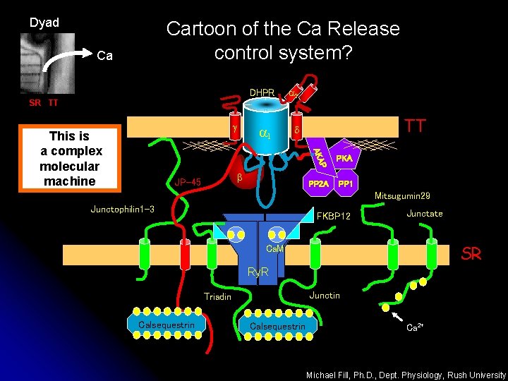
Dyad Cartoon of the Ca Release control system? Ca DHPR SR TT g a 1 TT d PKA PP 2 A PP 1 P AKA This is a complex molecular machine a 2 b JP-45 Mitsugumin 29 Junctophilin 1 -3 FKBP 12 Junctate SR Ca. M Ry. R Junctin Triadin Calsequestrin Ca 2+ Michael Fill, Ph. D. , Dept. Physiology, Rush University
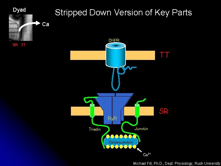
Dyad Stripped Down Version of Key Parts Ca DHPR SR TT TT SR Ry. R Junctin Triadin Calsequestrin Ca 2+ Michael Fill, Ph. D. , Dept. Physiology, Rush University
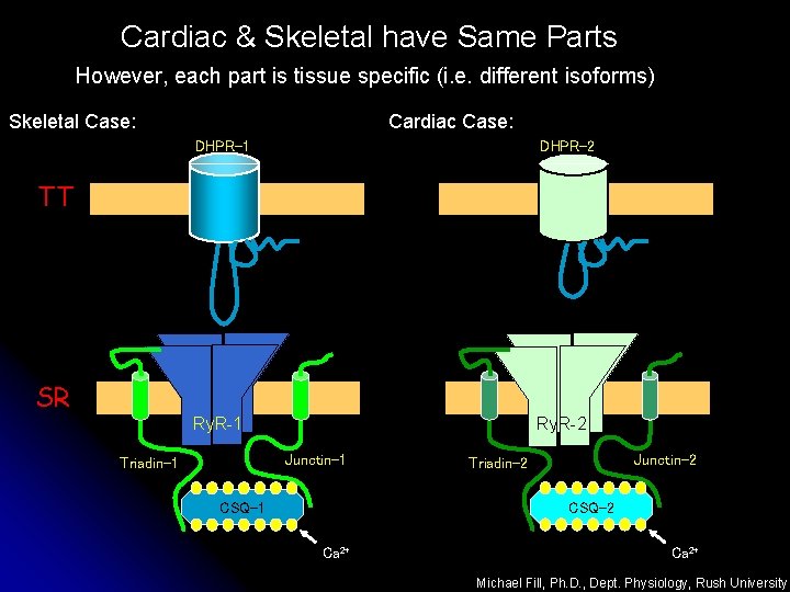
Cardiac & Skeletal have Same Parts However, each part is tissue specific (i. e. different isoforms) Cardiac Case: Skeletal Case: DHPR-1 DHPR-2 Ry. R-1 Ry. R-2 TT SR Junctin-1 Triadin-1 Junctin-2 Triadin-2 CSQ-1 Ca 2+ Michael Fill, Ph. D. , Dept. Physiology, Rush University
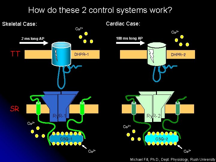
How do these 2 control systems work? Skeletal Case: Cardiac Case: Ca 2+ 100 ms long AP 2 ms long AP TT DHPR-1 DHPR-2 SR Ry. R-2 Ry. R-1 Ca 2+ CSQ-2 CSQ-1 Ca 2+ Michael Fill, Ph. D. , Dept. Physiology, Rush University
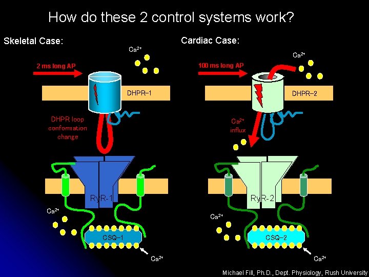
How do these 2 control systems work? Skeletal Case: Cardiac Case: Ca 2+ 100 ms long AP 2 ms long AP DHPR-1 DHPR-2 DHPR loop conformation change Ca 2+ influx Ry. R-2 Ry. R-1 Ca 2+ CSQ-2 CSQ-1 Ca 2+ Michael Fill, Ph. D. , Dept. Physiology, Rush University
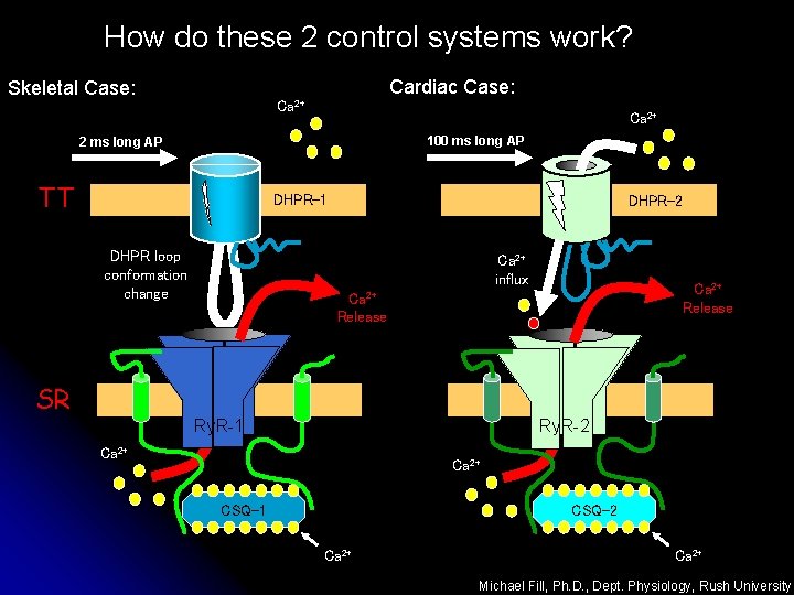
How do these 2 control systems work? Skeletal Case: Cardiac Case: Ca 2+ 100 ms long AP 2 ms long AP TT DHPR-1 DHPR loop conformation change DHPR-2 Ca 2+ influx Ca 2+ Release SR Ry. R-2 Ry. R-1 Ca 2+ CSQ-2 CSQ-1 Ca 2+ Michael Fill, Ph. D. , Dept. Physiology, Rush University
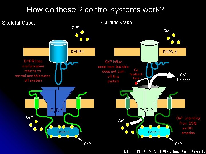
How do these 2 control systems work? Skeletal Case: Cardiac Case: Ca 2+ DHPR-1 DHPR loop conformation returns to normal and this turns off system DHPR-2 Ca 2+ influx ends here but this Ca does not turn feedback off this here system Ca 2+ Release Ry. R-2 Ry. R-1 Ca 2+ CSQ-2 CSQ-1 Ca 2+ unbinding from CSQ as SR empties Ca 2+ Michael Fill, Ph. D. , Dept. Physiology, Rush University
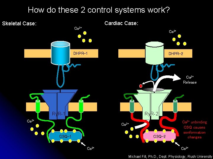
How do these 2 control systems work? Skeletal Case: Cardiac Case: Ca 2+ DHPR-1 DHPR-2 Ca 2+ Release Ry. R-2 Ry. R-1 Ca 2+ CSQ-2 CSQ-1 Ca 2+ unbinding CSQ causes sonformation changes Ca 2+ Michael Fill, Ph. D. , Dept. Physiology, Rush University
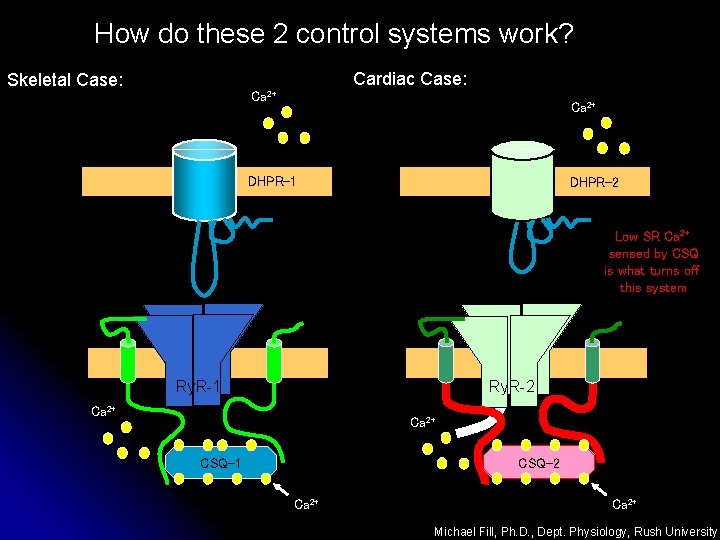
How do these 2 control systems work? Skeletal Case: Cardiac Case: Ca 2+ DHPR-1 DHPR-2 Low SR Ca 2+ sensed by CSQ is what turns off this system Ry. R-2 Ry. R-1 Ca 2+ CSQ-2 CSQ-1 Ca 2+ Michael Fill, Ph. D. , Dept. Physiology, Rush University
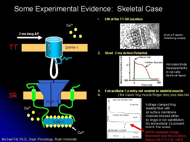
Some Experimental Evidence: Skeletal Case • EM of the TT-SR jucntion Ca 2+ 2 ms long AP (from a Franzini. Armstrong review) TT DHPR-1 2. Short 2 ms Action Potential microelectrode measurements in rat cells (text book figure) 3. 4. SR Ry. R-1 Extracellular Ca entry not needed in skeletal muscle ( the classic frog muscle Ringer story plus data like b Voltage clamped frog skeletal fiber with all surface membrane channels blocked either by drugs or ion substitution. No extracellular Ca present. Ca 2+ CSQ-1 (from E. Rios review) Ca 2+ Michael Fill, Ph. D. , Dept. Physiology, Rush University DHPR-mediated charge Movement (see Rios & Brum, Nature 325: 717 -720, 1987)
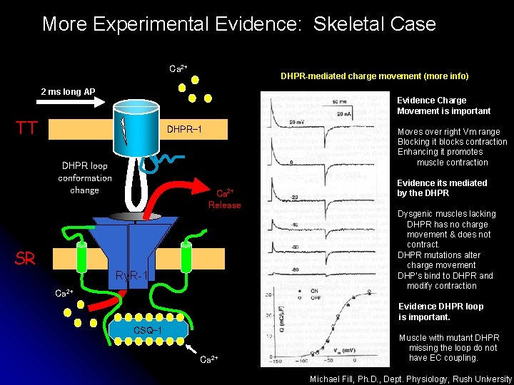
More Experimental Evidence: Skeletal Case Ca 2+ DHPR-mediated charge movement (more info) 2 ms long AP Evidence Charge Movement is important TT DHPR-1 DHPR loop conformation change Moves over right Vm range Blocking it blocks contraction Enhancing it promotes muscle contraction Ca 2+ Release SR Ry. R-1 Ca 2+ Evidence its mediated by the DHPR Dysgenic muscles lacking DHPR has no charge movement & does not contract. DHPR mutations alter charge movement DHP’s bind to DHPR and modify contraction Evidence DHPR loop is important. CSQ-1 Ca 2+ Muscle with mutant DHPR missing the loop do not have EC coupling. Michael Fill, Ph. D. , Dept. Physiology, Rush University
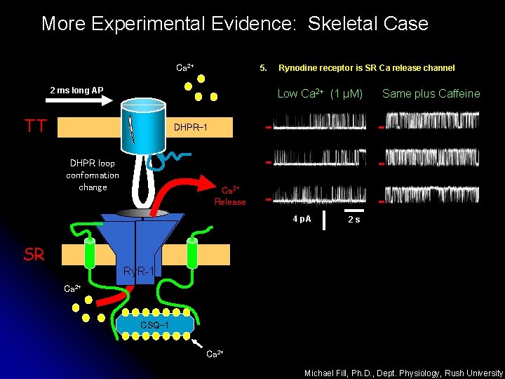
More Experimental Evidence: Skeletal Case Ca 2+ 5. 2 ms long AP Rynodine receptor is SR Ca release channel Low Ca 2+ (1 µM) TT Same plus Caffeine DHPR-1 DHPR loop conformation change Ca 2+ Release 4 p. A 2 s SR Ry. R-1 Ca 2+ CSQ-1 Ca 2+ Michael Fill, Ph. D. , Dept. Physiology, Rush University
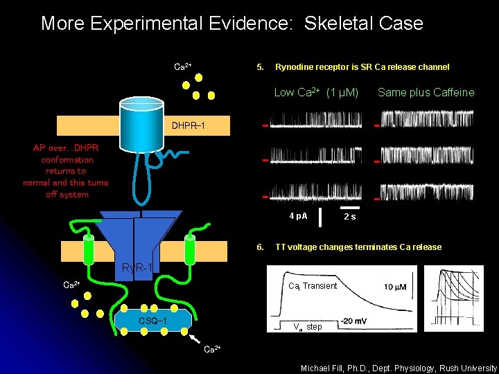
More Experimental Evidence: Skeletal Case Ca 2+ 5. Rynodine receptor is SR Ca release channel Low Ca 2+ (1 µM) Same plus Caffeine DHPR-1 AP over. . DHPR conformation returns to normal and this turns off system 4 p. A 6. 2 s TT voltage changes terminates Ca release Ry. R-1 Ca 2+ Cai Transient CSQ-1 Vm step Ca 2+ Michael Fill, Ph. D. , Dept. Physiology, Rush University
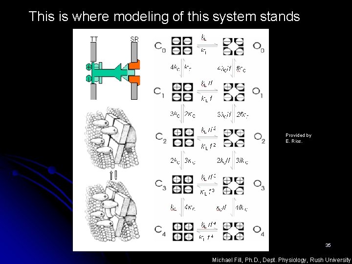
This is where modeling of this system stands Provided by E. Rios. 35 Michael Fill, Ph. D. , Dept. Physiology, Rush University
- Slides: 35