RENAL SYSTEM ASSESSMENT PYRAMID POINTS Renal and urinary


- Slides: 40

RENAL SYSTEM ASSESSMENT

PYRAMID POINTS • • • Renal and urinary tract assessment techniques Risk factors associated with renal disorders Normal renal function test results Procedure for collecting a 24 -hour urine test Preprocedure and postprocedure care for an intravenous pyelogram (IVP) and renal angiogram • Preprocedure care for a client scheduled for a cystoscopy and biopsy • Expected and unexpected findings in the postprocedure period following cystoscopy and biopsy

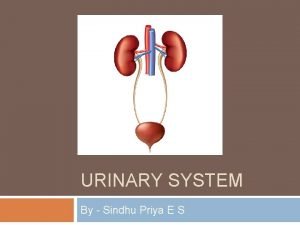
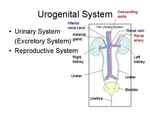
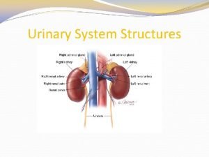
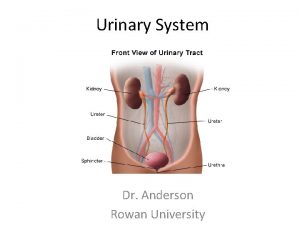
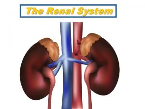
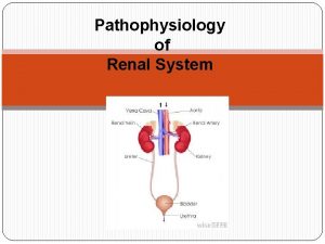
RENAL SYSTEM From Herlihy, B. & Maebius, N. (2000). The human body in health and illness. Philadelphia: W. B. Saunders.

LOCATION OF COSTOVERTEBRAL ANGLE From Beare PG, Myers JL (1998): Adult Health Nursing, ed. 3 St. Louis: Mosby.

RISK FACTORS OF RENAL DISORDERS • • Associated medical conditions Contact sports Family history of renal disease Frequent urinary tract infections High-sodium diet History of hypertension Medication use Trauma and injury

NORMAL RENAL FUNCTION TESTS • • • Blood urea nitrogen (BUN), 8 to 25 mg/dl Serum creatinine, 0. 6 to 1. 3 mg/dl Creatinine clearance, 100 to 120 ml/minute Uric acid serum, 2. 5 to 8. 0 ng/dl Uric acid urine, 250 to 750 mg/24 hours

URINALYSIS • DESCRIPTION – A urine test for evaluation of the renal system and for determining renal disease • IMPLEMENTATION – Wash perineal area and use a clean container – Obtain 10 to 15 ml of the first morning sample – Note that refrigerated samples may alter the specific gravity – If the client is menstruating, indicate this on the laboratory requisition form

SPECIFIC GRAVITY DETERMINATION • DESCRIPTION – A urine test that measures the kidney’s ability to concentrate urine – Normal value is 1. 016 to 1. 022 – An increase in specific gravity (more concentrated urine) occurs with insufficient fluid intake, decreased renal perfusion, or the presence of ADH – A decrease in specific gravity (less concentrated urine) occurs with increased fluid intake, diuretic administration, and diabetes insipidus

SPECIFIC GRAVITY DETERMINATION • IMPLEMENTATION – Can be measured by multiple-test dipstick (most common method), refractometer (an instrument used in the laboratory setting), or by a urinometer (least accurate method) – Factors that interfere with an accurate reading include radiopaque contrast agents, glucose, and proteins – Cold specimens may produce a false high reading

URINE CULTURE AND SENSITIVITY • DESCRIPTION – A urine test that identifies the presence of microorganisms and determines the specific antibiotics that will appropriately treat the existing microorganism

URINE CULTURE AND SENSITIVITY • IMPLEMENTATION – Clean perineal area and urinary meatus with bacteriostatic solution – Collect midstream sample in a sterile container – Send the collected specimen to the laboratory immediately – Note that urine from the client who forced fluids may be too dilute to provide a positive culture – Identify any sources of potential contaminants during the collection of the specimen, such as the hands, skin, clothing, hair, or vaginal or rectal secretions

CREATININE CLEARANCE TEST • DESCRIPTION – A blood and timed urine specimen that evaluates kidney function – Blood is drawn at the start of the test and the morning of the day that the 24 -hour urine specimen collection is complete

CREATININE CLEARANCE TEST • IMPLEMENTATION – Encourage adequate fluids before and during the test – Instruct the client to avoid tea, coffee, and medications during testing – If the client is taking corticosteroids or thyroid medication, check with the physician regarding the administration of these medications during testing – Maintain the specimen on ice or refrigerate, and check with the laboratory regarding the addition of a preservative to the specimen during collection

VANILLYLMANDELIC ACID (VMA) TEST • DESCRIPTION – A 24 -hour urine collection to diagnose pheochromocytoma, a tumor of the adrenal gland – The test identifies an assay of urinary catecholamines in the urine

VANILLYLMANDELIC ACID (VMA) TEST • IMPLEMENTATION – Instruct the client to avoid foods such as caffeine, cocoa, vanilla, cheese, gelatin, licorice, and fruits for at least 2 days prior to beginning the urine collection and during the collection, and to avoid taking medications for 2 to 3 days prior to beginning the test as prescribed – Instruct the client to avoid stress and to maintain adequate food and fluids during the test

VANILLYLMANDELIC ACID (VMA) TEST • IMPLEMENTATION – Save all urine, label the container, add preservative, and place the specimen on ice or refrigerate – Check with the laboratory regarding medication restrictions

URIC ACID TEST • DESCRIPTION – A 24 -hour urine collection to diagnose gout and kidney disease • IMPLEMENTATION – Encourage fluids and a regular diet during testing – Place the specimen on ice or refrigerate, and check with the laboratory regarding the addition of a preservative

KIDNEYS, URETERS, AND BLADDER RADIOGRAPH • DESCRIPTION – Known as a KUB – An x-ray film that views the urinary system and adjacent structures – Used to detect urinary calculi • IMPLEMENTATION – There is no specific preparation

BLADDER ULTRASONOGRAPHY • DESCRIPTION – A noninvasive method of measuring the volume of urine in the bladder – May be performed for evaluating urinary frequency or inability to urinate • IMPLEMENTATION – There is no specific preparation

INTRAVENOUS PYELOGRAM (IVP) • DESCRIPTION – The injection of a radiopaque dye that outlines the renal system – Performed to identify abnormalities in the system

INTRAVENOUS PYELOGRAM (IVP) • PREPROCEDURE – Obtain a consent form – Assess the client for allergies to iodine, seafood, and radiopaque dyes – Withhold food and fluids after midnight on the night before the test – Inform the client about possible throat irritation, flushing of the face, warmth, or a salty taste that may be experienced during the test

INTRAVENOUS PYELOGRAM (IVP) • POSTPROCEDURE – Monitor vital signs – Instruct the client to drink at least 1 liter of fluid unless contraindicated – Assess the venipuncture site for bleeding – Monitor urinary output

RENAL ANGIOGRAPHY • DESCRIPTION – The injection of a radiopaque dye through a catheter for examination of the renal arterial supply

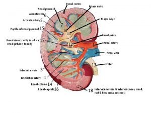
RENAL ARTERY AND OTHER INTERNAL STRUCTURES OF THE KIDNEY From Herlihy, B. & Maebius, N. (2000). The human body in health and illness. Philadelphia: W. B. Saunders.

RENAL ANGIOGRAPHY • PREPROCEDURE – Obtain a consent form – Assess the client for allergies to iodine, seafood, and radiopaque dyes – Inform the client about a possible burning feeling or a feeling of heat along the vessel when the dye is injected – Withhold food and fluids after midnight on the night before the test

RENAL ANGIOGRAPHY • PREPROCEDURE – Instruct the client to void immediately before the procedure – Administer enemas as prescribed – Shave injection sites as prescribed – Assess and mark the peripheral pulses

RENAL ANGIOGRAPHY • POSTPROCEDURE – Assess vital signs and peripheral pulses – Provide bed rest and use of a sandbag at the insertion site for 4 to 8 hours – Assess the color and temperature of the involved extremity – Inspect the catheter insertion site for bleeding or swelling – Force fluids unless contraindicated – Monitor urinary output

RENAL SCAN • DESCRIPTION – An IV injection of a radioisotope for visual imaging of renal blood flow

RENAL SCAN • PREPROCEDURE – Obtain a consent form – Assess for allergies – Assist with administering radioisotope as necessary – Instruct the client that he or she will be required to remain motionless – Instruct the client that imaging may be repeated at various intervals before the test is complete

RENAL SCAN • POSTPROCEDURE – Encourage fluids unless contraindicated – Assess the client for signs of delayed allergic reaction, such as itching and hives – Note that the radioactivity is eliminated in 24 hours – Follow standard precautions when caring for incontinent clients and double-bag client linens per agency policy

CYSTOMETROGRAM (CMG) • DESCRIPTION – A graphic recording of the pressures exerted at varying phases of the bladder • PREPROCEDURE – Inform the client of the voiding requirements during the procedure • POSTPROCEDURE – Monitor the client’s voiding after the procedure

CYSTOSCOPY AND BLADDER BIOPSY • DESCRIPTION – The bladder mucosa is examined for inflammation, calculi, or tumors by means of a cystoscope – Tissue may be obtained for biopsy

CYSTOSCOPE IN THE MALE BLADDER From Phipps WJ, Sands, J, Marek JF: Medical-surgical nursing: concepts and clinical practice, ed. 6, St. Louis, 1999, Mosby.

CYSTOSCOPY AND BLADDER BIOPSY • PREPROCEDURE – Obtain a consent form – Withhold food and fluids after midnight on the night before the test – Administer enemas and medications as prescribed

CYSTOSCOPY AND BLADDER BIOPSY • POSTPROCEDURE – Monitor vital signs – Monitor for postural hypotension – Force fluids as prescribed – Monitor intake and output – Encourage deep-breathing exercises to relieve bladder spasms – Administer analgesics as prescribed – Administer sitz baths for back and abdominal pain

CYSTOSCOPY AND BLADDER BIOPSY • POSTPROCEDURE – Note that leg cramps are common due to the lithotomy position maintained during the procedure – Assess the urine for color and consistency – Note that pink-tinged or tea-colored urine is common – Monitor for bright red urine or clots and notify the physician if this occurs

RENAL BIOPSY • DESCRIPTION – Insertion of a needle into the kidney to obtain a sample of tissue for examination

RENAL BIOPSY From Black, J. , Hawks, J, and Keene, A. (2001). Medical-surgical nursing, ed 6, Philadelphia: W. B. Saunders

RENAL BIOPSY • PREPROCEDURE – Assess vital signs – Assess baseline clotting studies – Obtain a consent form – Withhold food and fluids after midnight on the night before the test • DURING THE PROCEDURE – Position the client prone with a pillow under the abdomen and shoulders

RENAL BIOPSY • POSTPROCEDURE – Monitor vital signs – Monitor hemoglobin and hematocrit – Place the client in the supine position and on bed rest for 8 hours as prescribed – Provide pressure to the biopsy site for 30 minutes – Check the biopsy site for bleeding – Force fluids to 1500 to 2000 ml as prescribed – Instruct the client to avoid heavy lifting and strenuous activity for 2 weeks
 Ira pré renal renal e pós renal
 Sindrome nefrótica
 Renal corpuscle
 Lymphatic system and urinary system
 Renal pyramid
 Calyx minor
 Apex of renal pyramid
 Detrusal
 Bullseye brand positioning
 Point of difference and point of parity
 Physiology of urine formation
 Chapter 15 urinary system
 Polyrrhagia
 Interesting facts about the urinary system
 Pig reproductive anatomy
 Chapter 30 the urinary system
 Kidney pyramid labeled
 Chapter 20 urinary/excretory system
 Urinary system model
 Urinary system fun fact
 Alimentary canal
 Urinary system powerpoint
 Proximal convoluted tubule histology
 Function of antidiuretic hormone
 Urinary system histology
 Figure 15-4 is a diagram of a nephron
 Prone
Prone Defination of urine
 Excretory system
 Endocrine system of rat
 Rat urinary system
 Vena cava excretory system
 Kidneys location and structure figure 15-2
 Macula densa cells
 Renal tubule
 Urinary system
 Normal constituents of urine
 What is this called
 Normal and abnormal constituents of urine
 Layers of kidney
 Urinary system
