Phylogenetic tree of the major lineages phyla of
 of Bacteria based on 16 S ribosomal")
 • • • • • Purple phototrophic Bacteria The nitrifying")


")


 Found in the intestines of humans and animals, this bacterium is")

 versus Escherichia coli versus")
 The dynamic plague bacterium evolved from harmless stomach bug to blood-borne")
 This pathogen infects potatoes throughout the world. It causes the diseases")
 The bacterium that causes cholera, unlike other pathogens, has not one")


 Found in soil worldwide, this bacterium infects the roots")
- Slides: 34
Phylogenetic tree of the major lineages (phyla) of Bacteria based on 16 S ribosomal RNA sequence comparisons
Phylum 1: Proteobacteria (2086) • • • • • Purple phototrophic Bacteria The nitrifying Bacteria Sulphur and iron oxidizing Bacteria Hydrogen oxidizing Bacteria Methanotrophs and methylotrophs Pseudomonas and the Pseudomonads Acetic acid Bacteria Free living aerobic nitrogen fixing Bacteria Neisseria and Chromobacterium Enteric Bacteria Vibrio and photobacterium Rickettsia Spirilla Sheathed proteobacteria Budding and prosthecate/stalked Bacteria Gliding Myxobacteria Sulphate and sulphur reducing proteobacteria
Non-photosynthetic proteobacteria
Light microscopy of stained bacteria Escherichia coli, gram negative, glucose fermenting facultative aerobe TEM of E. coli with intact flagella. Courtesy of Peggy S. Hayes, Centers for Disease Control http: //www. geocities. com/Cape. Canaveral/3504/gallery. htm
Fermentative rods and vibrios Enteric bacteria Vibrio and related genera Pasteurellea and Hemophilus Zymomonas Chromobacterium
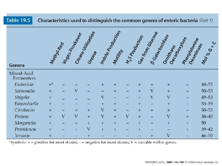
Enteric bacteria Relatedness among the enteric bacteria. Numbers indicate DNA/DNA reassociation values, a measure of the relatedness among the various genera.
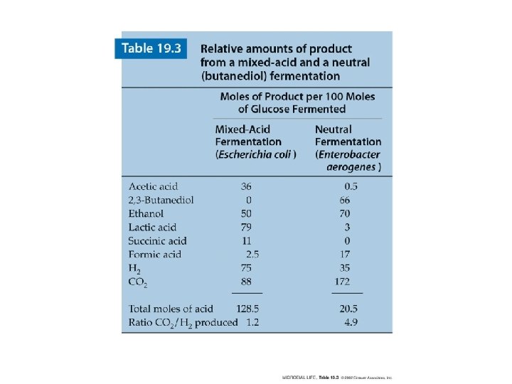
Mixed-acid fermentation of glucose by Escherichia coli. Pyruvate produced by glycolysis (the Embden-Meyerhof pathway) is catabolized to a mixture of products, high in acids. Acids are shown here and in Figure 19. 3 in their nonionized form.
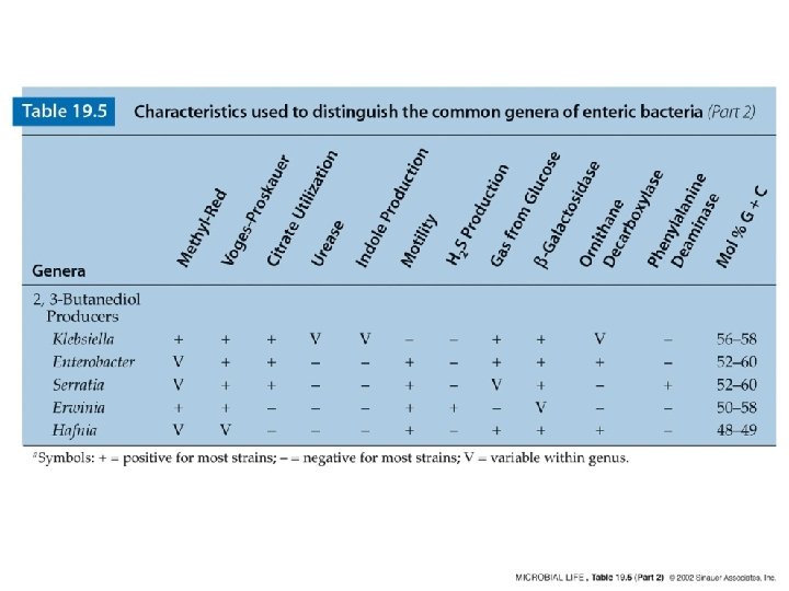
2, 3 -Butanediol fermentation In the butanediol variation of mixed-acid fermentation, more neutral products are formed, as well as a higher ratio of CO 2 to H 2.
bio. Merieux API tests API 20 E Species/subspecies identification of Enterobacteriacae and group/species identification of non-fermenting gramnegative bacteria These identification products can identify a wide variety of microorganisms. As a standard in microbiology, API's give accurate identification results, have extensive databases and are standardized, easy-to-use test systems. The products comprise strips that generally contain 20 miniature biochemical tests. Currently, there are 16 identification products covering almost all bacterial groups and over 550 different species. Identification is now quick, safe and easy.
The human digestive system
Escherichia coli (Bacteria) Found in the intestines of humans and animals, this bacterium is usually harmless, but some strains can cause food poisoning and more serious illnesses. Most outbreaks involve contaminated beef that was not cooked thoroughly. The strain known as O 157: H 7 is considered a potential biological weapon. » Sequenced by: University of Wisconsin E. coli K-12 Abstract University of Wisconsin E. coli O 157: H 7 EDL 933 Abstract Genome Information Research Center E. coli O 157: H 7 Abstract University of Wisconsin E. coli UPEC-CFT 073 Abstract » Related GNN article: Researchers sequence strain of E. coli linked to disease » Image: Courtesy The Donnenberg Laboratory.
Fig. 1. Scanning electron micrograph showing microcolonies of EPEC displaying the characteristic localized adherence pattern of adherence to HEp-2 cells. Fig. 2. High power scanning electron micrograph of EPEC displaying localized adherence to HEp-2 cells. Note the elongated microvilli to which the bacteria appear to attach.
This screenshot shows the complete genome of Yersinia pestis (top) versus Escherichia coli versus Salmonella typhimurium (bottom). The comparisons were done with tblastx.
Yersinia pestis (Bacteria) The dynamic plague bacterium evolved from harmless stomach bug to blood-borne pathogen in a relatively short amount of time. Y. pestis is transmitted to humans by fleas that feed on the blood of infected animals such as rats, causing bubonic plague. When transmitted by air through respiratory droplets, the bacterium causes the more severe and often fatal pneumonic plague. » Sequenced by: Sanger Institute Y. pestis CO 92 Abstract University of Wisconsin Y. pestis KIM Abstract
Erwinia carotovora (Bacteria) This pathogen infects potatoes throughout the world. It causes the diseases “blackleg” in developing tubers and “soft rot” in harvested potatoes. Infected potatoes have mushy spots with black borders and can have a foul odor. » Sequenced in 2004 by The Wellcome Trust Sanger Institute E. carotovora subsp. atroseptica Abstract » Image: Courtesy Gary D. Kleinschmidt, University of California Statewide IPM Program
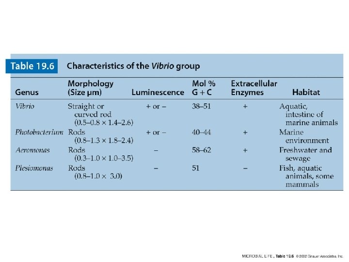
Vibrio cholerae (Bacteria) The bacterium that causes cholera, unlike other pathogens, has not one but two chromosomes. This may give it a competitive advantage in diverse environments. The rod-shaped bacterium was given the Latin name Vibrio because it possesses a flagellum and appears to vibrate. Vibrio parahaemolyticus (Bacteria) V. parahaemolyticus causes food-borne gastroenteritis, which inflames the stomach and small intestine. Most people recover completely from the illness by drinking plenty of fluids. The bacterial pathogen is a growing concern in North America, particularly in places where seafood is popular. Vibrio vulnificus (Bacteria) Found in warm coastal waters, this bacterium is related to the cholera pathogen and can cause a severe and potentially fatal illness. Infections tend to occur through eating raw or improperly cooked shellfish, particularly oysters, or exposing open wounds to seawater containing the organism
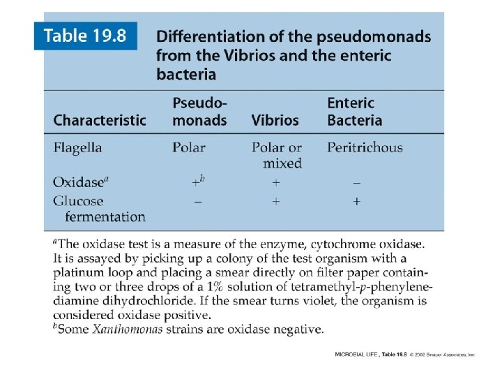
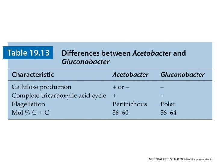
Oxidative rods and cocci Pseudomonads Azotobacter and other free living nitrogen fixing bacteria Rhizobium and Bradyrhizobium: Symbiotic nitrogen fixing bacteria Agrobacterium Vinegar bacteria: Acetobacter and Gluconobacter Legionella Neisseria and related bacteria Rickettsia Pseudomonas aeruginosa
Figure 19. 6 Pseudomonas
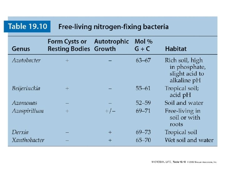
Figure 19. 10 Azotobacter
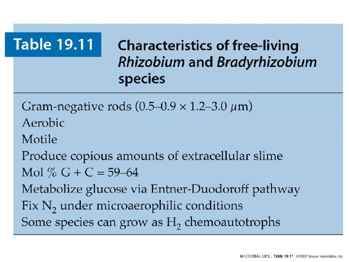
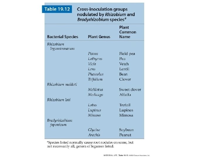
Development of root nodules in Rhizobiumplant symbiosis Diagram showing attachment and invasion in development of a root nodule in a leguminous plant. Rhizobium attaches to the root hair of a susceptible plant, enters, and forms an infection thread as it moves into the root cells. Factors contributed by both the bacterium and the root result in nodule formation.
Crown galls Agrobacterium tumefaciens (Bacteria) Found in soil worldwide, this bacterium infects the roots of plants and can cause fatal tumors in hundreds of species, including walnut trees and ornamental plants such as roses. It causes disease by transferring its own DNA into plant cells. In the laboratory, researchers use the bacterium experimentally to add genes to plants. » Sequenced by: Cereon Genomics A. tumefaciens C 58 (Cereon) Image: Courtesy Martha Hawes