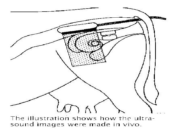
Palpation Chris Ellason Positions of tract The reproductive


 and")


- Slides: 28
Palpation Chris Ellason
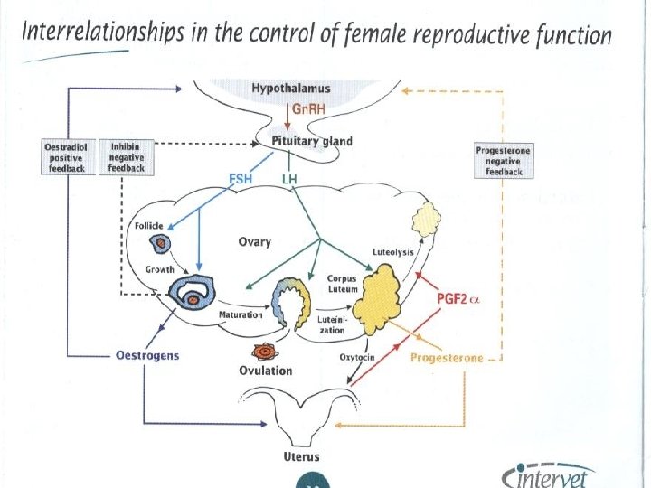
Positions of tract • The reproductive tract lies on the bottom of the pelvis suspended by the broad ligament with the ovaries lying on either side of the tract. • Ovaries may lie underneath in the gravid cow.
Entering the tract • Form an arrow with your fingers (nails clipped short) and enter the anal sphincter. • Fold your fingers back making a fist and enter further avoiding rectal contractions.
Position of other organs • Be careful to avoid the rumen which is on the left of the cow. • In late term pregnancies, the fetus will be down deep. • Rumen will indent and then slowly respond.
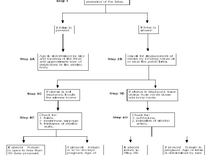
The open tract • On the ventral floor of the uterus. • Turgid to flaccid to the touch depending on stage of estrous cycle. • No fluid present. • Feel both horns.
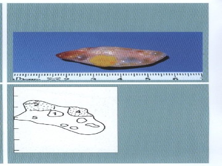
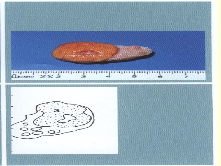
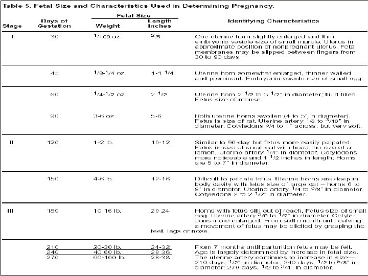
30 to 35 Days • Only experienced palpators with breeding records. • Uterus thin-walled with a small amount of fluid. Slight swell. • Embryo ½” long • Membrane slip. • Marble
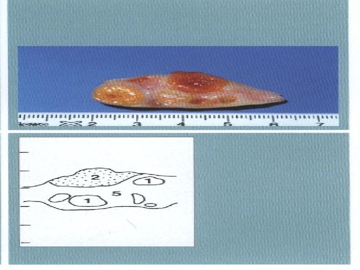
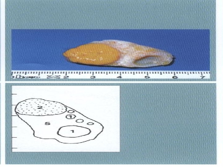
45 Days • • Implanted Uterus thin walled Embryo 1” long Vesicular membrane filled with fluid. • Gently pinch walls of uterus and feel membranes slip through the fingers.
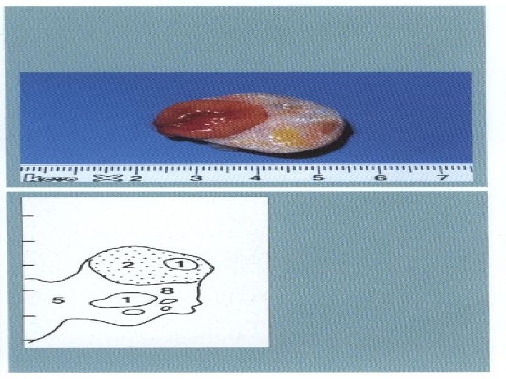
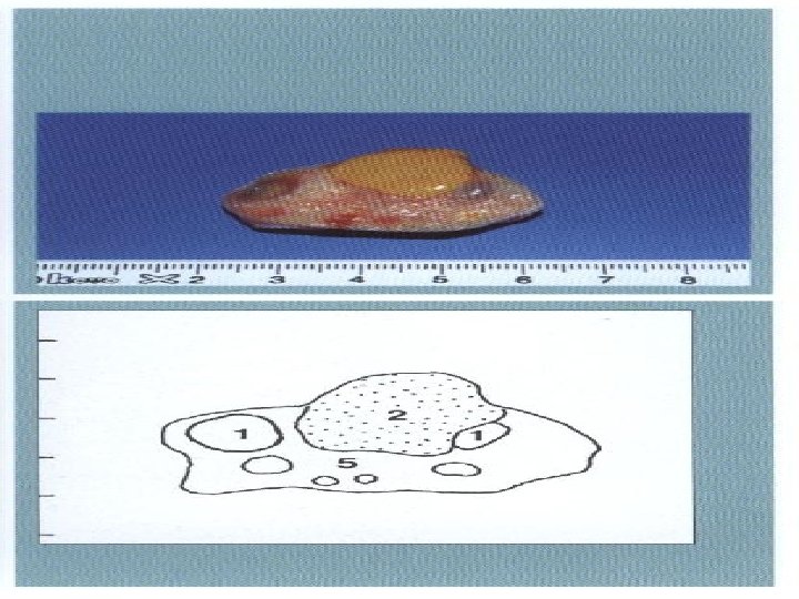
60 Days • Uterus enlarged-2 1/2” in diameter. • Fetus 2 1/2” to 3 1/2” long • Anterior end of pelvis.
90 Days • Fetus-6 ½” long • Uterus displaced • Uterine artery 1/8” to 3/16” diameter with gushing pulse. • Cotyledons-3/4” to 1” diameter. • Femoral artery.
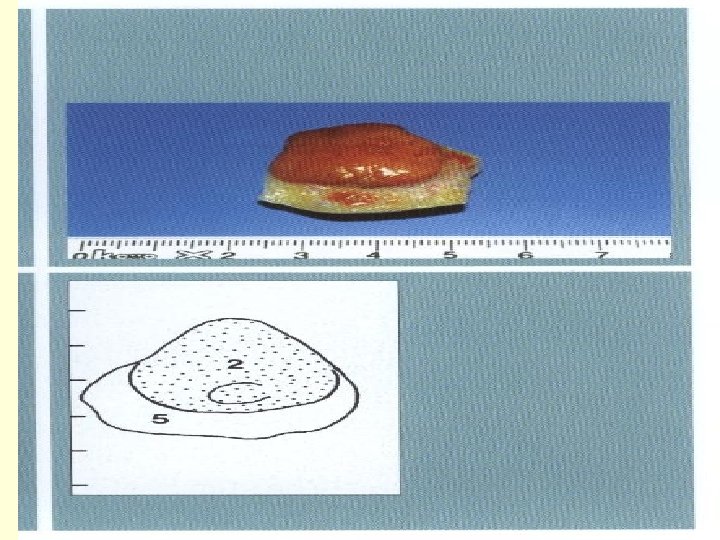
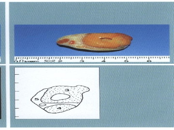
79 day • Placenta with cotyledons on surface. • Fetus appears as a bulge in a fluid filled sac.
120 days • Fetus 10 -12” long • Cotyledons 1 ½” diameter and firm • Mid-uterine artery enlarged and gushing.
5 -6 month • Deep in abdominal cavity. • Bounce the fetus • Mid-uterine artery • Cotyledons • Displaced cervix