Diastasis Recti THE ABDOMINAL MUSCLES The anterior and

Diastasis Recti

THE ABDOMINAL MUSCLES

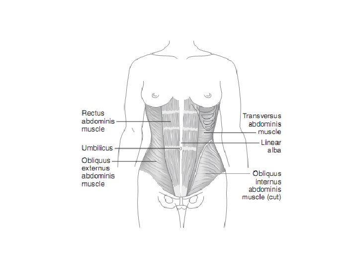
• The anterior and lateral abdominal wall is formed by the abdominal muscles • The deepest of the group is the transversus abdominis muscle, which lies internally to the internal and external oblique muscles • Aponeurosis is formed by these muscles intersection.

• The aponeurosis is reinforced by the two rectus abdominis muscles, which run in sheaths formed in the aponeurosis (Rectus Sheath) on either side of the linea alba. • Of particular relevance is the fact that the sheaths are elastic longitudinally and less so transversely. • Each rectus abdominis muscle has three transverse fibrous intersections, which are firmly attached to the anterior wall of the enclosing sheath. • The lowest intersection is about the level of the umbilicus, and the sheaths are deficient posteriorly in the lowest portion

Innervation of Muscles The oblique and transversus muscles are innervated by the • Lower six thoracic nerves, • Iliohypogastric and ilioinguinal nerves. The recti are innervated by the • lower six thoracic nerves

Abdominal Layers

A-fascial layers 1 - Superficial layer, referred to as the fatty layer, is a single layer with varying amounts of fat. 2 -Deep fascial layer, known as the membranous layer, is more membranous and contains elastic fibers.

B-Anterolateral muscle group of the abdomen consist of four pairs of muscles which include • 1 - External oblique muscle • 2 - Internal oblique muscle • 3 - Transversus abdominis • 4 - -Rectus abdominis

Rectus sheath The rectus abdominis is entirely enclosed in a sheath formed by the aponeuroses of the muscles of the lateral walls of the abdomen.

Functions of the abdominal muscles 1. Protect the abdominal viscera and keep them in their positions 2. Maintain a good erect posture 3. Stabilize pelvis during straight leg raising 4. Help in respiration and defecation 5. Concern with forward flexion of the trunk 6. Help expulsion of the fetus during second stage of labour. 7. Improving the function of the pelvic floor muscles

C- The aponeuroses • which are defined as flat sheets of densely collagen fibers usually consisting of several layers with few elastic fibers

D- The linea alba • which is the result of the fusion of the right and left aponeuroses of the three pairs of the anterolateral abdominal muscles in the midline from sternum to the pubis.

Diastasis Recti

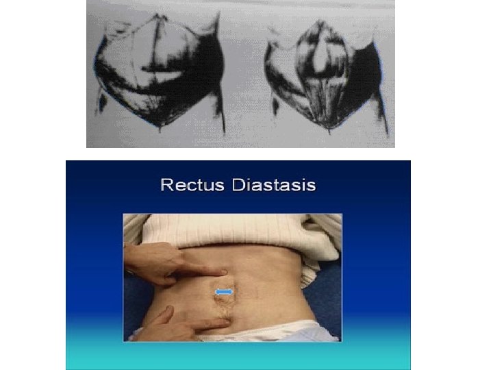
Definition It is the separation of the rectus abdominis muscle which involve • widening of the linea alba with gap greater than the normal distance between the rectus bellies (2 Cm or 2 fingers above umbilicus) • palpated above, below or at the level of the umbilicus

Normal development of diastasis recti • Infants: A diastasis recti looks like a ridge, which runs down the middle of the stomach from the bottom of the sternum to the belly button. It increases with muscle straining and is clearly seen when the child tries to sit up. In infants, the rectus abdominis muscles continue to grow and the diastasis recti gradually disappears. • Pregnancy: Pregnant women may develop the condition because of increased tension on the abdominal wall. The risk is higher if with multiple births or many pregnancies. Women who are 12 or more weeks pregnant should not do exercises that stress the abdomen. This may worsen the condition. Often seen after pregnancy and childbirth but should disappear after a few months. • Adults: Often seen in patients with strenuous jobs and weak trunk muscles. It can become very large and increases with muscle straining and is clearly seen when the patient lifts head to sit up.

• Diastasis recti is a separation between the left and right side of the rectus abdominis muscle, which covers the front surface of the belly area.

Etiology • Diastasis recti may occur in pregnancy as a result of 1. Hormonal effects on the connective tissue 2. Biomechanical changes of pregnancy; • It may also develop during labor, especially with excessive breath-holding during the second stage. • It can occur above, below, or at the level of the umbilicus but appears to be less common below the umbilicus. • It appears to be less common in women with good abdominal tone before pregnancy. • Clinically, a diastasis may be found in women well past their childbearing years and also in men. Routine assessment for this condition is highly recommended and can easily be done in conjunction with abdominal strength testing.

Significance / Effects Diastasis recti may produce 1. Musculoskeletal Problems 2. Activity imitation 3. Decreased fetal Protection 4. Potential for Hernia Musculoskeletal complaints such as low back pain, possibly as a result of decreased ability of the abdominal musculature and thoracolumbar fascia to stabilize the pelvis and lumbar spine.

Activity limitations. • Activity limitations can also occur, such as inability to perform independent supine-tositting transitions because of extreme loss of the mechanical alignment and function of the rectus muscle.

Decreased fetal protection • In severe separations, the remaining midline layers of abdominal wall tissue are skin, fascia, subcutaneous fat, and peritoneum. • The lack of muscular support provides less protection for the fetus.

Potential for herniation • Severe cases of diastasis recti may progress to herniation of the abdominal viscera through the separation at the linea alba. • This degree of separation requires surgical repair.

Examination for Diastasis Recti

Self Testing • Instruct patients to perform a self-test on or after the third postpartum day for optimal accuracy. • Why after third day? Because Until 3 days after delivery, the abdominal musculature has inadequate tone for valid test results.

Patient position and procedure • Hook-lying. Have the patient slowly raise her head and shoulders off the floor, reaching her hands toward the knees, until the spines of the scapulae leave the floor. • Place the fingers of one hand horizontally across the midline of the abdomen at the umbilicus • If a separation exists, 1. the fingers will sink into the gap between the rectus muscles, 2. a visible bulge between the rectus bellies may be appreciated. • The number of fingers that can be placed between the muscle bellies is then documented. Because this condition can occur above, below, or at the level of the umbilicus, test for it at all three areas.
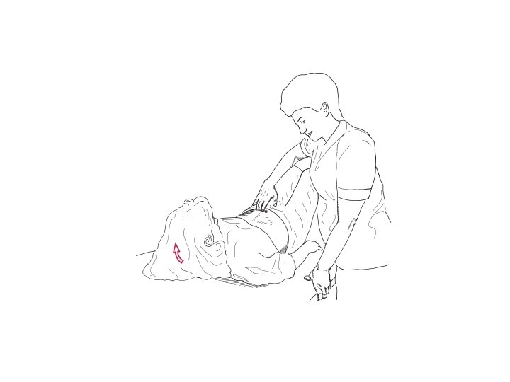

The palpation should assess the following 1 - Width and length of any recti separation 2 - Region of the greatest diastasis 3 - Bulge of the abdomen on recti contraction 4 - The women's ability to activate abdominal musculature (Strength) 5 - The endurance capacity of the abdominal musculature.

Predisposing factors 1 -Child bearing especially when there was succession of pregnancies 2 -Hernias the simplest form of a ventral or epigasteric hernia is the region of the linea alba and represents diastasis recti.

4 - Conditions that results in a persistent excessive increase in intra-abdominal pressure favors the development of a diastasis such as lifting or carrying heavy objects or chronic cough. 5 - Sudden strain or fall may also be the starting point of a diastasis 6 - Decrease in the tone of the tissues of the abdominal wall as a result of general weakness may also predispose to diastasis development

Incidence Diastasis recti are not a condition limited to pregnant or postpartum women, it also can be seen in • Obese males, • In patients with chronic lung disease • In children (whose linea alba is wider than the adult)

Incidence • Second trimester • Childbearing period at full term 66% • Immediate post-partum 50% • Multiple gestation • ↓Incidence in women with Good abdominal tone prior to pregnancy • Above umbilicus 37% - At umbilicus 52%- Below umbilicus 11%

INTERVENTIONS

Corrective Exercises for Diastasis Recti • corrective exercises (head lift or head lift with pelvic tilt) should be used until the separation is corrected to 2 cm (two finger widths) or less

Head Lift Patient position and procedure: • Hook-lying with her hands crossed over midline at the level of the diastasis for support. • Have the woman exhale and lift only her head off the floor. • At the same time, her hands should gently approximate the rectus muscles toward midline • Then have the woman lower head slowly and relax
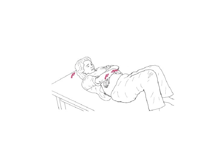

• This exercise emphasizes the rectus abdominis muscle and minimizes the obliques. • Some women may not be able to successfully reach over their abdomens. • In this case, the use of a sheet wrapped around the trunk at the level of the separation can be used to provide support and approximation

Head Lift with Pelvic Tilt • Patient position and procedure: Hook-lying. The arms are crossed over the diastasis for support as before. Have the patient slowly lift only her head off the floor while approximating the rectus muscles and performing a posterior pelvic tilt, then slowly lower head and relax. • All abdominal contractions should be performed with an exhalation so that intra-abdominal pressure is minimized.

Strengthening exercises

• Core contraction - In a seated position, place both hands on abdominal muscles. Take small controlled breaths. Slowly contract the abdominal muscles, pulling them straight back towards the spine. Hold the contraction for 30 seconds, while maintaining the controlled breathing. Complete 10 repetitions. • Seated squeeze - Again in a seated position, place one hand above the belly button, and the other below the belly button. With controlled breaths, with a mid-way starting point, pull the abdominals back toward the spine, hold for 2 seconds and return to the mid-way point. Complete 100 repetitions. • Head lift - In a lying down position, knees bent at 90° angle, feet flat, slowly lift the head, chin toward your chest, (concentrate on isolation of the abdominals to prevent hipflexors from being engaged), slowly contract abdominals toward floor, hold for two seconds, lower head to starting position for 2 seconds. Complete 10 repetitions.

• Upright push-up - A standup pushup against the wall, with feet • • together arms-length away from wall, place hands flat against the wall, contract abdominal muscles toward spine, lean body towards wall, with elbows bent downward close to body, pull abdominal muscles in further, with controlled breathing. Release muscles as you push back to starting position. Complete 20 repetitions. Squat against the wall - Also known as a seated squat, stand with back against the wall, feet out in front of body, slowly lower body to a seated position so knees are bent at a 90° angle, contracting abs toward spine as you raise body back to standing position. Optionally, this exercise can also be done using an exercise ball placed against the wall and your lower back. Complete 20 Repetitions. Squat with squeeze - A variation to the "Squat against the wall" is to place a small resistance ball between the knees, and squeeze the ball as you lower your body to the seated position. Complete 20 repetitions.

Diastasis Recti Tape

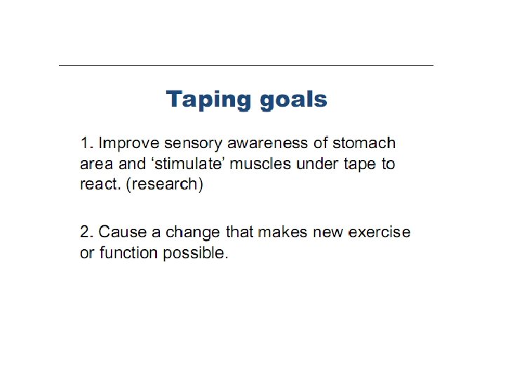
Why use tape? • As a stimulus to assist function and strengthen muscles. • After application we see and feel the muscle activation and the space between the muscle decrease. • The child becomes ‘alert’ and is able to do more with the tape on. • Adults can perform a given exercise with more ease. • The tape assists in sensory awareness.

Ligament technique
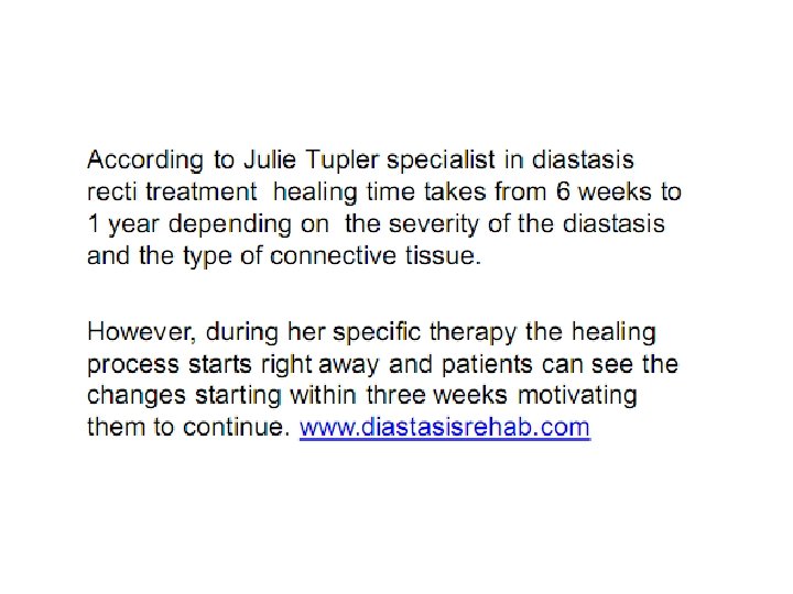

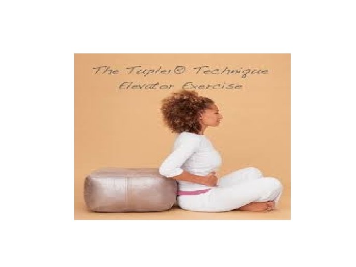
Tupler Techniques

- Slides: 49