Neuron Structure Synapse The Synapse 1 Synthesis of


 2. Storage and transport of NT within")


 • Makes use of radioactive tracer")
 • Uses electromagnets to")
: event-related potentials (momentary shifts in electrical activity of brain")
- Slides: 23
Neuron Structure
Synapse
The Synapse 1. Synthesis of neurotransmitter (NT) 2. Storage and transport of NT within vesicles 3. NT Release 4. Activation of postsynaptic receptors 5. Termination of transmitter effect (e. g. reuptake)
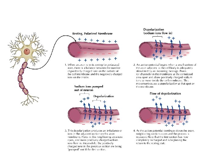
Resting Potential Sodium ions are concentrated on the outside of the axon membrane. Potassium ions are concentrated on the inside of the axon membrane. Ion channels are closed. The inside of the axon membrane is more negative that is the outside.
Action Potential • Action potential occurs when the membrane potential rapidly shifts from -70 to +40 m. V – Ion channels open in the membrane, allowing sodium ions to enter the axon – Sodium entry shifts the membrane potential toward a positive value – Potential is restored when other channels open, allowing potassium ions to exit the axon
Myelin • Myelin is a fatty, waxy substance coating the axon of some neurons. • Functions: – Speeds neurotransmission – Insulates neurons from each other – Makes neurotransmission more efficient
Neurotransmitters • • Serotonin Acetylcholine Dopamine Norepinephrine Epinephrine GABA Endorphins
Midline Brain View
Brainstem • Brainstem is a primitive portion of brain – Pons: involved in respiration, sleep regulation, dreaming – Medulla: involved in life support functions such as respiration and heart rate – Reticular activating system is an arousal system within the brainstem
Subcortical Brain Areas • Corpus callosum: band of axons that interconnects the hemispheres • Thalamus: sensory relay area • Limbic system: involved in emotionality • Hypothalamus: feeding, fleeing, mating, fighting, homeostasis • Cerebellum: involved in motor control
Limbic System: Seat of Motivation, Emotions
Cerebral Cortex • Cortex refers to the outer covering of the brain – Consists of left and right hemispheres – Cortex is divided into lobes • Frontal: Self-awareness, planning, voluntary movement, emotional control, speech, working memory • Parietal: Body sensations • Occipital: Vision • Temporal: Hearing, language comprehension – Localization of function: do discrete circuits carry out different functions?
Cortical Lobes
Cerebral Cortex
Motor and Somatosensory Cortex
Language areas: Broca & Wernicke
Primary Visual Pathways
Dorsal and Ventral Visual Pathways
Split brain: Hemispherical Specialization
Brain imaging technologies • Positron Emission Tomography (PET) • Makes use of radioactive tracer to measure cerebral blood flow.
Brain imaging technologies • Functional Magnetic Resonance Imaging (f. MRI) • Uses electromagnets to measure oxygen levels in brain.
Brain imaging technologies Electroencephalogram (EEG): event-related potentials (momentary shifts in electrical activity of brain associated with external signals) Transcranial magnetic stimulation (TMS): Produces ‘virtual’ lesions