Neural tube defect Encephalocele Meningocele Meylomeningocele Spina bifida


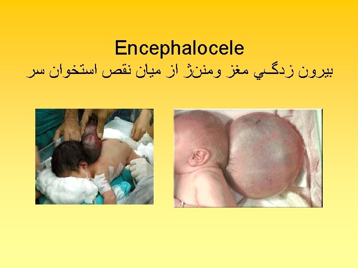
Neural tube defect • Encephalocele • Meningocele • Meylomeningocele
 • 2 -spina bifida cystica")
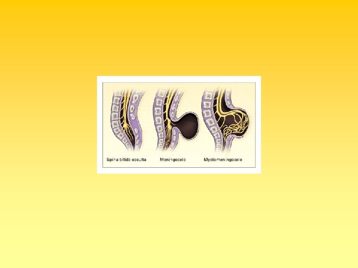
• Spina bifida • 1 - Spina bifida(occulta) • 2 -spina bifida cystica
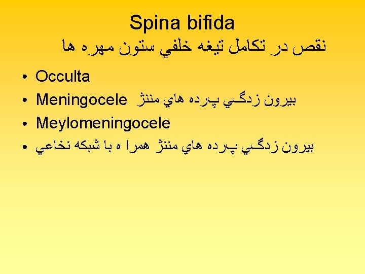

Meningocele

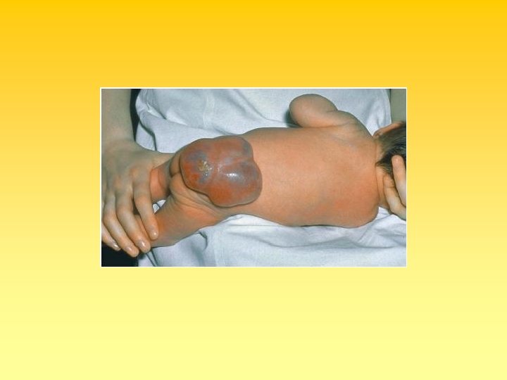
Meylomeningocele


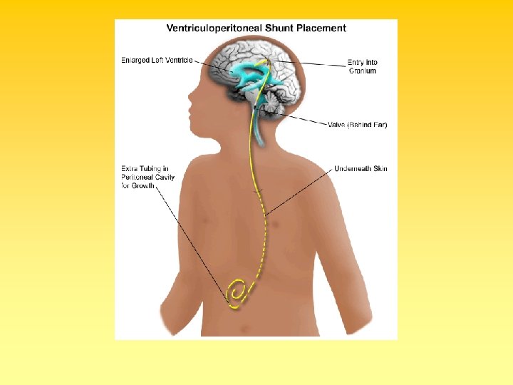
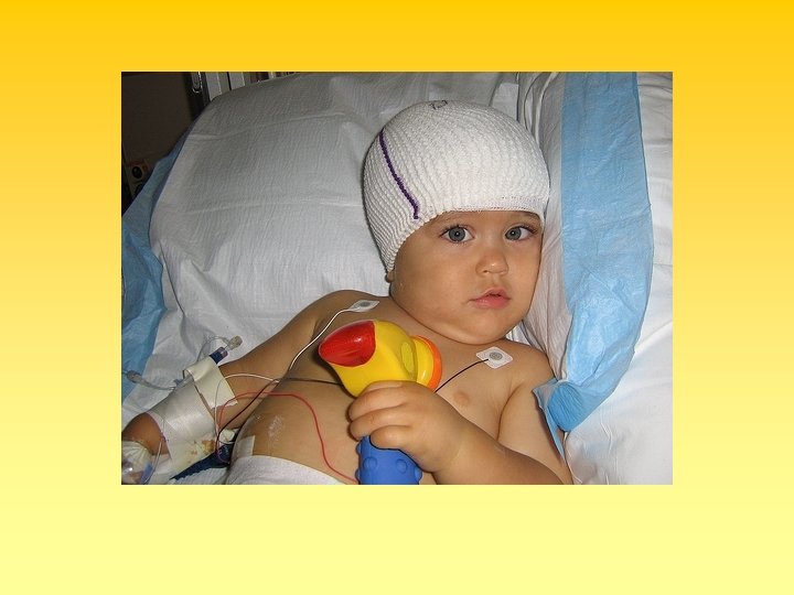
HYDROCEPHALUS


• • • Each shunt has 3 parts: 1 - Ventricular catheter: a small flexible tube which goes in the brain, in one of the cavities where the CSF is being retained. 2 - Reservoir : a small pump which regulates the amount of fluid that goes out. Through this the doctor can also check the working state of the shunt, as well as take CSF samples, when necessary, with a needle.

• 3 - Distal catheter: another flexible tube that will take the fluid to the place where it is going to be absorbed. It is usually left with sufficient length, thinking in the child's growth. . • The shunts regulates the draining pressure. There are different levels of pressure, as high, medium and low. There also some differences in the design but the means is always the same.


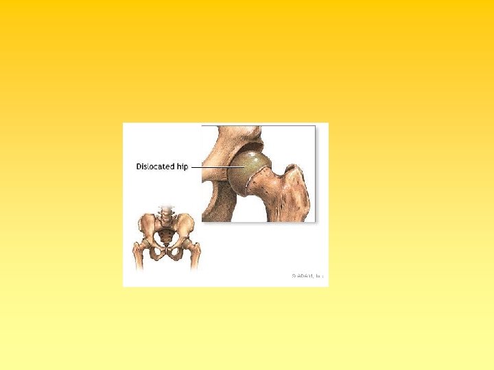
 • Acetabular Dysplasia( preluxation) • Subluxation • Dislocation")
Developmental Dysplasia of the Hip (DDH) • Acetabular Dysplasia( preluxation) • Subluxation • Dislocation

• • Figure 3. A positive Galeazzi sign in a seven-month-old girl with left hip dislocation. Note the apparent femoral shortening


• Figure 4. A three-yearold with a left hip dislocation. Note the limited abduction.

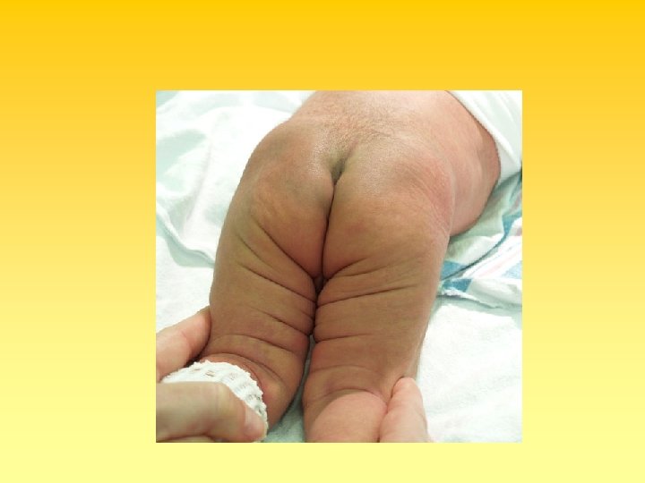
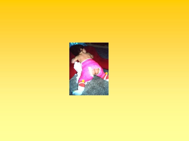
• • Figure 2. A 21 -month-old child with right hip dislocation. Note the asymmetric skinfolds in the upper thigh
 Ortolani maneuver.")
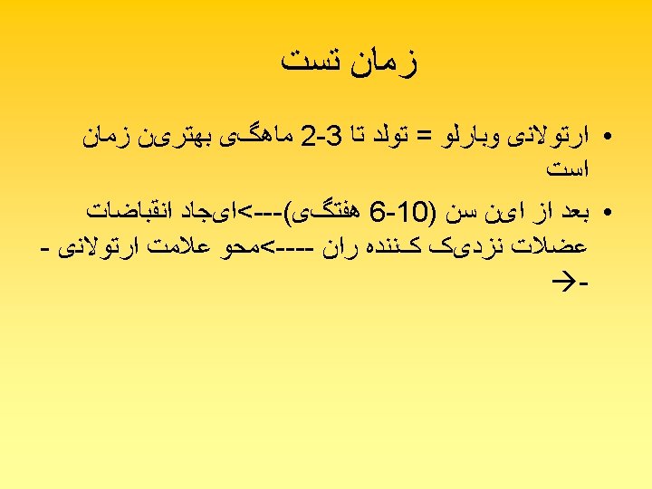
• Figure 1. Tests commonly used to assess hip stability. (A) Ortolani maneuver. A gentle upward force is applied while the hip is abducted. (B) Barlow maneuver. A gentle downward force is applied while the hip is adducted.

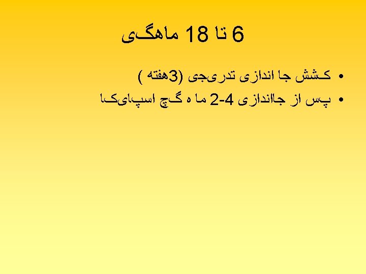


• Figure 7. A fourmonth-old child in a hip spica cast following bilateral closed reductions and adductor tenotomies

• Figure 6. A newborn with bilateral hip dislocations in a Pavlik harness. The harness prevents hip extension and adduction but allows flexion and abduction


- Slides: 48